
Lycenchelys maculata Toyoshima, 1985
|
publication ID |
https://doi.org/ 10.11646/zootaxa.4762.1.1 |
|
publication LSID |
lsid:zoobank.org:pub:BEBD8F0D-1347-4A44-86D4-2915433D2E7B |
|
DOI |
https://doi.org/10.5281/zenodo.3809741 |
|
persistent identifier |
https://treatment.plazi.org/id/006C5E1A-FF83-FF8A-3EC6-B41EFCD8A899 |
|
treatment provided by |
Plazi |
|
scientific name |
Lycenchelys maculata Toyoshima, 1985 |
| status |
|
Lycenchelys maculata Toyoshima, 1985 View in CoL
(Japanese name: Kurobuchi-hebigenge)
( Figs. 14–16 View FIGURE 14 View FIGURE 15 View FIGURE 16 ; Table 4)
Lycenchelys View in CoL sp.: Toyoshima, 1984: 293, pl. 274-D (brief description).
Lycenchelys maculatus Toyoshima, 1985: 149 View in CoL , figs. 6–9, 31, tables 1–2 (original description, type locality: off Onahama, Pacific coast of Fukushima Prefecture, Honshu Island, Japan); Hatooka, 1993: 904, unnumbered fig. (key to species); Imamura, 1998: 31, fig. 10 (brief description); Shinohara et al., 2009: 724 (species list).
Lycenchelys maculata Anderson, 1994: 117 View in CoL (species list); Hatooka, 2000: 1035, unnumbered fig. (key to species); Hatooka, 2002: 1035, unnumbered fig. (key to species); Anderson & Fedorov, 2004: 17 (species list); Shinohara & Anderson, 2007: 64 (key to species); Balushkin et al., 2011: 980 (catalog of specimens); Hatooka, 2013: 1229, unnumbered fig. (key to species); Nakabo & Hirashima, 2015: 217 (species list and etymology of scientific name).
Materials examined.
Holotype: HUMZ 71361 , male, 281.1 mm SL, off Onahama, Fukushima Prefecture, Tohoku District, northwestern Pacific , 200–300 m depth, 10 Nov. 1977 .
Paratypes (7 specimens, 264.4–287.5 mm SL, collection locality and depth same as holotype): HUMZ 71217, 1 male, 276.3 mm SL, 8 Nov. 1977; HUMZ 71362–64, 2 males and 1 female, 264.4–276.2 mm SL, 10 Nov. 1977; HUMZ 71391–92, 1 male and 1 female, 269.4–273.4 mm SL, 11 Nov. 1977; HUMZ 71537, 1 male, 282.9 mm SL, 13 Nov. 1977.
Diagnosis. Vertebrae 28–30 + 110–112 = 137–142; head length 11.7–13.7% SL; interorbital pore 1; occipital pores 3; postorbital pores 4; suborbital pores 7 + 1–2; preoperculomandibular pores 8; vomerine teeth 1–6; palatine teeth 4–9, arranged in single row; opercular flap well developed; pelvic-fin base positioned anterior to lower edge of gill opening; lateral line incomplete and positioned ventrally; scales present on pectoral fin and absent on its base; body grayish when fresh; blackish irregular blotches on most portions of dorsal fin and dorsal part of body.
Description. Counts and proportional measurements in Table 4.
Body very elongate, cross section oval anteriorly, compressed laterally near tail; its width at anal-fin origin 3.3–4.3 (3.3)% SL. Head short, ovoid; dorsal profile of head sloping extremely gently from above posterior edge of eye to above last postorbital pore. Snout moderately short, 127.7–179.3 (179.3)% of eye diameter. Eye ovoid, relatively small. Interorbital space moderately narrow, width 21.9–43.0 (43.0)% of eye diameter. Nostril tube short, not reaching upper lip when depressed. Mouth subterminal. Posterior edge of upper jaw reaching vertical through posterior part of eye in males (including holotype), reaching to below middle or anterior part of eye in females. Labial lobe of lower jaw moderately developed. Teeth on jaws, vomer and palatine small and conical; upper jaw with 2 rows anteriorly and single row posteriorly; lower jaw with 2 irregular rows anteriorly and single row posteriorly; vomerine teeth irregularly arranged; palatine teeth in single row. Lower edge of gill opening slightly above lower end of pectoral-fin base. Opercular flap well developed. Gill rakers short and triangular. Pseudobranch filaments long. Lateral line deciduous, incomplete and positioned ventrally; originating posterior to last postorbital pore and terminating above about middle of anal fin. Scales small and cycloid, present on body, nape, pectoral-fin rays basally, tail and about basal half of vertical fins. Head and pectoral-fin base without scales.
Dorsal-fin origin above pectoral-fin base; 1st dorsal-fin pterygiophore between neural spines of 2nd to 4th (between 2nd and 3rd) vertebrae. Anal-fin origin below 23rd to 25th (25th) dorsal-fin ray; 1st anal-fin pterygiophore posterior to parapophysis of ultimate to antepenultimate (penultimate) abdominal vertebra. Last dorsal-fin pterygiophore between neural spines of 2nd to 4th (between 3rd and 4th) preural vertebrae. Last anal-fin pterygiophore between hemal spines of 2nd to 4th (between 3rd and 4th) preural vertebrae. Caudal fin with 2 epural, 5 upper hypural and 5 lower hypural rays. Pectoral fin moderately short; its posterior margin reaching about middle of abdomen, and having notches. Upper end of pectoral-fin base about on lateral midline of body. Pelvic fin short; its base anterior to lower edge of gill opening; its posterior margin just reaching to below pectoral-fin base.
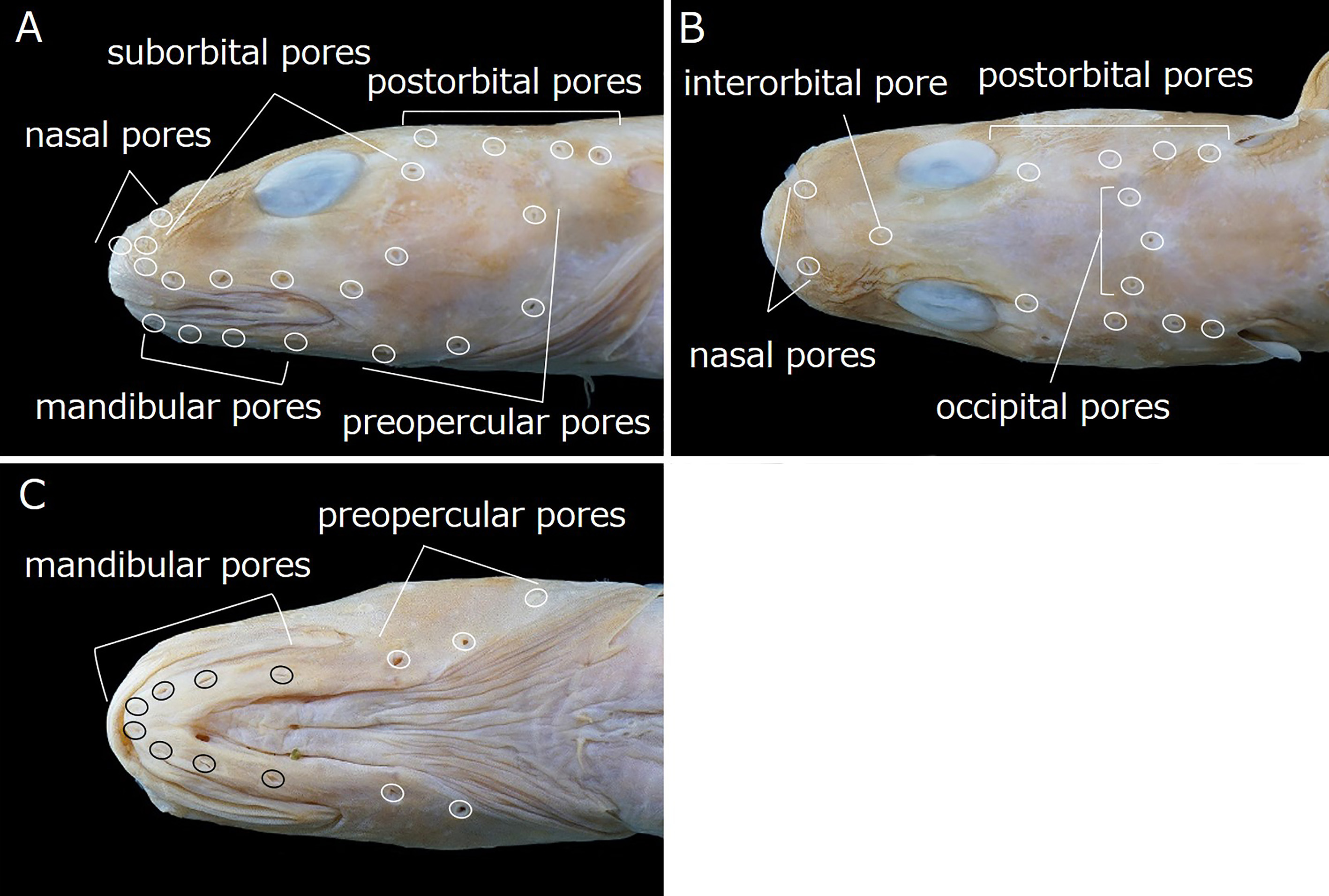
Head pores small and distinct. Nasal pores 2; anterior pore in front of nostril tube, posterior pore above and slightly anterior to vertical through 1st suborbital pore ( Fig. 15A, B View FIGURE 15 ). Postorbital pores 4; distance between 1st and 2nd pores longest of those between adjacent pores ( Fig. 15A, B View FIGURE 15 ). Suborbital pores 8–9 (9); 7 pores located below eye and 1 or 2 (2) on ascending part of suborbital canal behind eye; 4th pore below anterior margin of eye; 7th pore located posterior to posterior margin of eye ( Fig. 15A View FIGURE 15 ). Preoperculomandibular pores 8, 4 on lower jaw and 4 on preopercle; last preoperculomandibular pore located posterior to about lower margin of eye ( Fig. 15A, C View FIGURE 15 ). One interorbital pore located on dorsal midline anterior to eyes ( Fig. 15B View FIGURE 15 ). Occipital pores 3; 1 on dorsal midline of occiput, remaining 2 on either side; middle pore located slightly posterior to more lateral pores; all pores located anterior to 3rd postorbital pore ( Fig. 15B View FIGURE 15 ).
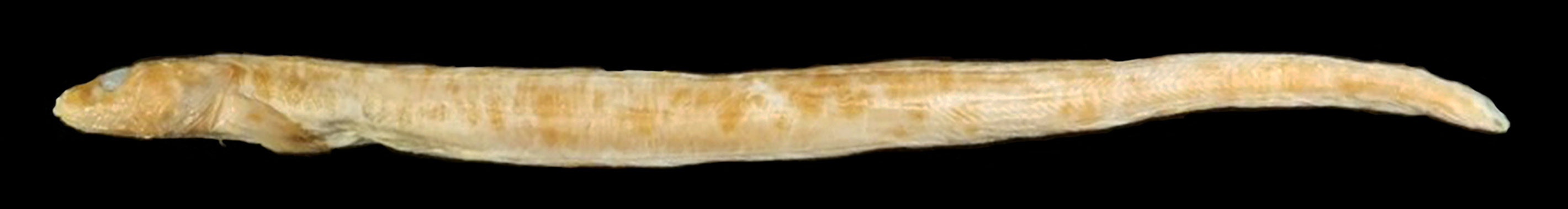
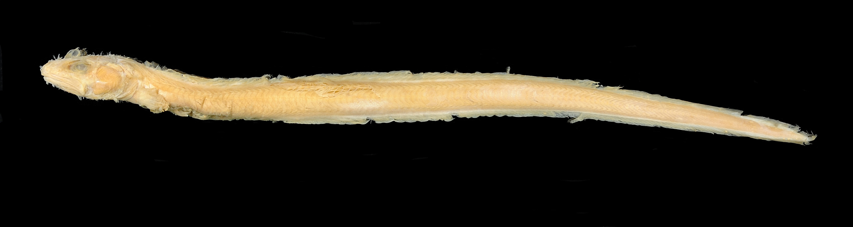
Color in alcohol. Holotype ( Fig. 16 View FIGURE 16 ) with pale gray head, body, pectoral-fin base, anterior part of pectoral fin and vertical fins, slightly darker posterior part of pectoral fin; grayish irregular blotches on most portions of dorsal fin and dorsal part of body. Paratypes more uniformly brownish than holotype.
Color when fresh [based on color photograph in Toyoshima (1984)]. Head, body, pectoral-fin base, anterior part of pectoral fin and vertical fins grayish, posterior part of pectoral fin and abdomen darker. Blackish irregular blotches on most portions of dorsal fin and dorsal part of body.
Distribution. Off northwestern Pacific coast of the Kuril Islands, and from Fukushima to Ibaraki prefectures, at depths of 200–380 m ( Toyoshima, 1983, 1984, 1985; Hatooka, 1993, 2000, 2002, 2013; Anderson, 1994; Imamura, 1997, 1998; Koyanagi, 1997; Anderson & Fedorov, 2004; Shinohara et al., 2009; Balushkin et al., 2011; this study).
Size. Maximum length 30 cm TL ( Hatooka, 1993, 2000, 2002, 2013). The largest specimen examined during this study was 287.5 mm SL (292.8 mm TL), and is probably the same specimen previously measured for maximum length.
Remarks. Lycenchelys maculata resembles Lycenchelys kolthoffi Jensen, 1904 in having more than 100 dorsal- and anal-fin rays, and total vertebrae, pelvic fins and irregular blotches on most portions of the dorsal fin to dorsal part of the body blackish (vs. lacking this combination of characters in other species of Lycenchelys ) (e.g., Jensen, 1904; Andriashev, 1955; Toyoshima, 1985; Koyanagi, 1995; this study). Lycenchelys maculata can be easily distinguished from L. kolthoffi by the numbers of postorbital pores (4 vs. 1), interorbital pore (1 vs. 0) and occipital pores (3 vs. 0), respectively ( Table 4) ( Koyanagi, 1995; this study). Furthermore, Lycenchelys maculata is also separable from L. kolthoffi in having higher numbers of dorsal-fin rays (134–138 vs. 114–117), anal-fin rays (115–120 vs. 102–104) and total vertebrae (137–142 vs. 116–124), respectively ( Table 4) ( Jensen, 1904; Andriashev, 1955; Toyoshima, 1985; Koyanagi, 1995; this study).
Some previous studies described the lateral line of L. maculata as only reaching to near the posterior end of the pectoral fin ( Toyoshima, 1985; Hatooka, 1993, 2000, 2002, 2013), but specimens examined for this study for the lateral line reaching to about the middle of the anal fin.
No known copyright restrictions apply. See Agosti, D., Egloff, W., 2009. Taxonomic information exchange and copyright: the Plazi approach. BMC Research Notes 2009, 2:53 for further explanation.
|
Kingdom |
|
|
Phylum |
|
|
ParvPhylum |
Osteichthyes |
|
Class |
|
|
Order |
|
|
Family |
|
|
Genus |
|
Kingdom |
|
|
Phylum |
|
|
ParvPhylum |
Osteichthyes |
|
Class |
|
|
Order |
|
|
Family |
|
|
Genus |
|
Kingdom |
|
|
Phylum |
|
|
ParvPhylum |
Osteichthyes |
|
Class |
|
|
Order |
|
|
Family |
|
|
Genus |
|
Kingdom |
|
|
Phylum |
|
|
ParvPhylum |
Osteichthyes |
|
Class |
|
|
Order |
|
|
Family |
|
|
Genus |
|
Kingdom |
|
|
Phylum |
|
|
ParvPhylum |
Osteichthyes |
|
Class |
|
|
Order |
|
|
Family |
|
|
Genus |
|
Kingdom |
|
|
Phylum |
|
|
ParvPhylum |
Osteichthyes |
|
Class |
|
|
Order |
|
|
Family |
|
|
Genus |
|
Kingdom |
|
|
Phylum |
|
|
ParvPhylum |
Osteichthyes |
|
Class |
|
|
Order |
|
|
Family |
|
|
Genus |
|
Kingdom |
|
|
Phylum |
|
|
ParvPhylum |
Osteichthyes |
|
Class |
|
|
Order |
|
|
Family |
|
|
Genus |
|
Kingdom |
|
|
Phylum |
|
|
ParvPhylum |
Osteichthyes |
|
Class |
|
|
Order |
|
|
Family |
|
|
Genus |
|
Kingdom |
|
|
Phylum |
|
|
ParvPhylum |
Osteichthyes |
|
Class |
|
|
Order |
|
|
Family |
|
|
Genus |
|
Kingdom |
|
|
Phylum |
|
|
ParvPhylum |
Osteichthyes |
|
Class |
|
|
Order |
|
|
Family |
|
|
Genus |
|
Kingdom |
|
|
Phylum |
|
|
ParvPhylum |
Osteichthyes |
|
Class |
|
|
Order |
|
|
Family |
|
|
Genus |
|
Kingdom |
|
|
Phylum |
|
|
ParvPhylum |
Osteichthyes |
|
Class |
|
|
Order |
|
|
Family |
|
|
Genus |
|
Kingdom |
|
|
Phylum |
|
|
ParvPhylum |
Osteichthyes |
|
Class |
|
|
Order |
|
|
Family |
|
|
Genus |
Lycenchelys maculata Toyoshima, 1985
| Kawarada, Shumpei, Imamura, Hisashi, Narimatsu, Yoji & Shinohara, Gento 2020 |
Lycenchelys maculata
| Nakabo, T. & Hirashima, Y. 2015: 217 |
| Hatooka, K. 2013: 1229 |
| Balushkin, A. V. & Sheiko, B. A. & Fedorov, V. V. 2011: 980 |
| Shinohara, G. & Anderson, M. E. 2007: 64 |
| Anderson, M. E. & Fedorov, V. V. 2004: 17 |
| Hatooka, K. 2002: 1035 |
| Hatooka, K. 2000: 1035 |
| Anderson, M. E. 1994: 117 |
Lycenchelys maculatus
| Shinohara, G. & Narimatsu, Y. & Hattori, T. & Ito, M. & Takata, Y. & Matsuura, K. 2009: 724 |
| Imamura, H. 1998: 31 |
| Hatooka, K. 1993: 904 |
| Toyoshima, M. 1985: 149 |
Lycenchelys
| Toyoshima, M. 1984: 293 |