
Lycenchelys makushok Fedorov & Andriashev, 1993
|
publication ID |
https://doi.org/ 10.11646/zootaxa.4762.1.1 |
|
publication LSID |
lsid:zoobank.org:pub:BEBD8F0D-1347-4A44-86D4-2915433D2E7B |
|
DOI |
https://doi.org/10.5281/zenodo.3809743 |
|
persistent identifier |
https://treatment.plazi.org/id/006C5E1A-FF87-FF86-3EC6-B3B4FCC5A99A |
|
treatment provided by |
Plazi |
|
scientific name |
Lycenchelys makushok Fedorov & Andriashev, 1993 |
| status |
|
Lycenchelys makushok Fedorov & Andriashev, 1993 View in CoL
(Japanese name: Wakataka-hebigenge)
( Figs. 17–20 View FIGURE 17 View FIGURE 18 View FIGURE 19 View FIGURE 20 ; Table 5)
Lycenchelys makushok Fedorov & Andriashev, 1993: 130 View in CoL , figs. 1–2 (original description, type locality: off Iturup, Pacific cost of Kuril Islands); Anderson, 1994: 117 (species list); Shinohara et al., 1996: 180, fig. 2A (description); Imamura, 1997: 60 (species list); Imamura, 1998: 32, fig. 11 (brief description); Hatooka, 2000: 1032, 1590, unnumbered fig. (key to species); Hatooka, 2002: 1032, 1581, unnumbered fig. (key to species); Anderson & Fedorov, 2004: 18 (species list); Shinohara & Anderson, 2007: 63 (key to species); Kitagawa et al., 2008: 95, unnumbered fig. (brief description); Shinohara et al., 2009: 723 (species list); Amaoka et al., 2011: 316, unnumbered fig. (brief description); Balushkin et al., 2011: 981, 1024 (catalog of specimens); Hatooka, 2013: 1226, 2078, unnumbered fig. (key to species); Nakabo & Hirashima, 2015: 217 (species list and etymology of scientific name).
Materials examined
Holotype: ZIN 42290 , 137.1 mm SL, off Iturup, Kuril Islands, northwestern Pacific (44°39.2’N, 149°02.2’E), 800 m depth, 10 Sep. 1968, R/V Vityaz, trawl GoogleMaps .
Other specimens (40 specimens, 111.2–168.7 mm SL): HUMZ 152378, 163847, 180633, 182293, 182295, 182383–89, 182395, 182478, 182480–82, 182484–85, 182487–88, 182519, 182636–44, 182646–47, 15 males and 19 females, 111.2–147.2 mm SL, Tohoku District, northwestern Pacific; HUMZ 192427 (4), 196342, 226078, 5 males and 1 females, 133.7–168.7 mm SL, eastern Hokkaido Island, northwestern Pacific.
Diagnosis. Vertebrae 24–26 + 107–113 = 132–139; head 10.0–12.9% SL; interorbital pore 1; occipital pore usually 1; postorbital pores 4; suborbital pores usually 7 + 1; preoperculomandibular pores usually 10; vomerine teeth 4–13; palatine teeth 5–12, arranged in single row; opercular flap well developed; pelvic-fin base positioned anterior to lower edge of gill opening; lateral line incomplete and positioned ventrally; scales present or absent on pectoral fin and absent on its base; body uniformly grayish brown when fresh.
Description. Counts and proportional measurements in Table 5.
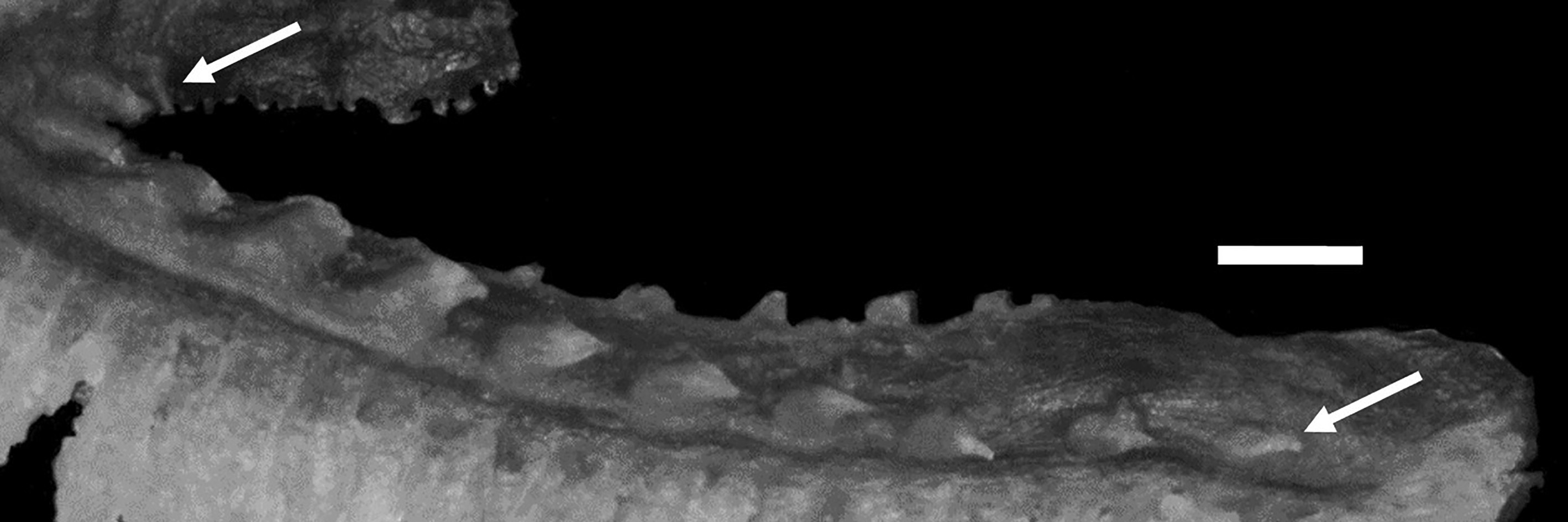
Body strongly elongate, cross section oval anteriorly, compressed laterally near tail; its width at anal-fin origin 2.1–3.6% SL (unknown for holotype). Head very short, ovoid, dorsal profile of head gently sloping from above posterior edge of eye to about above last postorbital pore. No sexual dimorphism in head recognized. Snout short, 103.3–162.8 (152.2)% of eye diameter. Eye ovoid, relatively small. Interorbital space narrow, width 10.0–43.5 (43.5)% of eye diameter. Nostril tube short, quite not reaching upper lip when depressed. Mouth subterminal. Posterior edge of upper jaw reaching about to about vertical through anterior margin of pupil in adult males, not reaching vertical through anterior margin of pupil in females and juveniles. Labial lobe of lower jaw weak. Teeth on jaws sharp, anterior teeth large and posterior teeth small; upper jaw with single row, sometimes having additional small teeth behind anteriormost tooth (unknown for holotype); lower jaw with 2–3 irregular rows anteriorly and single row posteriorly; vomerine teeth large and conical, arranged irregularly; palatine teeth smaller than vomerine teeth, arranged in single row. Lower edge of gill opening slightly above lower end of pectoral-fin base. Opercular flap well developed. Gill rakers short and triangular ( Fig. 18 View FIGURE 18 ). Pseudobranch filaments short. Lateral line deciduous, incomplete and positioned ventrally; originating posterior to 5th postorbital pore terminating its end area above about middle of anal fin. Scales small and cycloid, present on body and tail. Scales covering basal portions of dorsal and anal fins anteriorly; extent of scaled areas gradually increasing posteriorly, except at margins. Scales present or absent on pectoral axilla and basal portions of lower pectoral-fin rays (unknown for holotype). Head, nape and pectoral-fin base without scales.
Dorsal-fin origin above middle of pectoral fin; 1st dorsal-fin pterygiophore between neural spines of 3rd to 5th (between 3rd and 4th) vertebrae. Anal-fin origin below 20th to 22nd (20th) dorsal-fin ray; 1st anal-fin pterygiophore posterior to parapophysis of ultimate or penultimate (penultimate) abdominal vertebra. Last dorsal-fin pterygiophore between neural spines of 2nd to 5th (between 3rd and 4th) preural vertebrae. Last anal-fin pterygiophore between hemal spines of 2nd to 5th (between 3rd and 4th) preural vertebrae. Caudal fin with 2 epurals, 4 upper hypurals and 3–5 (5) lower hypural rays. Pectoral fin moderately short, not quite reaching middle of abdomen; its posterior margin having notches. Upper end of pectoral-fin base about on lateral midline of body. Pelvic fin short; its base anterior to lower edge of gill opening; its posterior margin not reaching vertical through pectoral-fin base.
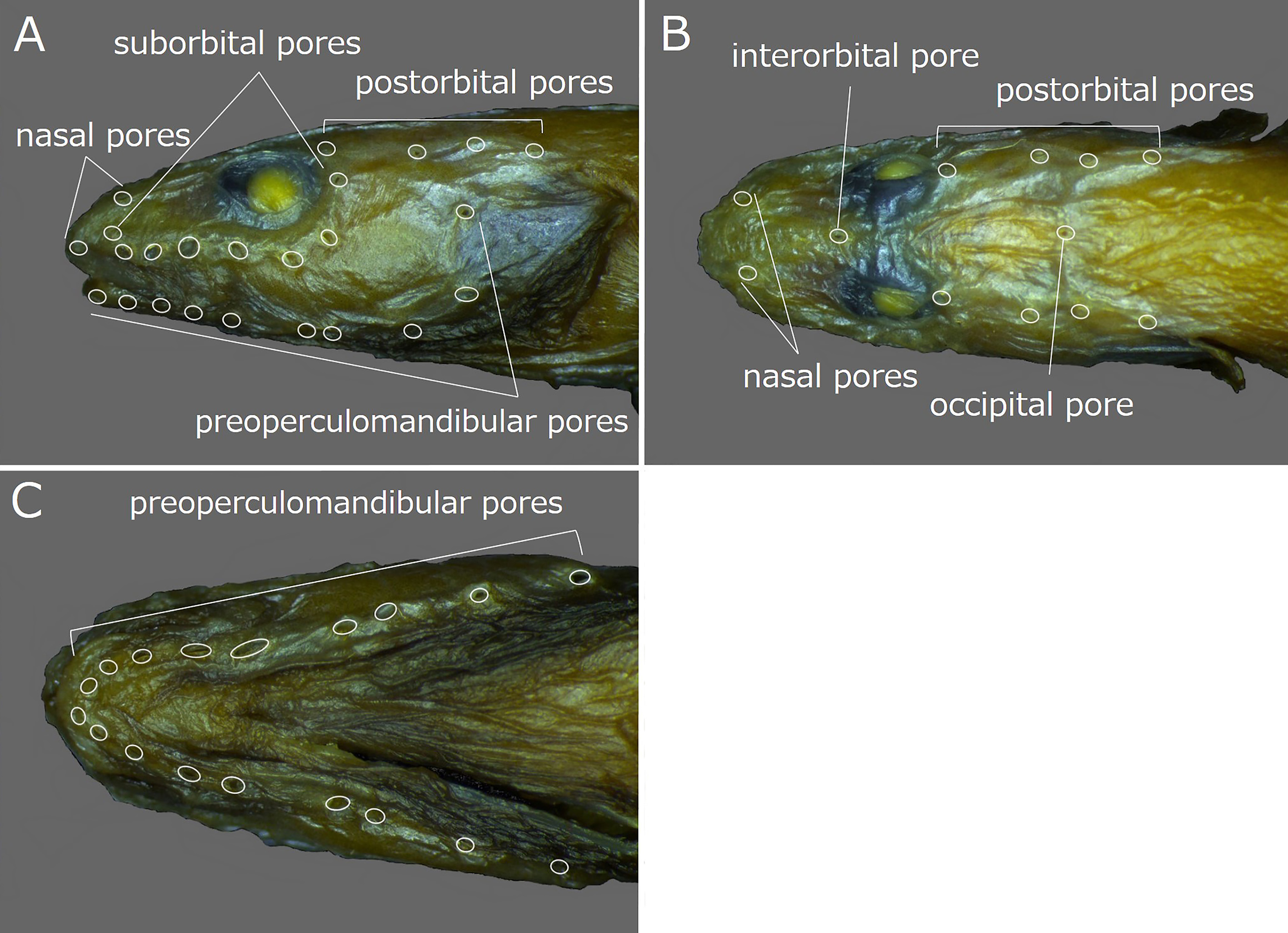
Head pores well developed and distinct. Nasal pores 2; anterior pore in front of nostril tube, posterior pore above 1st suborbital pore ( Fig. 20A, B View FIGURE 20 ). Postorbital pores 4; distance between 1st and 2nd pores longest of those between adjacent pores ( Fig. 20A, B View FIGURE 20 ). Suborbital pores usually 8 (8), rarely 9; 7 pores located below eye and last pore posterior to eye; when 9 pores, 8 pores below eye and 9th behind eye on left side in HUMZ 182639 and on right side in HUMZ 182293; 5th below anterior margin of pupil; last pore of those below eye located posterior to vertical posterior to margin of eye ( Fig. 20A View FIGURE 20 ). Preoperculomandibular pores usually 10 (10), rarely 9; 5 on lower jaw, 2 at junction of lower jaw and preopercle, and 3 on preopercle; 8th and 9th pores united into 1 pore on left side of HUMZ 182479 and counted as 9; last preoperculomandibular pore located posterior to lower margin of eye ( Fig. 20A, C View FIGURE 20 ). One interorbital pore located on dorsal midline anterior to anterior margin of pupils ( Fig. 20B View FIGURE 20 ). Usually 1 occipital pore located on dorsal midline at middle of occiput (1); 1 additional pore present on left side of middle pore in HUMZ 182640; 2 additional pores, one on either side of middle pore in HUMZ 182642; occipital pore(s) located anterior to 3rd postorbital pore ( Fig. 20B View FIGURE 20 ).
Color in alcohol. Holotype (based on color photograph; Fig. 19 View FIGURE 19 ) with uniformly light brown head, body and vertical fins, slightly paler pectoral fin; dark brown opercular region; purplish gray abdomen. Margin of vertical fins in non-type specimens dark brown, but coloration otherwise similar to holotype.
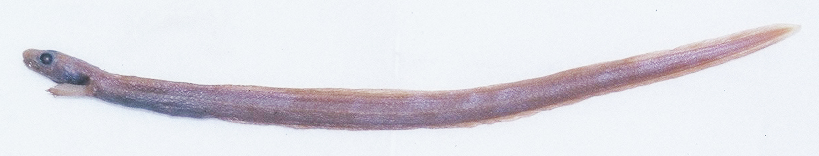
Color when fresh (based on color photograph of HUMZ 152378; Fig. 17 View FIGURE 17 ). Head and margin of vertical fins dark brown; body and vertical fins uniformly grayish brown; pectoral fins gray; abdomen dark purplish gray.
Distribution. Off northwestern Pacific coast of the Kuril Islands, eastern Hokkaido Island and Honshu Island from Miyagi to Ibaraki prefectures, at depths of 399–1219 m ( Fedorov & Andriashev, 1993; Anderson, 1994; Shinohara et al., 1996; Imamura, 1997, 1998; Hatooka, 2000, 2002, 2013; Anderson & Fedorov, 2004; Shinohara & Anderson, 2007; Kitagawa et al., 2008; Shinohara et al., 2009; Amaoka et al., 2011; Balushkin et al., 2011; this study).
Size. Maximum length 180 mm TL ( Amaoka et al., 2011; Hatooka, 2013). The largest specimen examined for this study measured 168.7 mm SL (172.2 mm TL).
Remarks. Lycenchelys makushok resembles L. hippopotamus , L. melanostomias and L. rassi in having more than 100 total vertebrae, 1 interorbital pore, 1–3 occipital pores, 4 postorbital pores, a single ventral lateral line and no distinct spots or blotches on the body (vs. lacking this combination of this characters in other species of Lycenchelys ) (e.g., Toyoshima, 1983, 1985; Fedorov & Andriashev, 1993; Hatooka, 1993, 2000, 2002, 2013; Anderson, 1995; Shinohara et al., 1996; Imamura et al., 2004; Shinohara & Anderson, 2007; this study). See Remarks for L. hippopotamus for a detailed comparison of L. makushok and L. hippopotamus . Lycenchelys makushok is separable from L. melanostomias and L. rassi by the number and position of occipital pores. Lycenchelys makushok always has an occipital pore on the dorsal midline at the middle of the occiput. When 2 or 3 occipital pores are present (2 of 41 specimens observed in this study), the additional pore or two are on the lateral side or sides of the middle pore. In contrast, L. melanostomias and L. rassi usually have 2 occipital pores (1 occipital pore on right side was examined in only 1 specimen of L. melanostomias in this study), that are located on the left and right sides of the midline of the occiput. Lycenchelys makushok is further separable from L. melanostomias in having a higher number of total vertebrae (132–139, vs. 117–124, respectively), and from L. rassi in having a shorter head (head length 10.0–12.9% SL, vs. 13.4–16.4% SL, respectively).

No known copyright restrictions apply. See Agosti, D., Egloff, W., 2009. Taxonomic information exchange and copyright: the Plazi approach. BMC Research Notes 2009, 2:53 for further explanation.
|
Kingdom |
|
|
Phylum |
|
|
ParvPhylum |
Osteichthyes |
|
Class |
|
|
Order |
|
|
Family |
|
|
Genus |
|
Kingdom |
|
|
Phylum |
|
|
ParvPhylum |
Osteichthyes |
|
Class |
|
|
Order |
|
|
Family |
|
|
Genus |
|
Kingdom |
|
|
Phylum |
|
|
ParvPhylum |
Osteichthyes |
|
Class |
|
|
Order |
|
|
Family |
|
|
Genus |
|
Kingdom |
|
|
Phylum |
|
|
ParvPhylum |
Osteichthyes |
|
Class |
|
|
Order |
|
|
Family |
|
|
Genus |
|
Kingdom |
|
|
Phylum |
|
|
ParvPhylum |
Osteichthyes |
|
Class |
|
|
Order |
|
|
Family |
|
|
Genus |
|
Kingdom |
|
|
Phylum |
|
|
ParvPhylum |
Osteichthyes |
|
Class |
|
|
Order |
|
|
Family |
|
|
Genus |
|
Kingdom |
|
|
Phylum |
|
|
ParvPhylum |
Osteichthyes |
|
Class |
|
|
Order |
|
|
Family |
|
|
Genus |
|
Kingdom |
|
|
Phylum |
|
|
ParvPhylum |
Osteichthyes |
|
Class |
|
|
Order |
|
|
Family |
|
|
Genus |
Lycenchelys makushok Fedorov & Andriashev, 1993
| Kawarada, Shumpei, Imamura, Hisashi, Narimatsu, Yoji & Shinohara, Gento 2020 |
Lycenchelys makushok
| Nakabo, T. & Hirashima, Y. 2015: 217 |
| Hatooka, K. 2013: 1226 |
| Amaoka, K. & Nakaya, K. & Yabe, M. 2011: 316 |
| Balushkin, A. V. & Sheiko, B. A. & Fedorov, V. V. 2011: 981 |
| Shinohara, G. & Narimatsu, Y. & Hattori, T. & Ito, M. & Takata, Y. & Matsuura, K. 2009: 723 |
| Kitagawa, D. & Imamura, H. & Goto, T. & Ishito, Y. & Fujiwara, K. & Ueda, Y. 2008: 95 |
| Shinohara, G. & Anderson, M. E. 2007: 63 |
| Anderson, M. E. & Fedorov, V. V. 2004: 18 |
| Hatooka, K. 2002: 1032 |
| Hatooka, K. 2000: 1032 |
| Imamura, H. 1998: 32 |
| Imamura, H. 1997: 60 |
| Shinohara, G. & Endo, H. & Matsuura, K. 1996: 180 |
| Anderson, M. E. 1994: 117 |
| Fedorov, V. V. & Andriashev, A. P. 1993: 130 |