
Lycenchelys rassi Andriashev, 1955
|
publication ID |
https://doi.org/ 10.11646/zootaxa.4762.1.1 |
|
publication LSID |
lsid:zoobank.org:pub:BEBD8F0D-1347-4A44-86D4-2915433D2E7B |
|
DOI |
https://doi.org/10.5281/zenodo.3809747 |
|
persistent identifier |
https://treatment.plazi.org/id/006C5E1A-FF8E-FFB9-3EC6-B284FCACAAE5 |
|
treatment provided by |
Plazi |
|
scientific name |
Lycenchelys rassi Andriashev, 1955 |
| status |
|
Lycenchelys rassi Andriashev, 1955 View in CoL
(Japanese name: Rasu-hebigenge)
( Figs. 26–31 View FIGURE 26 View FIGURE 27 View FIGURE 28 View FIGURE 29 View FIGURE 30 View FIGURE 31 ; Table 7)
Lycenchelys rassi Andriashev, 1955: 359 View in CoL , figs. 2, 5, 6 (original description, type locality: east coast of Sakhalin Island, Sea of Okhotsk); Andriashev, 1958:172 (description); Peden, 1973: 115, fig. 1, table 1 (description); Toyoshima, 1983: 269, 332, pl. 155 (description); Toyoshima, 1984: 293, pl. 274-B (brief description); Toyoshma, 1985: 149, 173, figs. 6–7, 28, 31, tables 1, 4 (description); Hatooka, 1993: 902, unnumbered fig. (key to species); Anderson, 1994: 113, 117 (species list); Amaoka et al., 1995: 240, pl. 403 (brief description); Anderson, 1995: 98, fig. 15 (description); Koyanagi, 1997: 538, fig. 4 (brief description); Hatooka, 2000: 1033, unnumbered fig. (keys to species); Mecklenburg et al., 2002: 703, unnumbered figs. (brief description); Hatooka, 2002: 1033, unnumbered fig. (keys to species); Anderson & Fedorov, 2004: 19 (species list); Shinohara & Anderson, 2007: 64 (key to species); Amaoka et al., 2011: 316, unnumbered fig. (brief description); Balushkin et al., 2011: 981, 1024 (catalog of specimens); Hatooka, 2013: 1226, unnumbered fig. (key to species); Nakabo & Hirashima, 2015: 217 (species list and etymology of scientific name).
Materials examined
Holotype: ZIN 32962 , female, 190.6 mm SL, off eastern Sakhalin Island , Okhotsk Sea (54°28’N, 145°21.6’E), 1500 m depth, R/V Vityaz. GoogleMaps
Other specimens (57 specimens): HUMZ 77747, 119939, 120328, 120330, 120347 –49, 121156, 121451, 121458 –59, 121464, 126117–22, 126185, 126187–93, 126195–96, 126198, 126200–05, 126211, 126252, 126257, 126367–75, 126377–86, 28 males and 29 females, 93.5–239.7 mm SL, northeastern Hokkaido Island, Okhotsk Sea.
Diagnosis. Vertebrae 23–25 + 98–109 = 122–134; head length 13.3–16.4% SL; interorbital pore 1; occipital pores 2; postorbital pores usually 4; suborbital pores usually 7 + 1; preoperculomandibular pores usually 8; vomerine teeth 3–11; palatine teeth 3–10, usually arranged in single row (sometimes 1–2 rows); opercular flap absent; pelvic-fin base positioned below lower edge of gill opening; lateral line complete and positioned ventrally; scales present or absent on pectoral fin and absent on its base; body uniformly grayish brown when fresh.
Description. Counts and proportional measurements in Table 7.
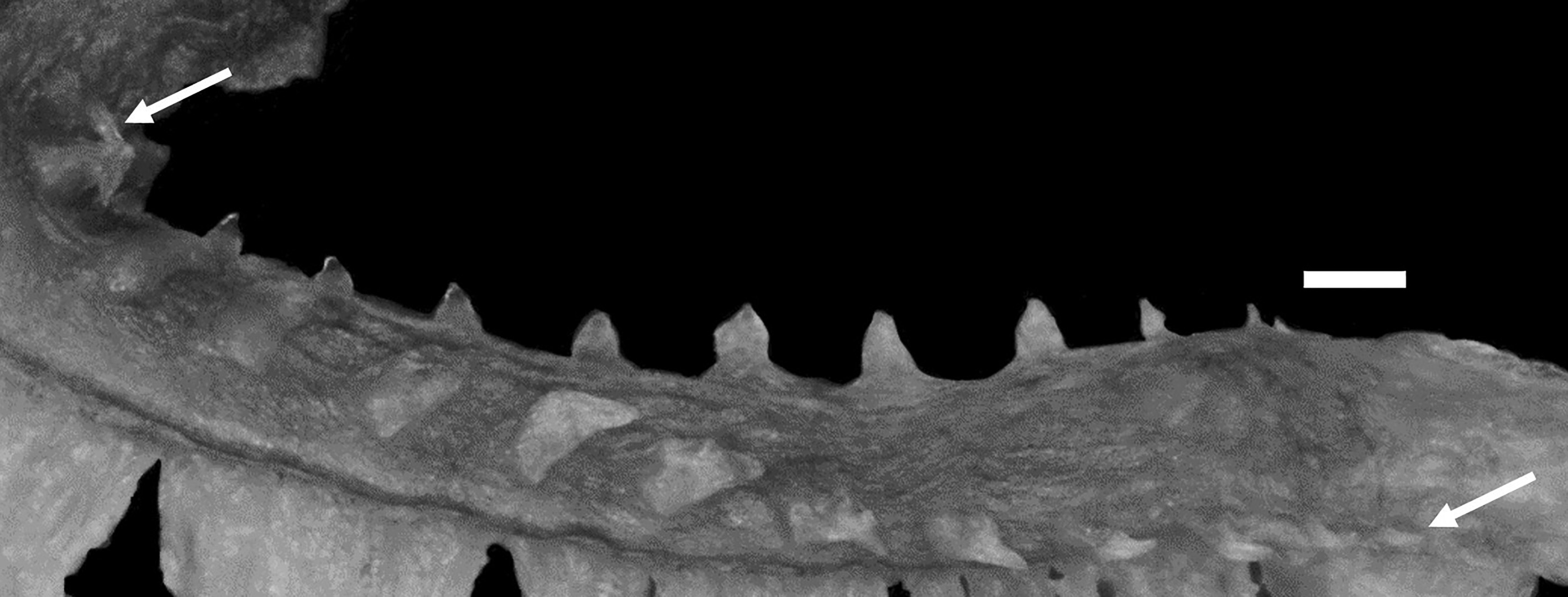
Body very elongate, cross section oval anteriorly, compressed laterally near tail; its width at anal-fin origin 2.5–4.8% SL (unknown for holotype). Head moderately long, ovoid; dorsal profile of head sloping extremely gently from posterior edge of eye to above about last postorbital pore. Cheek swollen in some males. Head in adults slightly longer in males than females. Snout short, 90.0–176.1 (141.3)% of eye diameter. Eye ovoid, moderately large. Interorbital space narrow, width 17.9–36.4 (19.0)% of eye diameter. Nostril tube long, reaching upper lip when depressed. Mouth subterminal. Posterior edge of upper jaw reaching vertical through middle to posterior part of eye in adult males, reaching vertical through anterior margin to middle of eye in females and juveniles (middle of eye). Labial lobe of lower jaw developed. Teeth on jaws sharp; upper jaw usually with 2 rows, rarely 3 rows (unknown for holotype) anteriorly, single row posteriorly; anteriormost teeth larger than other teeth; lower jaw with 2–5 irregular rows anteriorly, 1 or 1–2 rows posteriorly (unknown for holotype); vomerine and palatine teeth small and conical; vomerine teeth irregularly arranged; palatine teeth usually in single row, sometimes 1–2 rows (no data for holotype). Lower edge of gill opening slightly above lower end of pectoral-fin base. Opercular flap absent ( Fig. 31A, B View FIGURE 31 ). Gill rakers short; those on upper limb triangular, many triangular and some blunt on lower limb ( Fig. 27 View FIGURE 27 ). Pseudobranch filaments short. Lateral line deciduous, complete and positioned ventrally; originating posterior to last postorbital pore and terminating on tail. Scales small and cycloid, present on body and tail, except head, nape, pectoral-fin base and area around pelvic fin. Scales covering basal portions of dorsal and anal fins anteriorly; extent of scaled areas gradually increasing posteriorly, except at margins. Scales present or absent on pectoral axilla and basal portions of lower pectoral-fin rays (absent).
Dorsal-fin origin above middle of pectoral fin; 1st dorsal-fin pterygiophore between neural spines of 4th to 6th (between 5th and 6th) vertebrae. Anal-fin origin below 17th to 20th (19th) dorsal-fin ray; 1st anal-fin pterygiophore posterior to parapophysis of ultimate or penultimate (penultimate) abdominal vertebra. Last dorsal-fin pterygiophore between neural spines of 3rd to 5th (between 3rd and 4th) preural vertebrae. Last anal-fin pterygiophore between hemal spines of 2nd to 5th (between 2nd and 3rd) preural vertebrae. Caudal fin with 1–2 (2) epural, 4–5 (4) upper hypural and 3–4 (3) lower hypural rays. Pectoral fin moderately short, reaching to about middle of abdomen; its posterior margin notched. Upper end of pectoral-fin base about on lateral midline of body. Pelvic fin short; its base at about lower edge of gill opening; its posterior margin not quite reaching pectoral-fin base.
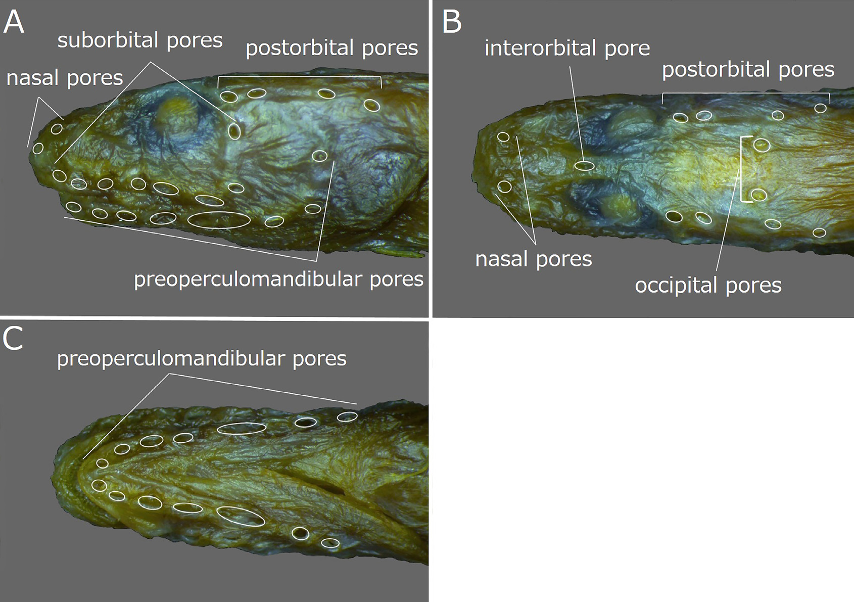
Head pores well developed and distinct. Nasal pores 2; anterior pore in front of nostril tube, posterior pore above 1st suborbital pore ( Fig. 29A, B View FIGURE 29 ). Postorbital pores usually 4 (4), rarely 3; when 4, distance between 2nd and 3rd pores longest of those between adjacent pores; when 3, 2nd pore absent ( Fig. 29A, B View FIGURE 29 ). Suborbital pores usually 8 (8), rarely 7 or 9; when 8, 7 pores below eye and last behind eye; when 9, 8 pores below eye and remaining pore behind eye, or 7 pores below eye and last 2 pores on ascending part of suborbital canal behind eye; 6th and 7th pores united into 1 pore on right side of HUMZ 126384 and counted as 7; 4th pore below vertical through anterior margin of eye; last pore of those below eye posterior to posterior margin of eye ( Fig. 29A View FIGURE 29 ). Preoperculomandibular pores usually 8 (8), rarely 7 or 9; 4 on lower jaw, 1 at junction of lower jaw and preopercle, and 3 on preopercle; pore at junction of lower jaw and preopercle separated into 2 pores in some specimens and counted as 9; 1st and 2nd pores united into 1 pore on right side of HUMZ 126382, 126378 and 126211, 3rd and 4th pores united into 1 pore on left side of HUMZ 126205, and 4th and 5th pores united into 1 pore on left side of HUMZ 126382 and counted as 7; last preoperculomandibular pore posterior to lower margin of eye ( Fig. 29A, C View FIGURE 29 ). One interorbital pore on dorsal midline anterior to middle of eyes ( Fig. 29B View FIGURE 29 ). Occipital pores 2, positioned on either side of dorsal midline; occipital pores located anterior to 3rd postorbital pore ( Fig. 29B View FIGURE 29 ).
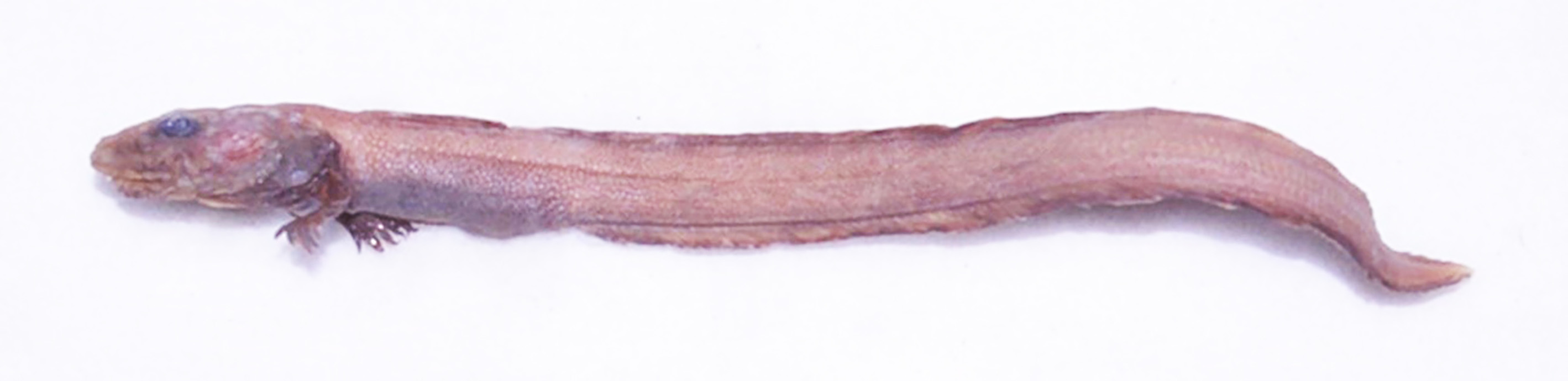
Color in alcohol. Holotype (based on color photograph; Fig. 28 View FIGURE 28 ) with uniformly brown head, body and vertical fins, slightly darker pectoral fin and margin of vertical fins, dark brown opercular region and purplish gray abdomen. Most other specimens similar to holotype, and some uniformly paler than holotype.
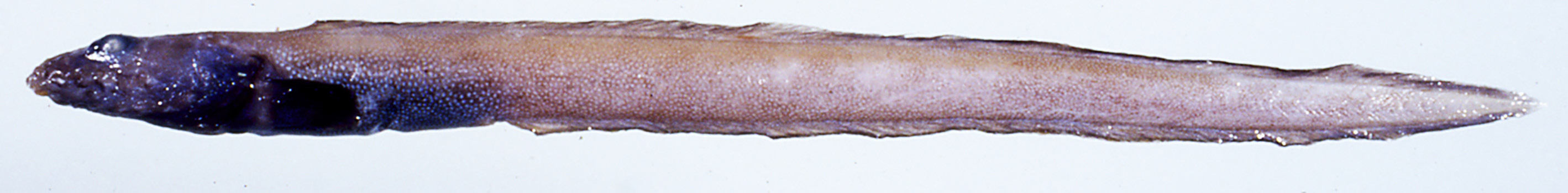
Color when fresh (based on color photograph of HUMZ 119939; Fig. 26 View FIGURE 26 ). Head purplish brown; body and vertical fins uniformly grayish brown; margin of vertical fins dark brown; opercular region and pectoral fin blackish; abdomen purplish.
Distribution. Okhotsk Sea to the eastern Bering Sea, at depths of 895–1805 m ( Andriashev, 1955, 1958; Peden, 1973; Toyoshima, 1983, 1984, 1985; Anderson, 1994, 1995; Amaoka et al., 1995, 2011; Koyanagi, 1997; Hatooka, 2000, 2002, 2013; Mecklenburg et al., 2002; Anderson & Fedorov, 2004; Shinohara & Anderson, 2007; Balushkin et al., 2011; this study).
Size. The largest specimen examined during this study measured 239.7 mm SL (242.9 mm TL), slightly exceeding the previously recorded maximum length of 24 cm TL ( Amaoka, 2011; Hatooka, 2013).
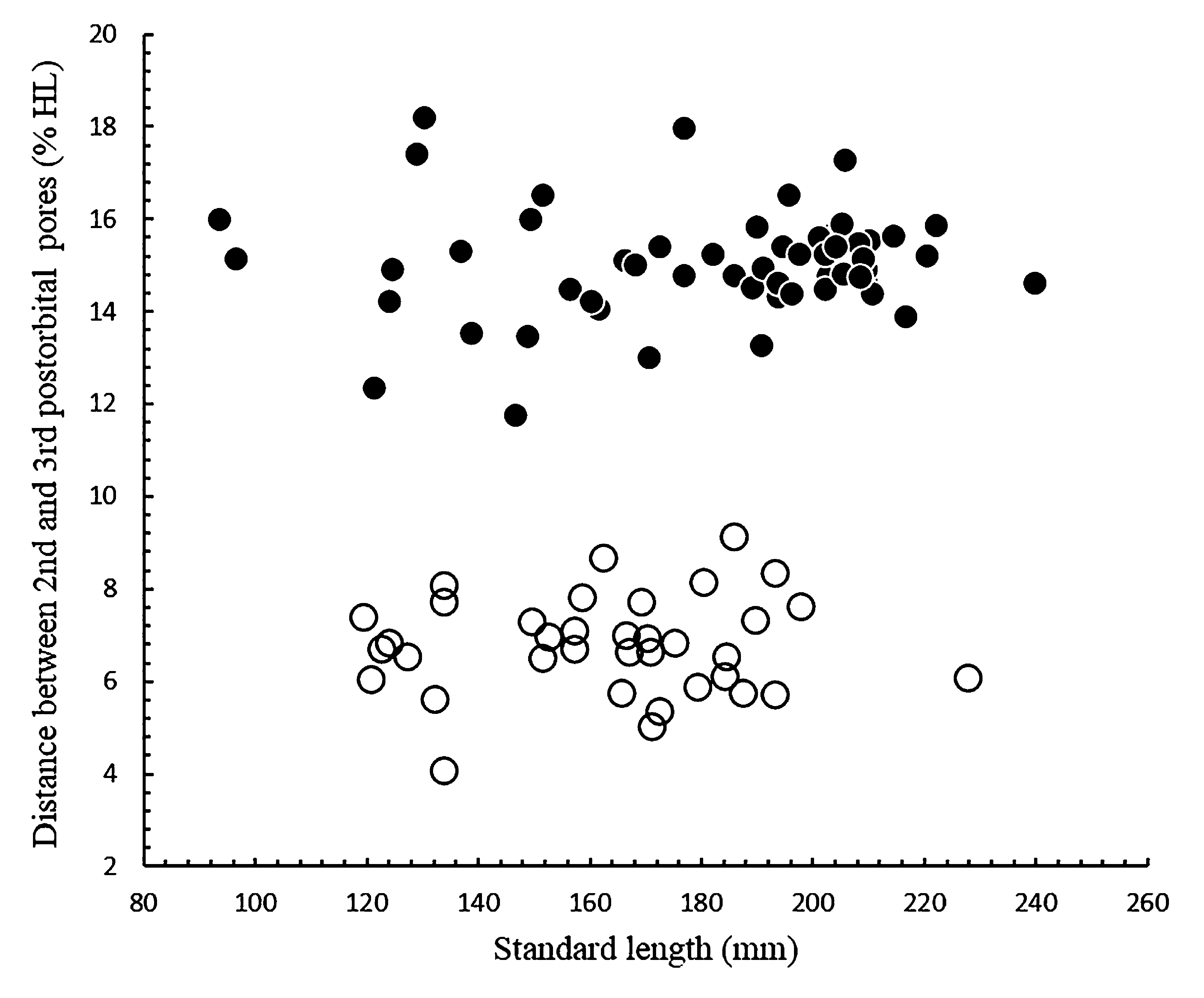
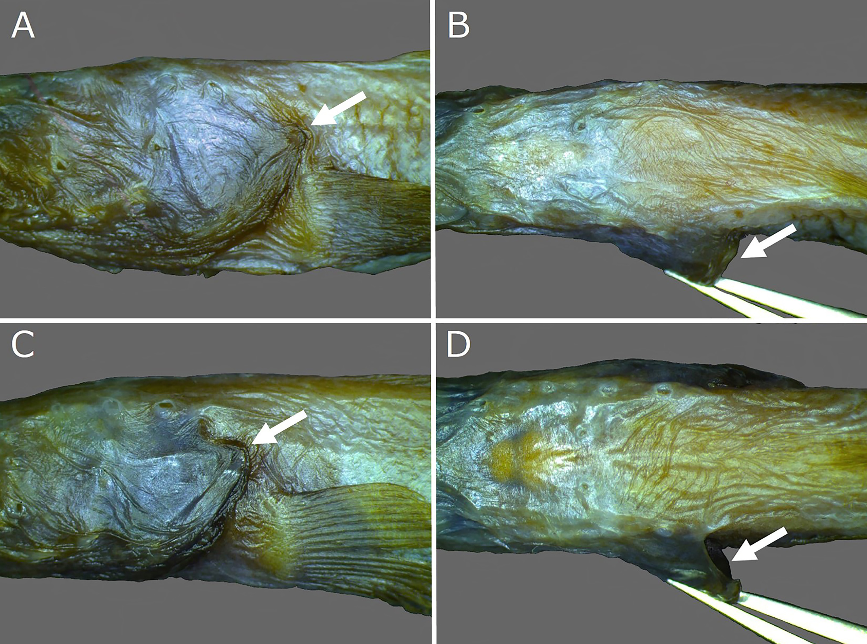
Remarks. Lycenchelys rassi resembles L. hippopotamus , L. makushok and L. melanostomias in having more than 100 total vertebrae, 1 interorbital pore, 2 occipital pores, 3–4 postorbital pores, a single ventrally positioned lateral line and no distinct spots or blotches on the body (vs. lacking this combination of characters in other species of Lycenchelys ) (e.g., Andriashev, 1955; Toyoshima, 1983, 1985; Fedorov & Andriashev, 1993; Hatooka, 1993, 2000, 2002, 2013; Anderson, 1995; this study). See Remarks under accounts for L. hippopotamus and L. makushok for detailed comparisons of L. melanostomias with those two species. Although L. rassi has been previously compared with L. melanostomias ( Toyoshima, 1983, 1985; Hatooka, 1993, 2000, 2002, 2013; Anderson, 1995; Shinohara & Anderson, 2007), this study found most characters considered to be useful for separating them as not valid for doing so. For example, Anderson (1995) redescribed L. rassi based on 6 specimens and claimed that L. rassi is readily separable from L. melanostomias by the following 5 characters: 3–4 postorbital, 7 + 1 or 8 + 1 suborbital and 8 preoperculomandibular pores (vs. 5, 7 + 2 and 9 pores in L. melanostomias ), dorsal-fin origin associated with 5th vertebra (vs. 2nd), and stomach pale (vs. black). However, this study found the interspecific variation in the 5 characters mentioned above to be 3–4 postorbital, 7–8 + 1–2 suborbital and 7–9 preoperculomandibular pores, and 1st dorsal-fin pterygiophore located between neural spines of 4th to 6th vertebrae in L. rassi vs. 4–5, 6–7 + 2–3 and 8–9 pores, and 2nd to 5th vertebrae respctively in L. melanostomias , and the stomach in some alcohol preserved specimens of L. melanostomias is pale. See also Imamura et al. (2004) for color of stomach in the holotype of L. melanostomias . In addition, the number of total vertebrae and the length of the head, which were described as being different between L. rassi and L. melanostomias in recently published papers ( Shinohara & Anderson, 2007; Hatooka, 2013), are also insufficient to clearly separate them (total vertebrae 122–134 vs. 117–124 and head length 13.3–16.4% SL vs. 11.6–15.0% SL, respectively) ( Anderson, 1995; Imamura et al., 2004, 2005; this study). Therefore, these two species cannot always be separated using previously recognized diagnostic characters. This study distinguishes L. rassi from L. melanostomias by the presence or absence of the opercular flap. All specimens of L. rassi observed in this study lack the opercular flap ( Fig. 31A, B View FIGURE 31 ), while it is present in all specimens of L. melanostomias examined ( Fig. 31C, D View FIGURE 31 ). The absence of the opercular flap is rare in species of Lycenchelys , and Andriashev (1955) and Toyoshima (1985) started that the absence of the opercular flap is one of the characters differing between L. rassi and its congeners [vs. L. hippopotamus in Andriashev (1955) and vs. L. brevimaxillaris in Toyoshima (1985) ]. Until now, the condition of the opercular flap has not been compared between L. rassi and L. melanostomias . This study concludes that the absence of the opercular flap is very valuable for separating L. rassi from L. melanostomias because it is easy to observe and there is no intraspecific variation in both species. Lycenchelys rassi is further distinguished from L. melanostomias by their different arrangements of the postorbital pores. In specimens having 4 postorbital pores, the typical condition in both species, the distance between the 2nd and 3rd pores is much greater in L. rassi (11.8–18.2% HL) than in L. melanostomias (4.1–9.2% HL) ( Fig. 30 View FIGURE 30 ). This difference is due to the positions of the 2nd pores; the 2nd pore emanates from the sphenotic and 3rd pore from the pterotic in L. rassi , while both pores emanates from the pterotic in L. melanostomias .
No known copyright restrictions apply. See Agosti, D., Egloff, W., 2009. Taxonomic information exchange and copyright: the Plazi approach. BMC Research Notes 2009, 2:53 for further explanation.
|
Kingdom |
|
|
Phylum |
|
|
ParvPhylum |
Osteichthyes |
|
Class |
|
|
Order |
|
|
Family |
|
|
Genus |
|
Kingdom |
|
|
Phylum |
|
|
ParvPhylum |
Osteichthyes |
|
Class |
|
|
Order |
|
|
Family |
|
|
Genus |
|
Kingdom |
|
|
Phylum |
|
|
ParvPhylum |
Osteichthyes |
|
Class |
|
|
Order |
|
|
Family |
|
|
Genus |
|
Kingdom |
|
|
Phylum |
|
|
ParvPhylum |
Osteichthyes |
|
Class |
|
|
Order |
|
|
Family |
|
|
Genus |
|
Kingdom |
|
|
Phylum |
|
|
ParvPhylum |
Osteichthyes |
|
Class |
|
|
Order |
|
|
Family |
|
|
Genus |
|
Kingdom |
|
|
Phylum |
|
|
ParvPhylum |
Osteichthyes |
|
Class |
|
|
Order |
|
|
Family |
|
|
Genus |
|
Kingdom |
|
|
Phylum |
|
|
ParvPhylum |
Osteichthyes |
|
Class |
|
|
Order |
|
|
Family |
|
|
Genus |
Lycenchelys rassi Andriashev, 1955
| Kawarada, Shumpei, Imamura, Hisashi, Narimatsu, Yoji & Shinohara, Gento 2020 |
Lycenchelys rassi
| Nakabo, T. & Hirashima, Y. 2015: 217 |
| Hatooka, K. 2013: 1226 |
| Amaoka, K. & Nakaya, K. & Yabe, M. 2011: 316 |
| Balushkin, A. V. & Sheiko, B. A. & Fedorov, V. V. 2011: 981 |
| Shinohara, G. & Anderson, M. E. 2007: 64 |
| Anderson, M. E. & Fedorov, V. V. 2004: 19 |
| Mecklenburg, G. W. & Mecklenburg, T. A. & Thorsteinson, L. K. 2002: 703 |
| Hatooka, K. 2002: 1033 |
| Hatooka, K. 2000: 1033 |
| Koyanagi, M. 1997: 538 |
| Amaoka, K. & Nakaya, K. & Yabe, M. 1995: 240 |
| Anderson, M. E. 1995: 98 |
| Anderson, M. E. 1994: 113 |
| Hatooka, K. 1993: 902 |
| Toyoshima, M. 1984: 293 |
| Toyoshima, M. 1983: 269 |
| Peden, A. E. 1973: 115 |
| Andriashev, A. P. 1955: 359 |