
Lycenchelys remissaria Fedorov, 1995
|
publication ID |
https://doi.org/ 10.11646/zootaxa.4762.1.1 |
|
publication LSID |
lsid:zoobank.org:pub:BEBD8F0D-1347-4A44-86D4-2915433D2E7B |
|
DOI |
https://doi.org/10.5281/zenodo.3809759 |
|
persistent identifier |
https://treatment.plazi.org/id/006C5E1A-FFB4-FFB5-3EC6-B060FEBFAB55 |
|
treatment provided by |
Plazi |
|
scientific name |
Lycenchelys remissaria Fedorov, 1995 |
| status |
|
Lycenchelys remissaria Fedorov, 1995 View in CoL
(Japanese name: Kawari-hebigenge)
( Figs. 32–35 View FIGURE 32 View FIGURE 33 View FIGURE 34 View FIGURE 35 ; Table 8)
Lycenchelys remissaria Fedorov, 1995b: 135 View in CoL , figs. 1–2 (original description, type locality: off Ibaraki Prefecture, Pacific coast of Honshu Island, Japan); Shinohara et al., 1996: 181, fig. 2B (description); Imamura, 1997: 60 (species list); Imamura, 1998: 32, fig. 12 (brief description); Hatooka, 2000: 1032, 1590, unnumbered fig. (key to species); Hatooka, 2002: 1032, 1581, unnumbered fig. (key to species); Anderson & Fedorov, 2004: 20 (species list); Shinohara & Anderson, 2007: 63, table 1 (comparison with Lycenchelys ryukyuensis and key to species); Kitagawa et al., 2008: 95, unnumbered fig. (brief description); Shinohara et al., 2009: 724 (species list); Amaoka et al., 2011: 317, unnumbered fig. (brief description); Balushkin et al., 2011: 982 (catalog of specimens); Hatooka, 2013: 1225, 2078, unnumbered fig. (key to species); Nakabo & Hirashima, 2015: 217 (species list and etymology of scientific name).
Materials examined
Holotype: ZIN 50586 , male, 200.5 mm SL, off Miyagi Prefecture, Tohoku District, northwestern Pacific (36°54.4’N, 141°55.7’E), 1020 m depth, 4 Feb. 1981, R/V Mys Dal’niy. GoogleMaps
Other specimens (4 specimens): HUMZ 180877, NSMT-P 47278, NSMT-P 49022 (2), 1 male and 3 females, 216.1–304.4 mm SL, Tohoku District, northwestern Pacific.
Diagnosis. Vertebrae 25–26 + 99–105 = 124–130; head length 14.1–15.3% SL; interorbital pores and occipital pores absent; postorbital pores 3–4; suborbital pores 7 + 1; preoperculomandibular pores 9; vomerine teeth 3–5; palatine teeth 2–4, arranged in single row; opercular flap well developed; pelvic-fin base positioned below 7th suborbital pore; two incomplete lateral lines, positioned mediolaterally and ventrally; scales present on pectoral fin and its base; body uniformly grayish when fresh.
Description. Counts and proportional measurements in Table 8.
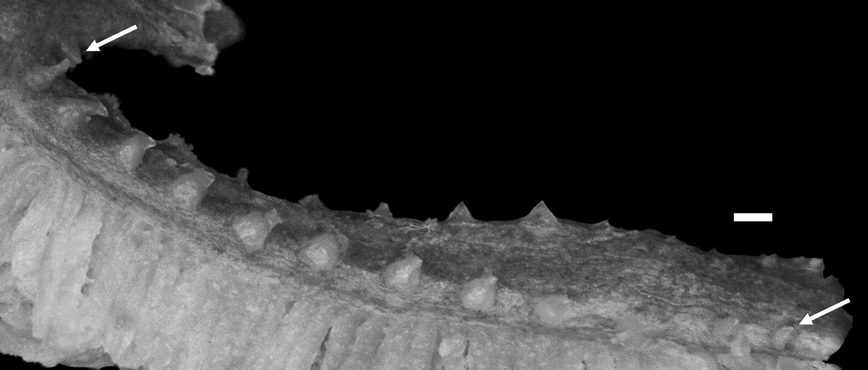
Body elongate, cross section oval anteriorly, compressed laterally near tail; its width at anal-fin origin 4.4–5.3% SL (unknown for holotype). Head moderately long, ovoid; dorsal profile of head sloping extremely gently to dorsal-fin origin. Snout short, 95.5–106.8% of eye diameter (unknown for holotype). Eye ovoid, moderately large. Interorbital space narrow, width 14.1–21.1 (17.1)% of eye diameter. Nostril tube short, not reaching upper lip when depressed. Mouth terminal. Posterior edge of upper jaw reaching to about vertical through anterior edge of pupil or middle of eye (anterior edge of pupil); no significant sexual dimorphism recognized in upper jaw length. Labial lobe of lower jaw weak. Teeth on jaws, vomer and palatine small and conical; upper jaw with 2 rows anteriorly, and 1 or 1–2 rows posteriorly; lower jaw with 2–4 irregular rows anteriorly, and 1 or 1–2 rows posteriorly (tooth arrangement in jaws unknown for holotype); vomerine teeth irregularly arranged; palatine teeth in single row. Lower edge of gill opening slightly above lower end of pectoral-fin base. Opercular flap weakly developed. Gill rakers short and triangular ( Fig. 33 View FIGURE 33 ). Pseudobranch filaments short. Two lateral lines present, deciduous, incomplete; mediolateral line more deciduous, only visible above pectoral fin; ventral line originating posterior to last postorbital pore and terminating anterior to anus. Scales small and cycloid, present on nape, body, except around pelvic fin, pectoral axilla, about 10–30% of pectoral fin basally, pectoral-fin base, tail and about 70–80 % vertical fins basally (scaled areas unknown for holotype). Head and margins of vertical fins without scales.
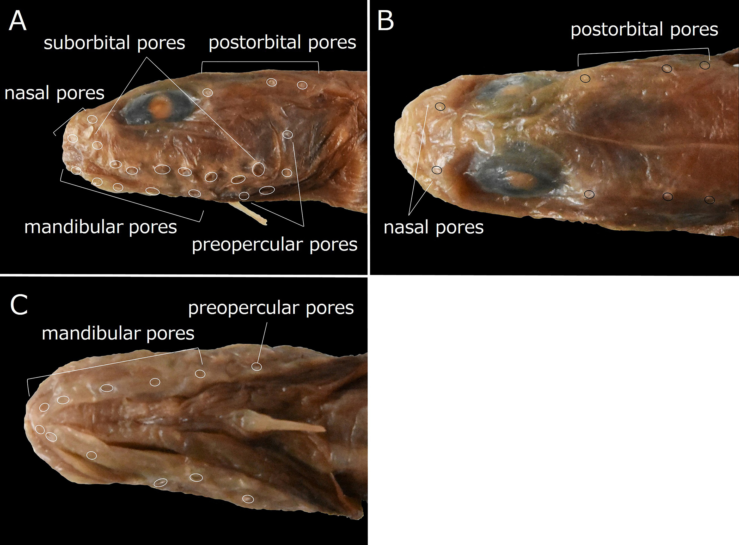
Dorsal-fin origin above middle of pectoral fin; 1st dorsal-fin pterygiophore between neural spines of 7th to 9th vertebrae (between 7th and 8th). Anal-fin origin below 16th or 17th (17th) dorsal-fin ray; 1st anal-fin pterygiophore posterior to parapophysis of ultimate abdominal vertebra. Last dorsal-fin pterygiophore between neural spines of 3rd to 5th (between 3rd and 4th) preural vertebrae. Last anal-fin pterygiophore between hemal spines of 3rd to 5th (between 3rd and 4th) preural vertebrae. Caudal fin with 2 epural, 3–4 (4) upper hypural and 4–5 (5) lower hypural rays. Pectoral fin moderately short, reaching to middle of abdomen; its posterior margin rounded dorsally and notched ventrally. Upper end of pectoral-fin base on about lateral midline of body. Pelvic fin relatively long; its base below 7th suborbital pore ( Fig. 35A View FIGURE 35 ); its posterior margin reaching to or slightly beyond lower edge of gill opening (position of posterior margin of pelvic fin unknown for holotype).
Head pores well developed and distinct. Nasal pores 2; anterior pore in front of nostril tube, posterior pore above and slightly anterior to vertical through 1st suborbital pore ( Fig. 35A, B View FIGURE 35 ). Postorbital pores 3 ( Fig. 35A, B View FIGURE 35 ); distance between 1st and 2nd pores longest of those between adjacent pores. Suborbital pores 8; 7 pores below eye and last on ascending part of suborbital canal behind eye; 7th pore above pelvic-fin base; 8th pore slightly higher than other 7 suborbital pores and set posterior edge of cheek ( Fig. 35A View FIGURE 35 ). Preoperculomandibular pores 9; 5 on lower jaw and 4 on preopercle; last preoperculomandibular pore anterior to upper end of pectoral-fin base ( Fig. 35A, C View FIGURE 35 ). Interorbital pores and occipital pores absent ( Fig. 35B View FIGURE 35 ).
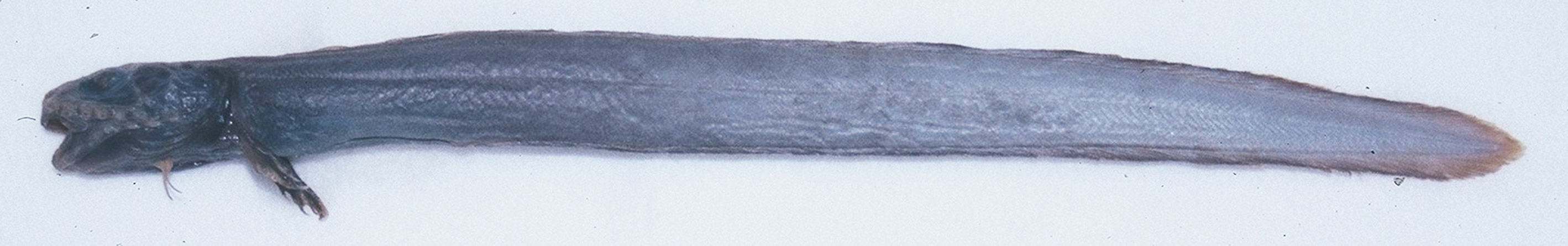
Color in alcohol. Holotype (based on color photograph; Fig. 34 View FIGURE 34 ) with dark brown head, pectoral fin and margin of vertical fins and bluish gray body, vertical fins and abdomen. Other specimens with pale chocolate brown head, pectoral fin and margins of vertical fins, pale brown body and vertical fins, and dark brown abdomen.
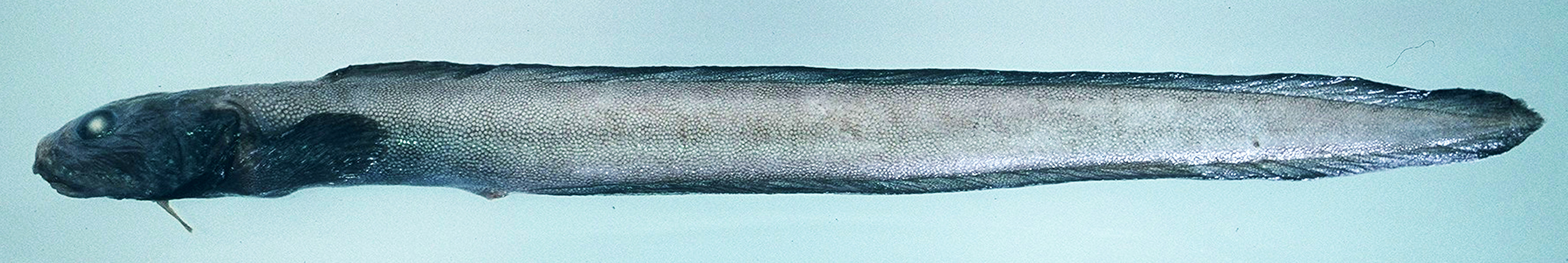
Color when fresh (based on color photograph of HUMZ 180877; Fig. 32 View FIGURE 32 ). Head, pectoral fin and margin of vertical fins blackish, body and vertical fins uniformly grayish, and abdomen dark gray.
Distribution. Off northwestern Pacific coast of Honshu Island from Iwate to Ibaraki prefectures, at depths of 1020–2034 m ( Fedorov, 1995b; Shinohara et al., 1996; Imamura, 1997, 1998; Hatooka, 2000, 2002, 2013; Anderson & Fedorov, 2004; Shinohara & Anderson, 2007; Kitagawa et al., 2008; Shinohara et al., 2009; Amaoka et al., 2011; Balushkin et al., 2011; this study).
Size. Maximum length 32 cm TL ( Hatooka, 2013; Amaoka et al., 2011). The largest specimen examined during this study measured 304.4 mm SL (311.0 mm TL).
Remarks. Lycenchelys remissaria resembles Lycenchelys cicatrifer (Garman, 1899) and Lycenchelys novaezealandiae Anderson & Møller, 2007 in having no interorbital pores and occipital pores, 3–4 postorbital pores, 2 lateral lines and scales on the nape in large specimens (vs. lacking this combination of characters in other species of Lycenchelys ) (e.g., Anderson, 1995; Fedorov, 1995b; Shinohara et al., 1996; Hatooka, 2000, 2002, 2013; Anderson & Møller, 2007; Shinohara & Anderson, 2007; this study). Lycenchelys remissaria is easily distinguished from L. novaezealandiae by its fewer numbers of dorsal-fin rays (116–123 vs. 128–132), anal-fin rays (103–107 vs. 119), and total vertebrae (124–130 vs., 139–141), respectively ( Anderson & Møller, 2007; this study). Lycenchelys remissaria is allied to L. cicatrifer in having similar counts and proportional measurements (e.g., 108–115 dorsal-fin rays, 97–104 anal-fin rays and 116–124 total vertebrae in L. cicatrifer ) ( Table 8). However, L. remissaria is clearly separable from L. cicatrifer by the position of the pelvic fin. In L. remissaria , the pelvic fin is positioned extremely anteriorly with its base below the the 7th suborbital pore ( Fig. 35A View FIGURE 35 ). In contrast, the pelvic-fin base of L. cicatrifer is located posterior to the suborbital pores. The position of the pelvic-fin base is also useful for separating L. remissaria from all species of Lycenchelys (except for 6 the species without pelvic fins), which have the more posterior position of the fin.
No known copyright restrictions apply. See Agosti, D., Egloff, W., 2009. Taxonomic information exchange and copyright: the Plazi approach. BMC Research Notes 2009, 2:53 for further explanation.
|
Kingdom |
|
|
Phylum |
|
|
ParvPhylum |
Osteichthyes |
|
Class |
|
|
Order |
|
|
Family |
|
|
Genus |
|
Kingdom |
|
|
Phylum |
|
|
ParvPhylum |
Osteichthyes |
|
Class |
|
|
Order |
|
|
Family |
|
|
Genus |
|
Kingdom |
|
|
Phylum |
|
|
ParvPhylum |
Osteichthyes |
|
Class |
|
|
Order |
|
|
Family |
|
|
Genus |
|
Kingdom |
|
|
Phylum |
|
|
ParvPhylum |
Osteichthyes |
|
Class |
|
|
Order |
|
|
Family |
|
|
Genus |
|
Kingdom |
|
|
Phylum |
|
|
ParvPhylum |
Osteichthyes |
|
Class |
|
|
Order |
|
|
Family |
|
|
Genus |
|
Kingdom |
|
|
Phylum |
|
|
ParvPhylum |
Osteichthyes |
|
Class |
|
|
Order |
|
|
Family |
|
|
Genus |
|
Kingdom |
|
|
Phylum |
|
|
ParvPhylum |
Osteichthyes |
|
Class |
|
|
Order |
|
|
Family |
|
|
Genus |
|
Kingdom |
|
|
Phylum |
|
|
ParvPhylum |
Osteichthyes |
|
Class |
|
|
Order |
|
|
Family |
|
|
Genus |
|
Kingdom |
|
|
Phylum |
|
|
ParvPhylum |
Osteichthyes |
|
Class |
|
|
Order |
|
|
Family |
|
|
Genus |
|
Kingdom |
|
|
Phylum |
|
|
ParvPhylum |
Osteichthyes |
|
Class |
|
|
Order |
|
|
Family |
|
|
Genus |
|
Kingdom |
|
|
Phylum |
|
|
ParvPhylum |
Osteichthyes |
|
Class |
|
|
Order |
|
|
Family |
|
|
Genus |
|
Kingdom |
|
|
Phylum |
|
|
ParvPhylum |
Osteichthyes |
|
Class |
|
|
Order |
|
|
Family |
|
|
Genus |
|
Kingdom |
|
|
Phylum |
|
|
ParvPhylum |
Osteichthyes |
|
Class |
|
|
Order |
|
|
Family |
|
|
Genus |
Lycenchelys remissaria Fedorov, 1995
| Kawarada, Shumpei, Imamura, Hisashi, Narimatsu, Yoji & Shinohara, Gento 2020 |
Lycenchelys remissaria
| Nakabo, T. & Hirashima, Y. 2015: 217 |
| Hatooka, K. 2013: 1225 |
| Amaoka, K. & Nakaya, K. & Yabe, M. 2011: 317 |
| Balushkin, A. V. & Sheiko, B. A. & Fedorov, V. V. 2011: 982 |
| Shinohara, G. & Narimatsu, Y. & Hattori, T. & Ito, M. & Takata, Y. & Matsuura, K. 2009: 724 |
| Kitagawa, D. & Imamura, H. & Goto, T. & Ishito, Y. & Fujiwara, K. & Ueda, Y. 2008: 95 |
| Shinohara, G. & Anderson, M. E. 2007: 63 |
| Anderson, M. E. & Fedorov, V. V. 2004: 20 |
| Hatooka, K. 2002: 1032 |
| Hatooka, K. 2000: 1032 |
| Imamura, H. 1998: 32 |
| Imamura, H. 1997: 60 |
| Shinohara, G. & Endo, H. & Matsuura, K. 1996: 181 |
| Fedorov, V. V. 1995: 135 |