
Cylindrobulla schuppi, Laetz, Elise, Christa, Gregor, Händeler, Katharina & Wägele, Heike, 2014
|
publication ID |
https://doi.org/10.11646/zootaxa.3893.3.2 |
|
publication LSID |
lsid:zoobank.org:pub:38B39F8B-9218-439B-8C5B-5A68DA99E180 |
|
DOI |
https://doi.org/10.5281/zenodo.5612592 |
|
persistent identifier |
https://treatment.plazi.org/id/0312EC7F-FFDB-FFAC-FF37-F9FB3E47AF32 |
|
treatment provided by |
Plazi |
|
scientific name |
Cylindrobulla schuppi |
| status |
sp. nov. |
Cylindrobulla schuppi View in CoL sp. nov.
Type material. Five specimens from Coco’s Lagoon, Guam, USA territory, associated with Halimeda macroloba and its underlying sediment. Paratype and one additional specimen were used for radula preparation, one specimen sectioned, one used for molecular analyses and radula preparation. Type material (holotype and one paratype) deposited at the Zoologische Staatssammlung München (SNSB-Bavarian State Collection of Zoology, Munich; ZSM), Germany. Holotype ( ZSM Moll 20130057, shell length 5.7 mm, shell width 2.7 mm). Paratype ( ZSM Moll 20130058, shell length 3 mm, shell width 1.7 mm). One specimen used for molecular analyses and radula preparation ( ZSM Moll 20130059), one specimen used for histological analysis, one specimen lost during radula preparations.
Distribution. Only recorded at Cocos Lagoon, Guam, USA.
Etymology. This species is dedicated to Prof. Dr. Peter Schupp (now University of Oldenburg, Germany), who supported us to a great extent during our stay on Guam and who helped us in collecting other heterobranch material.
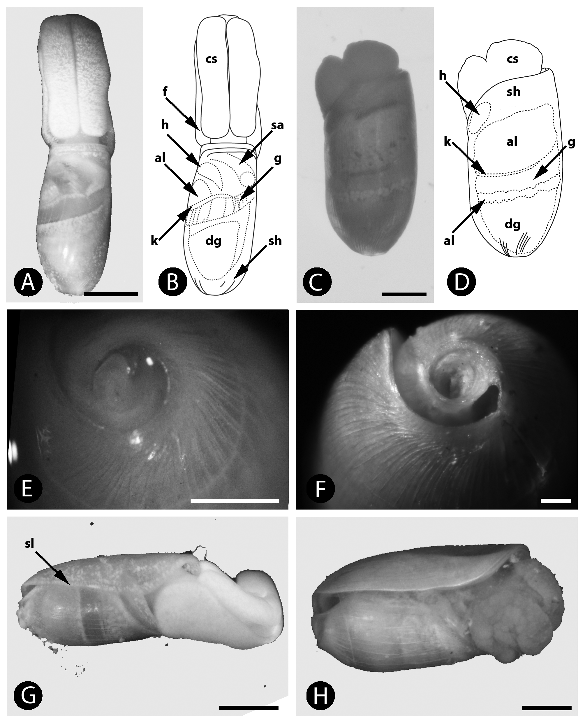
Description. External anatomy of living specimen. Head comprising large, mobile cephalic shield, almost as long as shell, having two large lobes that are lightly cleft along dorsal midline. Cephalic shield distinctly separated from foot by deep groove, attached to visceral mass by broad neck (two-thirds width of shell) visible in relaxed specimens ( Fig. 1 View FIGURE 1 A). Stressed specimens unable to retract cephalic shield completely. Eyes not visible in dorsal view. Foot extending anteriorly (slightly) and posteriorly past cephalic shield, opaque white. Shell oblong, anteriorly flattened, posteriorly rounded, translucent whitish with many hairline cracks covering entire surface and obscuring growth lines. Many internal structures visible through shell in living specimens ( Fig. 1 View FIGURE 1 A, B): light yellowish heart at anterior left side of visceral sac/cavity; transparent gill encircling visceral mass across shell midline, opaque white diaphragm muscle at anterior right side of body, translucent whitish kidney visible directly anterior to gill along shell midline, yellowish-white digestive gland visible posteriorly at center, pale yellow albumen gland visible at anterior left of gill and kidney. Posterior shell surface appearing slightly conical, shell whorl deeply keeled, protoconch not visible ( Fig. 1 View FIGURE 1 E). Shell lip straight ( Fig. 1 View FIGURE 1 G).
Anatomy of preserved specimen (inferred from histological slides). Visceral cavity densely packed with muscles and connective tissue, no distinct space between organs present. Highly muscular diaphragm separating anterior part of digestive tract and penial sheath from rest of organs.
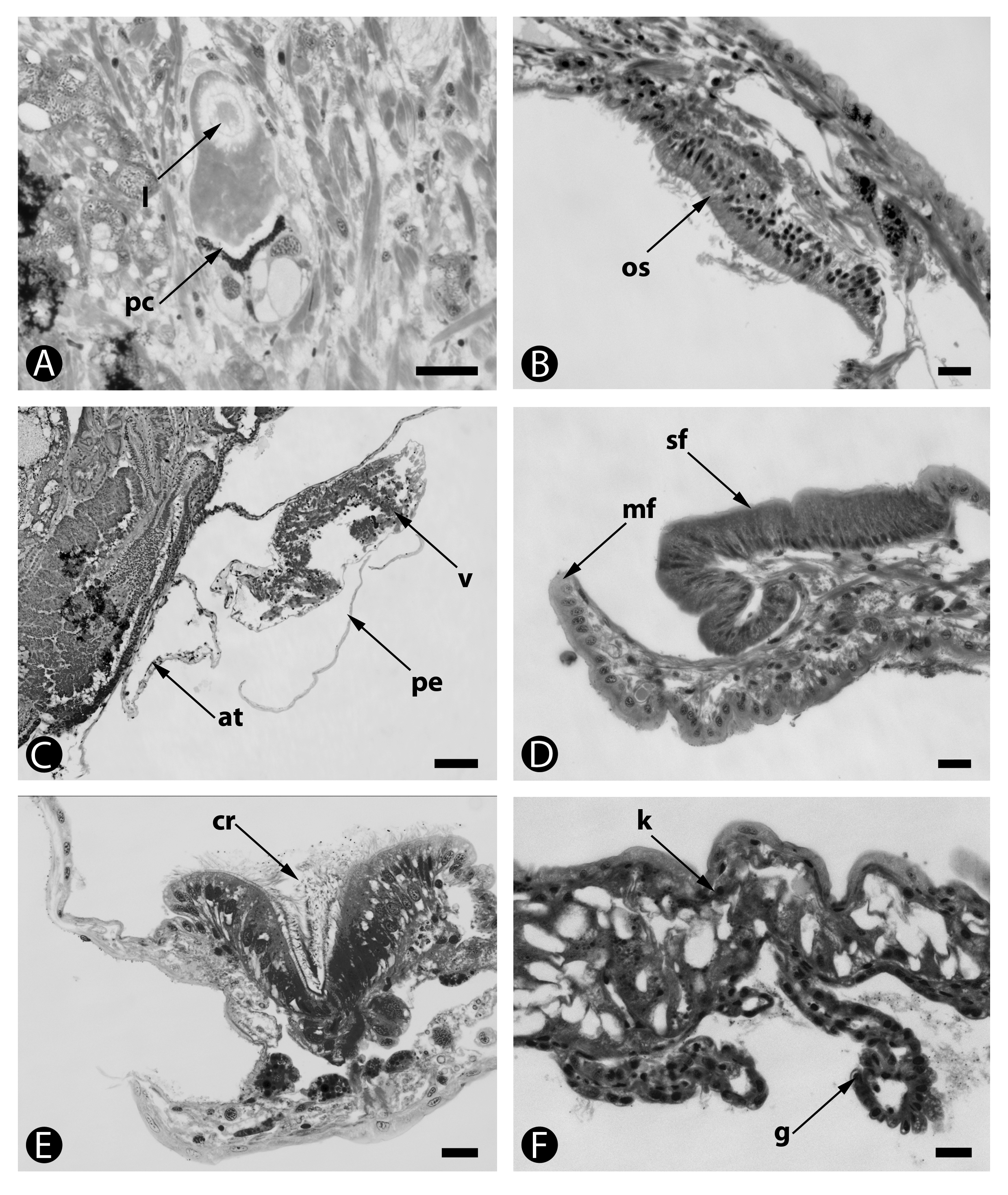
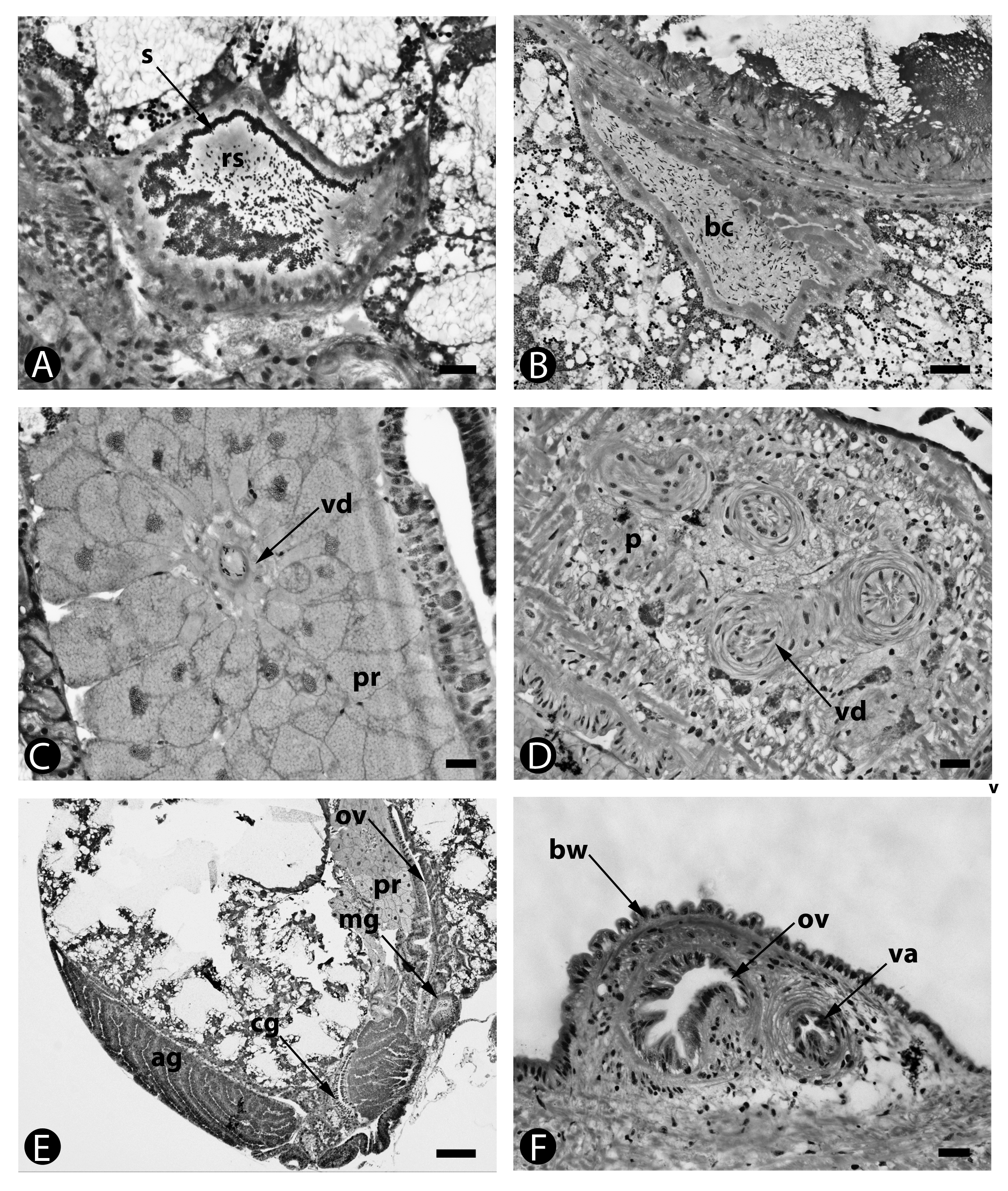
Mantle cavity, sensory organs and nervous system. Ciliated ridge on mantle edge, directed into mantle cavity ( Fig. 2 View FIGURE 2 E). Eyes located medio-laterally, comprised of irregularly shaped lens and pigment cup ( Fig. 2 View FIGURE 2 A). Osphradium ( Fig. 2 View FIGURE 2 B) and shell forming gland distinct, on mantle lip ( Fig. 2 View FIGURE 2 D). Nerve ring post-pharyngeal. Cerebral ganglia connected by rather elongate commissure. Cerebral ganglia and pleural ganglia closely attached; pedal ganglia with short commissure and short connectives towards cerebral and pleural ganglia. Buccal ganglia lying ventrally of esophagus, with short connectives towards cerebral ganglia. Visceral commissure with swellings, but number of ganglia could not be verified. Statocysts with one statolith.
Circulatory, respiratory and excretory systems. Kidney elongate and flat, with internal lamellae forming dense compartments. Cells containing large unstained vacuoles. Gill mainly situated on kidney, but extending toward edge of mantle, composed of lamellae directly attached to mantle or kidney wall ( Fig. 2 View FIGURE 2 F). Heart complex situated on left, with thin-walled atrium and muscular ventricle surrounded by pericardium ( Fig. 2 View FIGURE 2 C). No glandular structures observed along pericardial wall. Connection to kidney not detected.
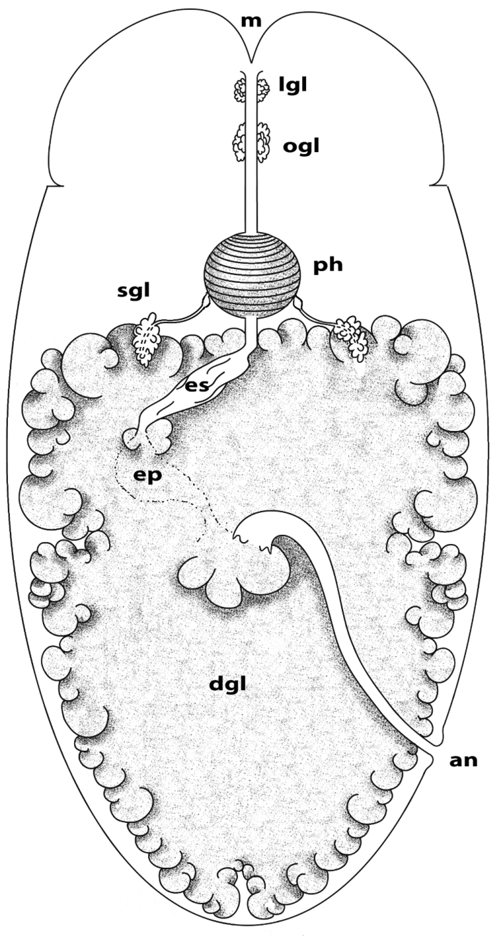
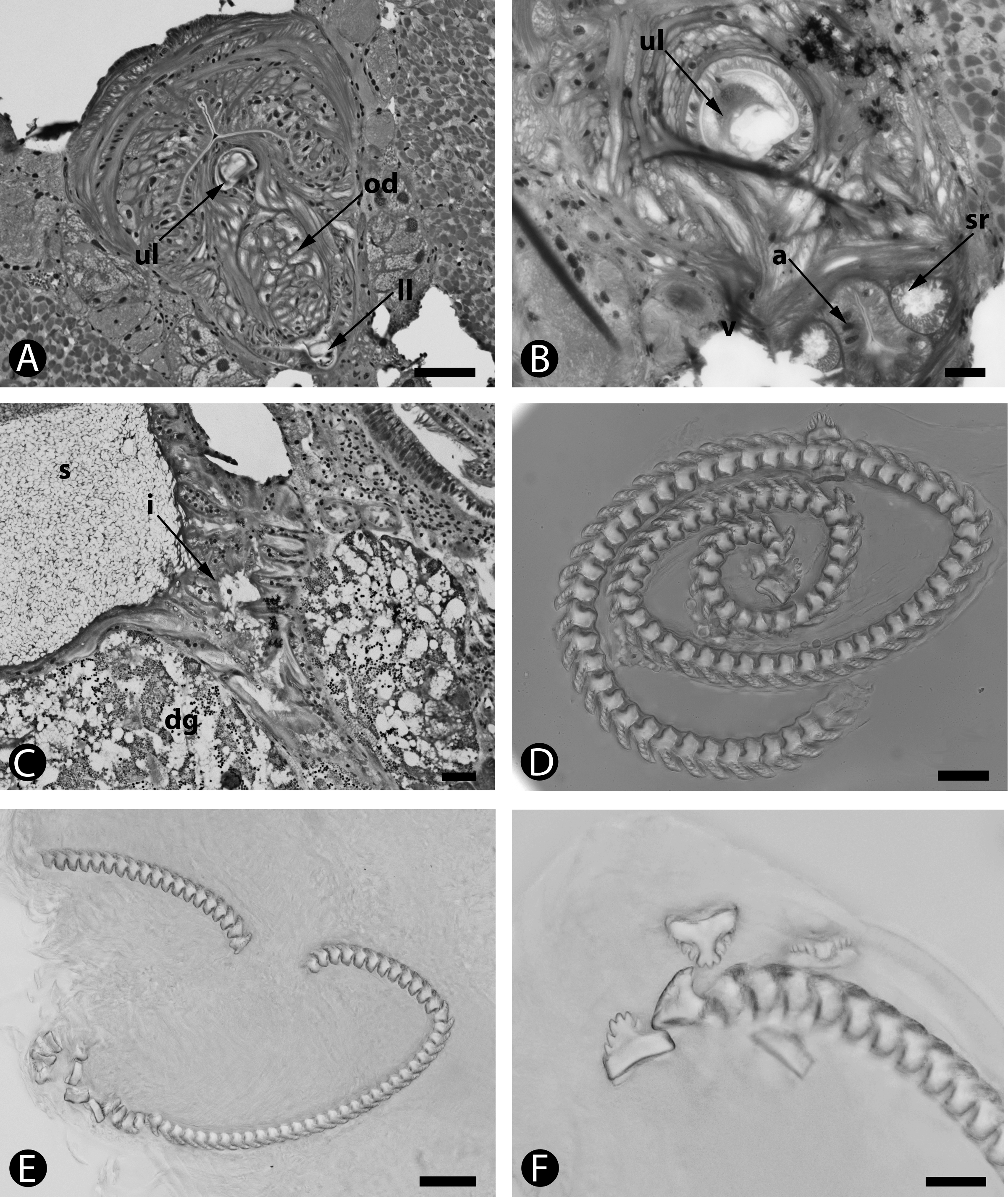
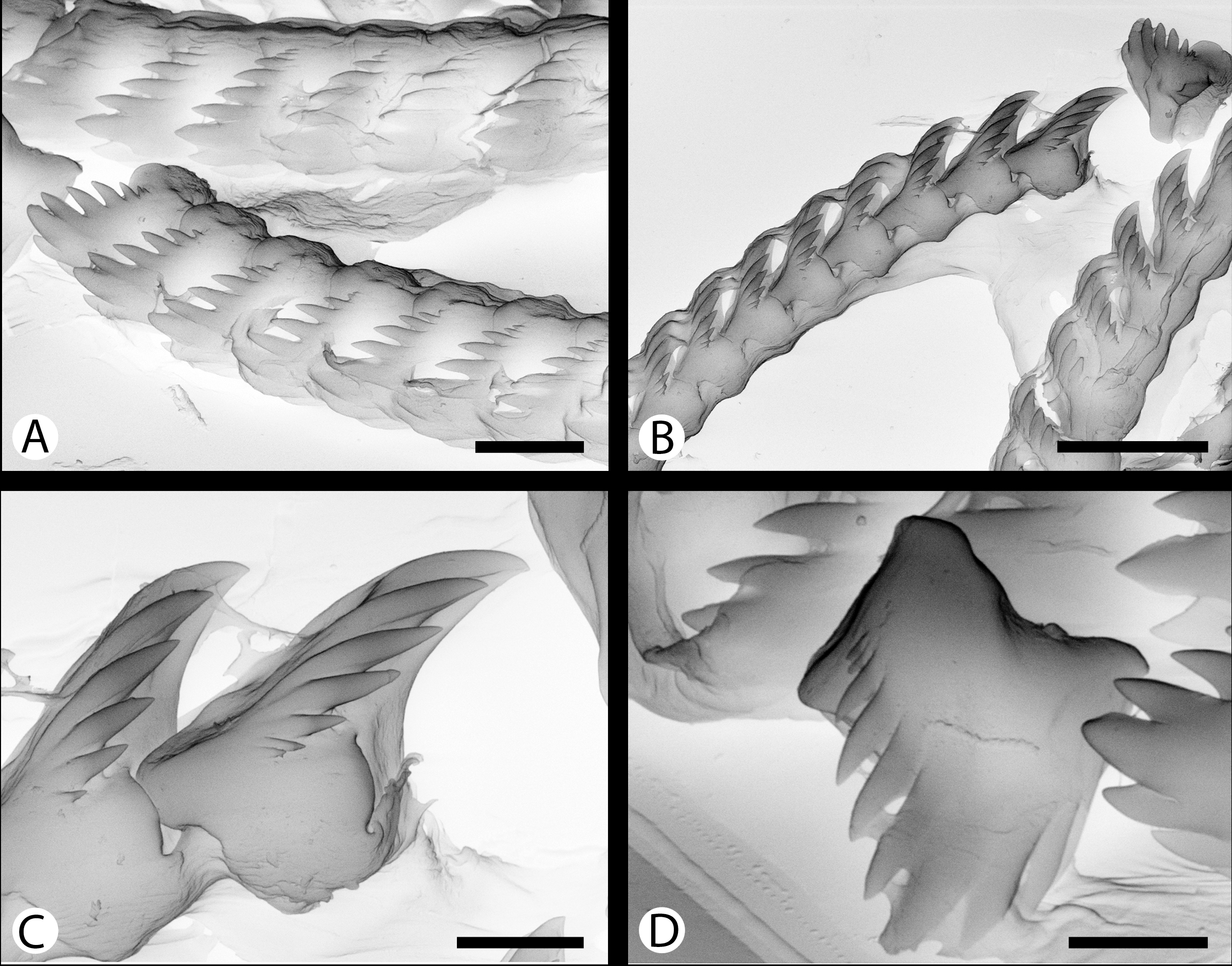
Digestive system. Schematic drawing in Figure 3 View FIGURE 3 , summarizing system. Mouth located between foot and cephalic shield, surrounded by lip glands. Oral tube very long, running along almost entire length of cephalic shield, surrounded by small oral glandular follicles staining violet and connected to large, heavily muscled pharynx ( Fig. 4 View FIGURE 4 A). Odontophore with radula suspended by muscles attached laterally. Long radula with equal length of upper and lower limbs; upper limb beginning at odontoblasts and lower limb ending at ascus ( Fig. 4 View FIGURE 4 B). No preradular teeth present. Ascus lying within musculature of pharynx. Radula with about 100 broad smooth teeth of similar size, each roughly pentagonal in shape with 5–6 denticles increasing in size toward median cusp ( Fig. 5 View FIGURE 5 A–D). Three denticles in center somewhat larger, but not protruding or sticking out from other denticles ( Fig. 5 View FIGURE 5 D). Salivary glands sac-like, adjacent to esophagus, leading into long thin salivary ducts that widen anteriorly into salivary reservoirs, which then lead into pharynx. Long esophagus with narrow beginning that widens into esophageal pouch, lying asymmetrically. Distal esophagus narrowing again before entering stomach. Typical ciliated stomach epithelium folded and only present on dorsal side, remainder fused with digestive gland. Large intestine emerging dorsally from stomach; with many internal folds, but lacking a typhlosole ( Fig. 4 View FIGURE 4 C). Anus opening ventrally into mantle cavity. Large digestive gland compact in outer appearance, but tubular in structure with large cavities; filling most of visceral cavity from anterior to posterior end, even in interior coils. Digestive gland cells containing many vacuoles, staining bright red, blue and purple in toluidine blue.
Genital system. Schematic drawing in Figure 6 View FIGURE 6 , summarizing system. Small, sac-like, male-phase (spermproducing) ovotestis, located ventrally along transverse midline within visceral mass. No oogonians observed. Ovotestis connected to elongated, thin pre-ampullar duct, proceeding anteriorly to posterior end of ampulla. Beanshaped ampulla containing sperm, anterior end connected to short, thin post-ampullar duct, and splitting into vas deferens and oviduct. Very long and winding vas deferens passing anteriorly from post-ampullar duct through visceral cavity to right side; accompanied by an extensive prostate gland ( Fig. 6 View FIGURE 6 , 7 View FIGURE 7 C). Vas deferens following distal oviduct in pallial wall, then leading towards head, mainly within pallial musculature. Entrance into penis next to pharynx, but last part of vas deferens before entering penis not observable due to a rupture in animal. Broad, long, cylindrical penis with coiled internal vas deferens ( Fig. 7 View FIGURE 7 D) directed posteriorly, although penial sac opening is anterior; no stylet present. Penial sac retracted deeply anterior body cavity ( Fig. 6 View FIGURE 6 ); connecting to male opening, located in groove between cephalic shield and foot on right side in dorsal view. No ciliated sperm groove present. Thin, short oviduct passing from post-ampullar duct to underdeveloped nidamental glands, containing large albumen and smaller capsule and mucus glands. Albumen gland forming large, crescent-shaped bundle of tubules spanning entire middle third of visceral mass on left side in dorsal view ( Fig. 7 View FIGURE 7 E). Capsule gland smaller, also crescent-shaped. Mucus gland separate, but lacking typical mucus cells, thus not yet differentiated from distal oviduct. Distal oviduct entering female genital opening together with vaginal duct on small papilla ( Fig. 7 View FIGURE 7 F). Vaginal duct separating into two ducts, one leading into receptaculum seminis ( Fig. 7 View FIGURE 7 A) and one into bursa copulatrix ( Fig. 7 View FIGURE 7 B). Receptaculum seminis containing allosperm attached to nutrition cells. Bursa copulatrix filled with irregularly arranged sperm. No distinct fertilization chamber or atrium found.

| ZSM |
Bavarian State Collection of Zoology |
No known copyright restrictions apply. See Agosti, D., Egloff, W., 2009. Taxonomic information exchange and copyright: the Plazi approach. BMC Research Notes 2009, 2:53 for further explanation.
|
Kingdom |
|
|
Phylum |
|
|
Class |
|
|
SubClass |
Heterobranchia |
|
SuperOrder |
Sacoglossa |
|
Order |
|
|
Family |
|
|
Genus |