
Hylarana nigrovittata ( Blyth, 1856 )
|
publication ID |
https://doi.org/10.5281/zenodo.186587 |
|
DOI |
https://doi.org/10.5281/zenodo.6217280 |
|
persistent identifier |
https://treatment.plazi.org/id/033A9525-465D-FFE9-77DB-A5F6B9C5A6F3 |
|
treatment provided by |
Plazi |
|
scientific name |
Hylarana nigrovittata ( Blyth, 1856 ) |
| status |
|
Hylarana nigrovittata ( Blyth, 1856) View in CoL from northern Thailand
Series examined. Tadpoles of Hylarana nigrovittata (ZFMK 87473-87480: n = 8), captive bred from adult specimens from southern Doi Suthep, near Chiang Mai, northern Thailand, in the Aquarium of the Cologne Zoo.
SVL 36.0 – 38.5 (37.67 ± 1.44) SL 6.0 – 9.0 (7.17 ± 1.61)
NL 2.5 – 3.2 (2.90 ± 0.36)
EN 3.0 – 3.5 (3.17 ± 0.29)
HW 13.0 – 14.0 (13.67 ± 0.58) HL 13.5 – 15.0 (14.17 ± 0.76) EE 7.0 – 8.5 (7.67 ± 0.76)
ED 4.5 – 5.0 (4.83 ± 0.29)
TD 2.5 – 3.2 (2.90 ± 0.36)
HaL 10.0 – 12.0 (11.17 ± 1.04) LL 63.0 – 72.0 (68.67 ± 4.93) FL 19.5 – 23.2 (21.40 ± 1.85) IMT 2.0 – 2.5 (2.17 ± 0.29)
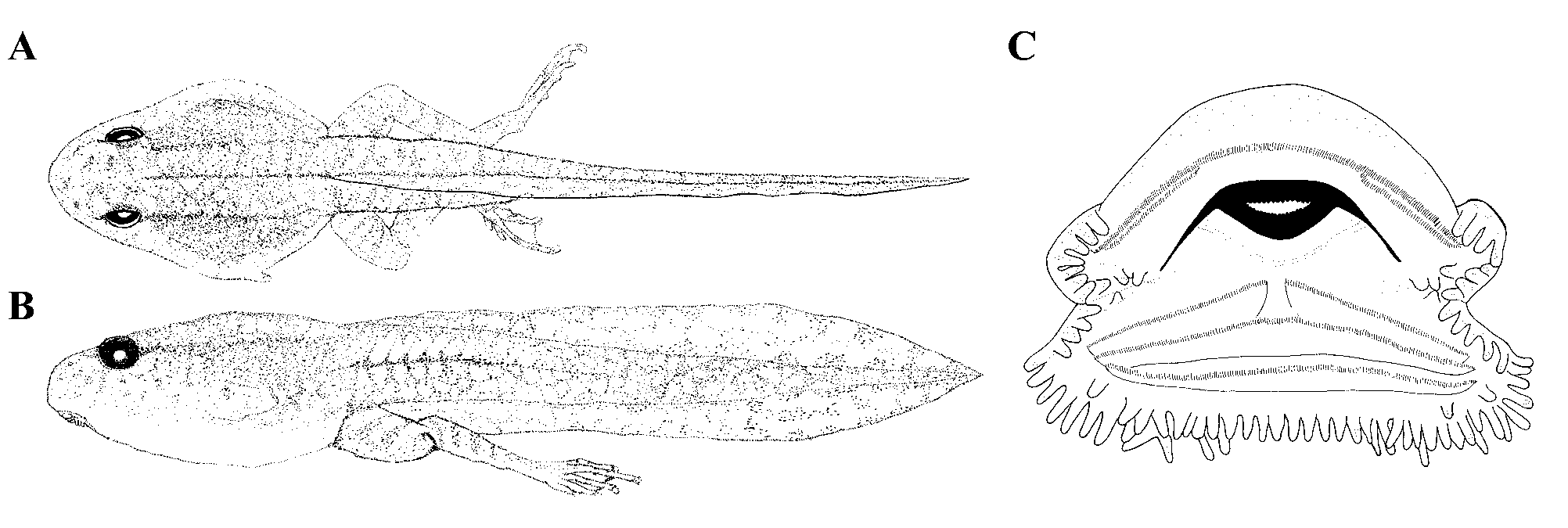
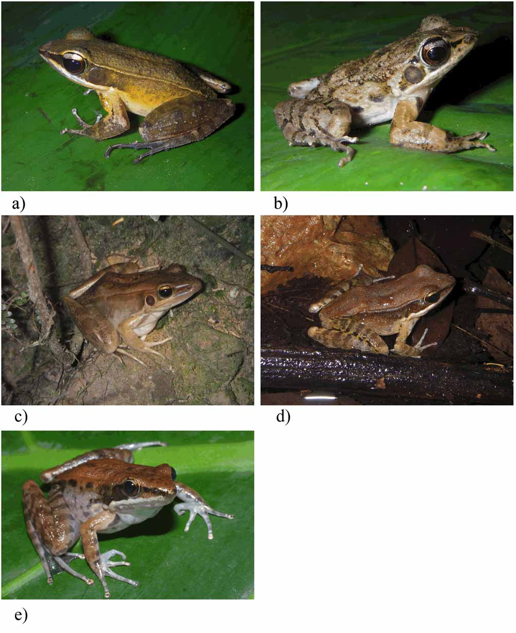
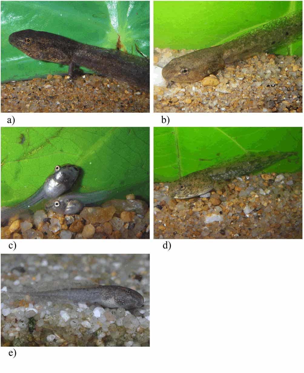
Identification. Adult male specimens ( Fig. 1 View FIGURE 1 a – d e; for measurements see Table 9) that were raised from aforementioned tadpoles ( Fig. 2 View FIGURE 2 a – d e) largely agreed with the description of Hylarana nigrovittata provided by Bourret (1942), Taylor (1962), and Ziegler (2002). Characteristic nigrovittata features are as follows: presence of distinct, broad dorso-lateral glandular folds; dorsum smooth to slightly granulated; flanks usually with some larger wart-like structures; large, distinct tympanum; vomer teeth in two small, elongated rows; males with oval glandular field above arm insertion; finger and toe tips somewhat broadened, with more or less discernible marginal grooves; first finger slightly longer than second; external metatarsal tubercle present; usually, last two phalanges of fourth toe and last two inner phalanges of second and third toe without webbing; adpressed hind limbs reach between eyes and snout tip (see compilation in Ziegler 2002). The following larval description is based on a single tadpole in Gosner stage 41 ( Fig. 7 View FIGURE 7 ; for measurements see Table 10).
Stage BH BL BW ED IND IP LF MTH NK NP 24/25 1.2 2.6 2.1 0.3 - 1.0 0.3 1.3 - - 25 1.1 4.0 3.0 0.5 0.7 1.4 0.5 1.7 33 37 29 2.5 5.4 3.8 0.7 1.3 1.8 0.6 2.3 33 51 30 3.2 6.5 4.4 0.8 1.5 1.8 0.6 3.0 36 62 35 4.8 9.2 6.0 1.1 1.8 3.1 1.0 3.5 34 70 36/37 6.0 11.9 7.5 1.3 2.3 3.5 1.1 4.4 29 - 40/41 5.0 11.0 7.4 1.6 2.0 3.6 1.0 4.7 32 69 41 6.8 13.5 9.5 1.8 2.2 4.8 1.8 6.0 28 78 Colour pattern. Colouration of tadpoles in preservative: body lightly fawn to white, dorsolaterally covered with brown to green pigments. Dorsal and dorsolateral body pigmentation is more dense than on the ventral side. Pigmentation behind the eyes and in the region of the intestinal coils strongest. Single pigments situated on the throat and the chest. Belly is slightly transparent and without pigments. Musculature of tail lightly fawn to yellow, covered with grey to green pigments. Upper and lower fin of tail are transparent, marbled and with irregular, single dark brown spots behind the second half of tail. Pigmentation of tail forms a line along the myotomes up to the end of the first quarter of tail. Hind limbs white to yellow, dorsolaterally covered with transverse grey stripes.
Colouration in life: body dorsally and laterally grey to olive, covered with dark brown blotches. Ventral side of body is transparent with small pigments, heart and intestinal spiral well visible. Tail fin marbled with grey to olive-coloured blotches. Upper and lower fin of tail are grey to transparent.
Description in dorsal view: Body at stage 41 egg-shaped (maximum body width 0.70 of body length, body length 0.43 of tail length) with a slightly tapered snout. Oral disc anteroventrally positioned. The eyes are of moderate size (maximum diameter of eye 0.13 of body length), dorsolaterally positioned at the end of first third of body and laterally directed. Interpupilar distance 0.50 of body width. Nares anterodorsally positioned and directed, closer to the tip of the snout than to the pupil (rostro-narial distance 0.35 of naropupilar distance). Internarial distance 0.46 of interpupilar distance. The musculature of tail is considerably developed (width of tail musculature at base 0.44 of maximum body width).
Description in lateral view: In lateral view the body at stage 41 is slightly depressed to round-bellied (maximum body height 0.71 of maximum body width). Spiracle sinistral, laterally positioned at the second third of body, conical and orientated in posterodorsal direction. Vent tube is completely reduced at this stage. The length of tail 2.29 of body length. Musculature of tail is remarkably developed (height of tail musculature at base 0.67 of tail musculature width at base). The myotomes of the tail musculature are V-shaped and from the proximal to second third of tail length half parallel, then gradually tapering to the tip of tail. Upper tail fin is situated behind the base of tail and gradually elevated. Highest point of upper fin is at the end of second third of tail fin. Maximum height of lower tail fin 0.75 of maximum height of upper fin.
Oral disc: Oral disc anteroventrally positioned (oral disc width 0.33 of maximum body width), emarginated and nearly framed by marginal, finger-shaped, white papillae with rounded ends. Upper labium with a large medial gap without papillae. Marginal papillae extended at lower labium. Two or three submarginal papillae situated in the corner of the mouth, lower labium with one row of submarginal papillae at the lower labium under the last keratodont row. Keratodont row formula 2(2)/3(1), with 28 keratodonts per 0.5 mm. Upper and lower beaks black and finely serrated. Upper beak curved and with long appendices, lower beak V-shaped.
Glandular Zones. Tadpoles in stages 30–41 revealed areas of concentrated whitish secretory glands dorsally at midbody and ventrolaterally at the end of body.
Measurements (in mm). BH 6.8; BL 13.5; BW 9.5; ED 1.8; IND 2.2; IP 4.8; LF 1.8; MTH 6.0; NK 28; NP 78; NPD 2.3; ODW 3.2; RND 0.8; TAL 31.0; TMH 4.0; TMW 4.2; UF 2.4.
Variation within the series. Series with seven tadpoles in stages 24–41. Proportions vary as follows: BW 0.63–0.81 of BL, ED 0.11–0.15 of BL, IP 0.41–0.52 of BW, RND 0.24–0.55 of NPD, IND 0.46–0.83 of IP, TMW 0.19–0.44 of BW, BH 0.37–0.80 of BW, TAL 1.70–2.30 of BL, TMH 0.47–0.64 of BH, TMH 0.41–0.69 of MTH, UF 0.34–0.43 of MTH, LF 0.55–0.75 of UF, ODW 0.33–0.41 of BW. In early developmental stages, the skin of body is transparent with weak pigmentation only, in older stages fawn with dense pigmentation.
Previous descriptions. Our tadpole description largely correspond with the data provided by Smith (1916) and Manthey & Grossmann (1997). However, secretory glands were neither mentioned by Smith (1916) nor by Manthey & Grossmann (1997).
No known copyright restrictions apply. See Agosti, D., Egloff, W., 2009. Taxonomic information exchange and copyright: the Plazi approach. BMC Research Notes 2009, 2:53 for further explanation.