
Haplaxius fornicus Bahder & Bartlett, 2023
|
publication ID |
https://doi.org/ 10.11646/zootaxa.5230.2.6 |
|
publication LSID |
lsid:zoobank.org:pub:BDD8E99D-CA48-4DD1-BDF6-48ADD7C185E1 |
|
DOI |
https://doi.org/10.5281/zenodo.7564674 |
|
persistent identifier |
https://treatment.plazi.org/id/03805B1D-1728-9259-FF1D-82B1A8072CB5 |
|
treatment provided by |
Plazi |
|
scientific name |
Haplaxius fornicus Bahder & Bartlett |
| status |
sp. nov. |
Haplaxius fornicus Bahder & Bartlett sp. n.
( Figures 3–10 View FIGURE 3 View FIGURE 4 View FIGURE 5 View FIGURE 6 View FIGURE 7 View FIGURE8 View FIGURE 9 View FIGURE 10 )
Type locality. Castleton Botanic Garden (18.170892, -76.823239), Saint Mary Parish, Jamaica ( Fig. 1 View FIGURE 1 ).
Diagnosis. Strongly sexually dimorphic; males pale yellow with fuscous wash over dorsal surface, forewings clear. females heavily infuscate over entire body, forwings mostly infuscate. Aedeagus with two large, serrated flanges near apex connecting to flagellum and a row of small spines on right-ventral margin.
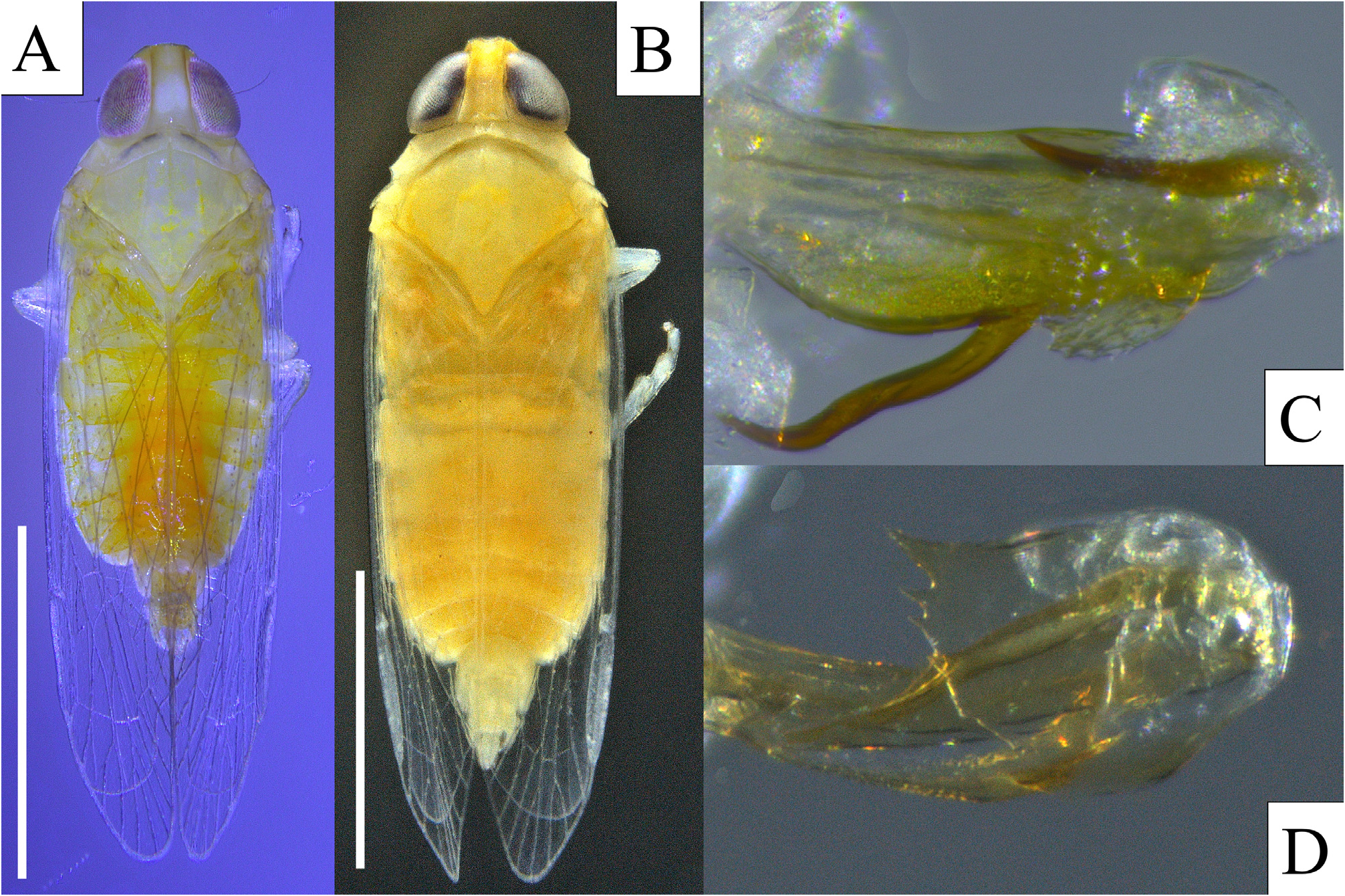
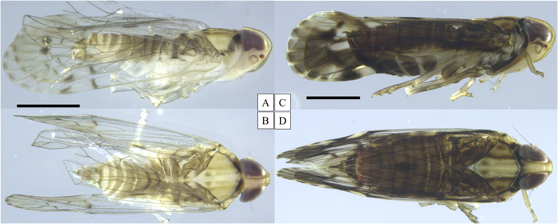
Description. Color. Body generally pale gray-yellow, head of uniform coloration (ocellus red), face unmarked. Males pale yellow on dorsum (dorsally washed with fuscous posterior to mesonotum, slightly paler between lateral carinae of mesonotum), ivory white on venter; disc of pronotum with irregular nearly black marking; wings mostly clear, tinged with fuscous in distal third past nodal line; crossveins tinged fuscous, a strong spot at im-crossvein, additional markings tracing Sc vein, at apex of pterostigma, at the apices of marginal cells and on the marginal vein of the clavus (A 2) proximad of composite Pcu+A 1 vein reaching wing margin. Females with same base color and pattern as males, except more strongly infuscate over entire body including venter and wings ( Fig. 3 View FIGURE 3 ).
Structure. Body length (including wings), males: 4.32 mm (n = 1), females 5.05–5.06 mm (n = 3). Body weakly compressed.
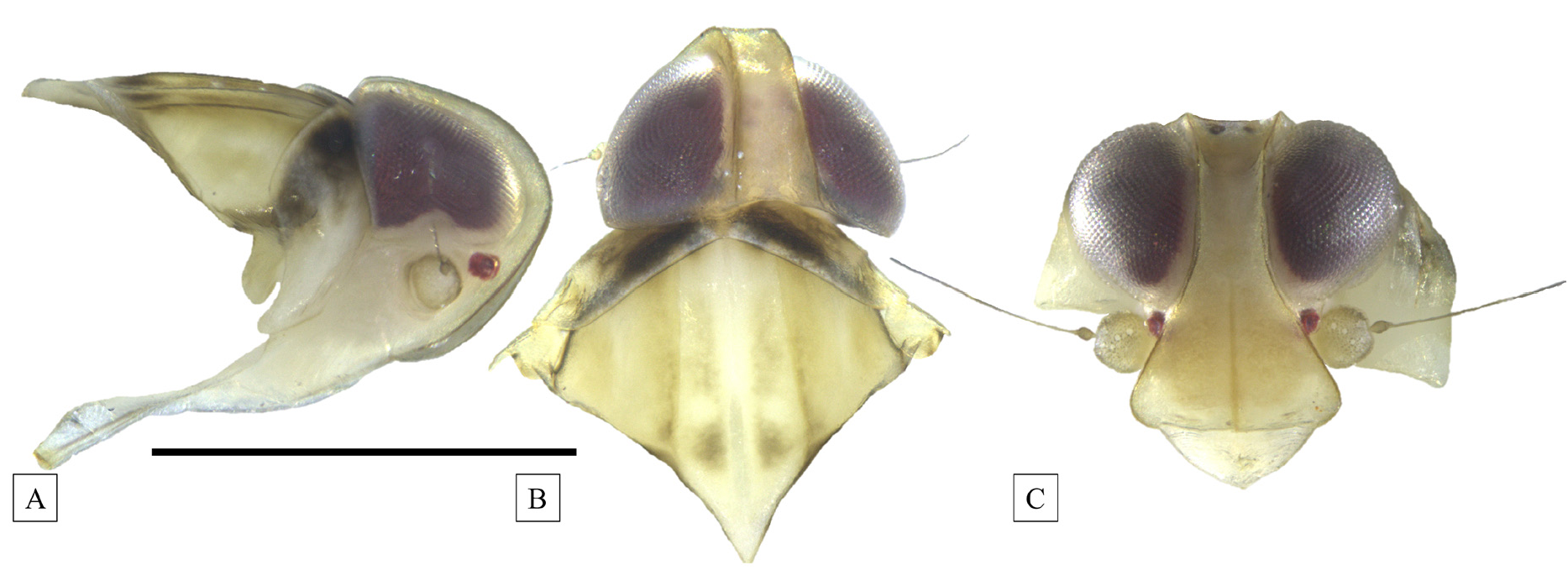
Head. Head in dorsal view narrower than pronotum; in lateral view, evenly rounded from back of head to frontoclypeal suture, head weakly projected in front of eyes ( Fig. 4A View FIGURE 4 ). In dorsal view ( Fig. 4B View FIGURE 4 ), vertex nearly parallel-sided, widest at posterior margin, narrowing slightly to anterior margin; posterior margin slightly concave, anterior margin truncate at fastigium, transverse carinae present at fastigium; vertex approximately twice as long as wide (at posterior margin). In frontal view ( Fig. 4C View FIGURE 4 ), face (frons plus clypeus) rhomboid, frons narrowest at dorsal margin, expanding ventrad to about lower level of antennae before constricting to frontoclypeal suture, median carina evident, reaching transverse suture at fastigium, median ocellus obsolete; frontoclypeus straight, clypeus inverse triangular.
Thorax. Pronotum narrow in dorsal view, convex on anterior margin, concave on posterior margin, median carina complete, lateral (postocular) carinae evident, terminating at ventrolateral apex ( Fig. 4B View FIGURE 4 ). Mesonotum longer at midline than vertex plus pronotum, tricarinate, lateral carinae subparallel, weakly diverging posteriorly ( Fig. 4B View FIGURE 4 ).
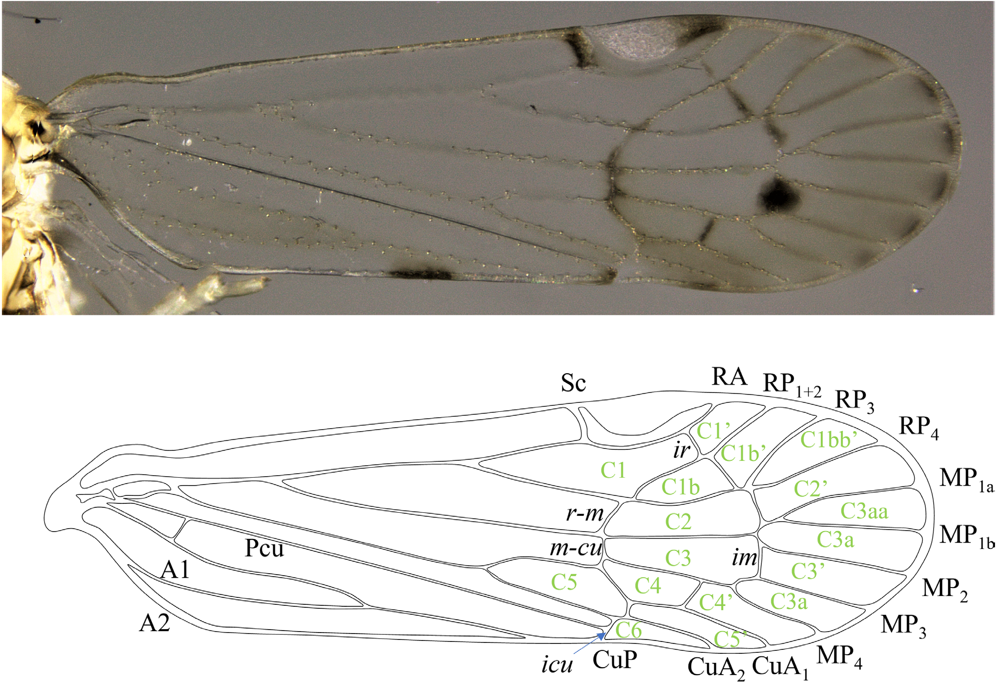
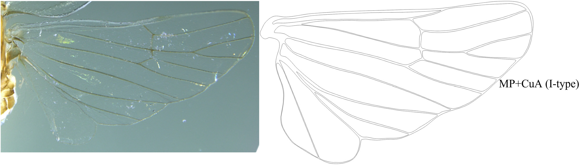
Forewing elongate oval, weakly expanding distad, apex rounded; apex of clavus just beyond midlength, fork of MP from ScP+R in proximal quarter of wing, fusion of Pcu+A 1 distad of branching of MP near midlength of clavus, branching of RP from ScP+RA (forming cell C1) and branching of CuA (forming cell C5) at same level, preceding apex of clavus. Vein branching pattern RA unbranched, RP 3-branched, MP 5-branched and CuA 2-branched ( Fig. 5 View FIGURE 5 ). Hindwing with I-type connection between MP and CuA veins ( Fig. 6 View FIGURE 6 ).
Spinulation of hind tibiae and tarsomeres is 6-7-8.
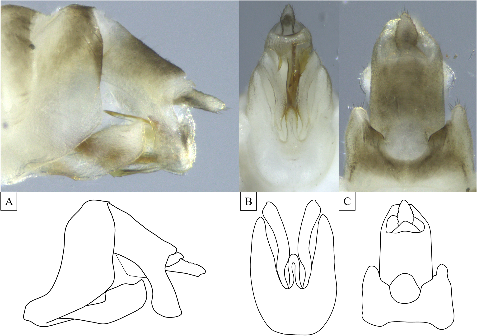
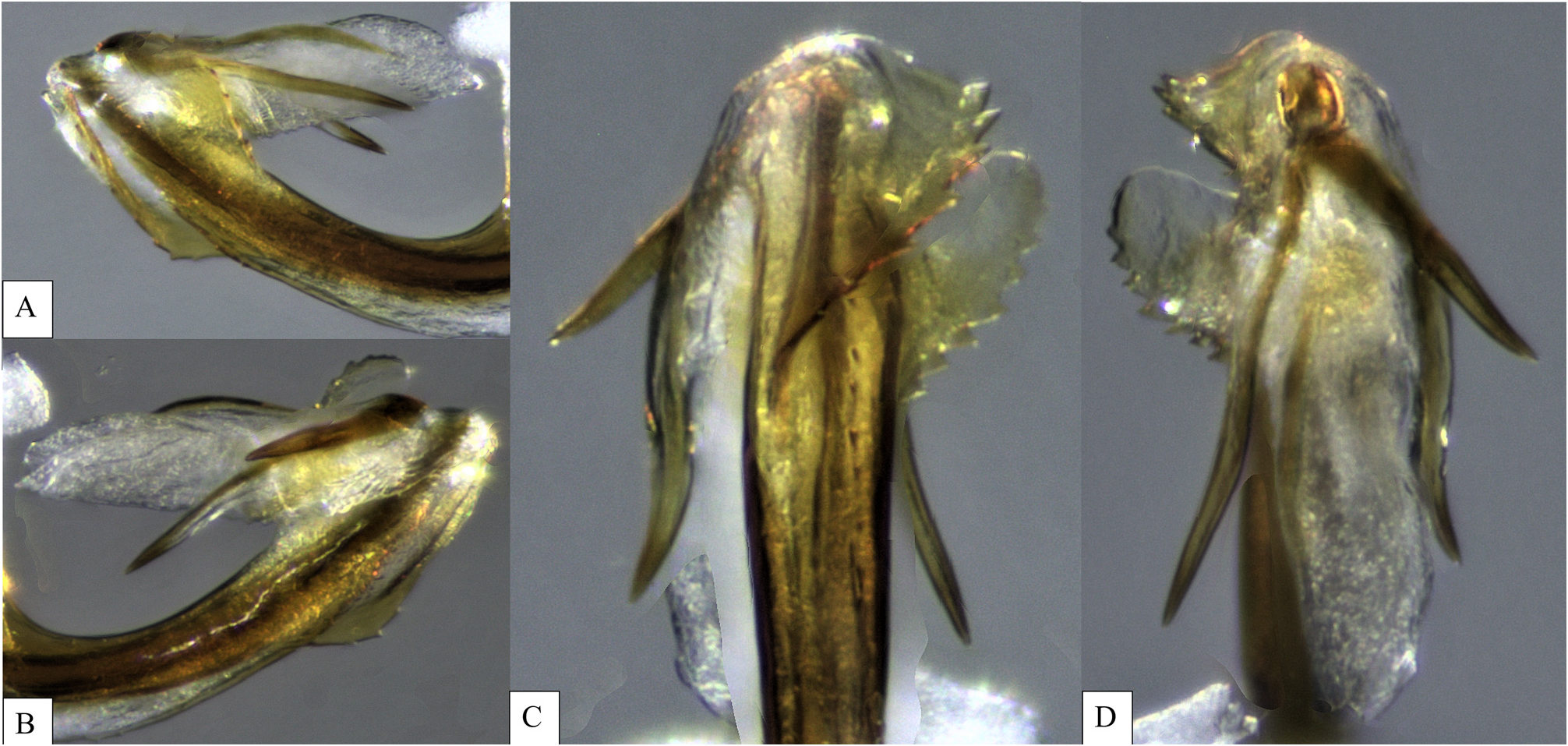
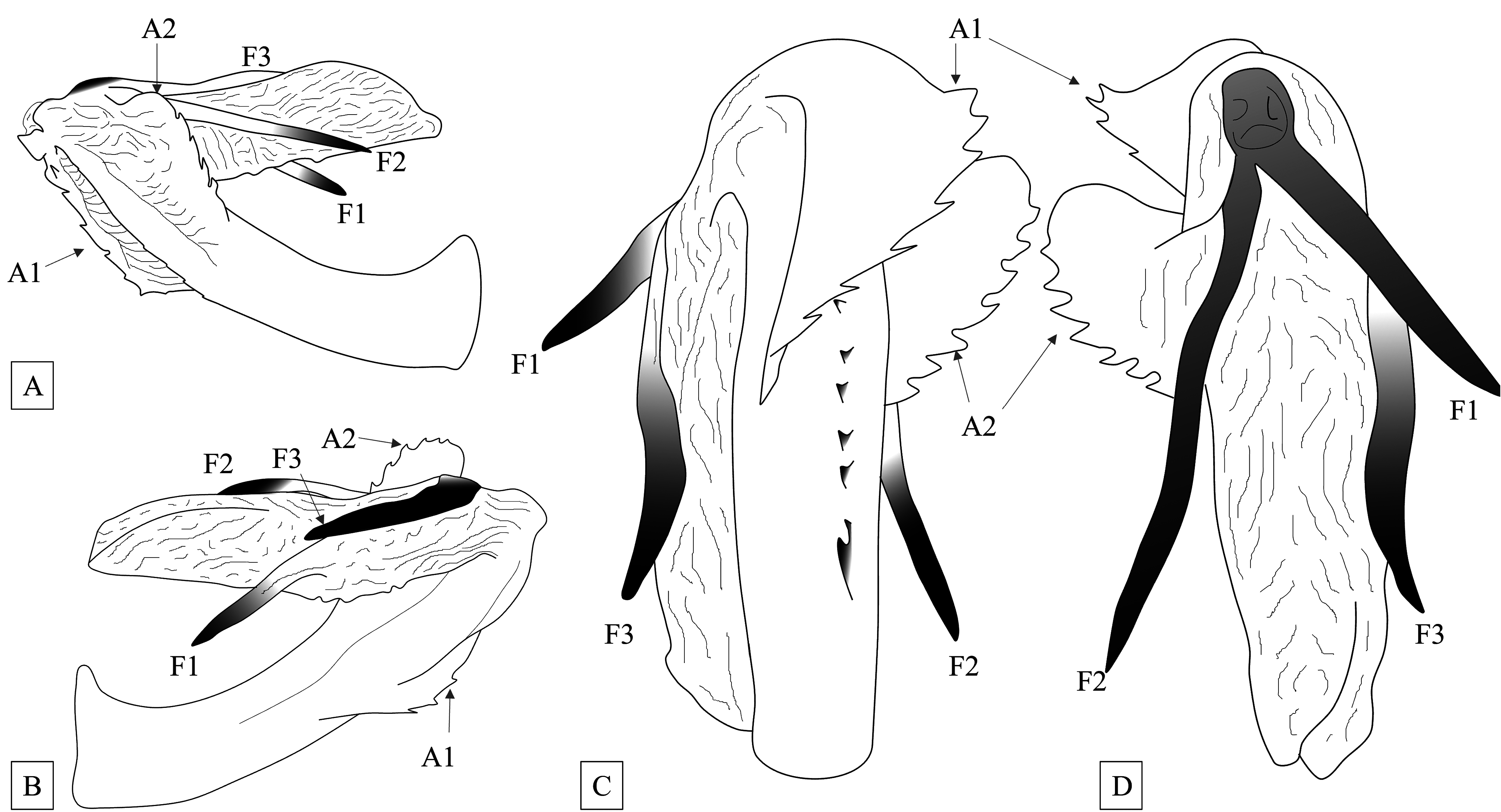
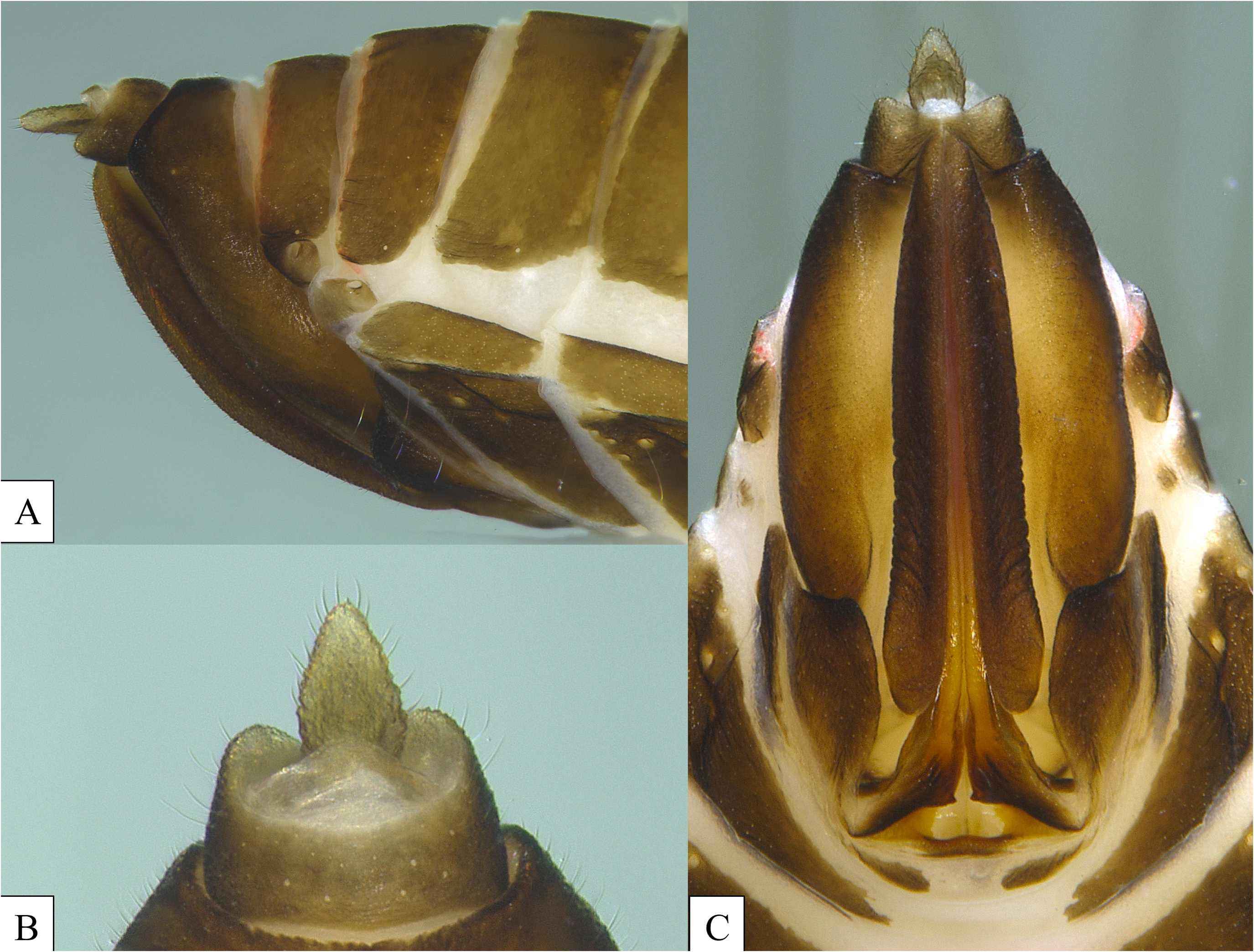
Terminalia. Pygofer irregular in lateral view, concavely sinuate on anterior margin, convexly sinuate on posterior margin, widest near midlength, narrowest at dorsal and ventral margins ( Fig. 7A View FIGURE 7 ). In ventral view, medioventral process elongate, approximately 3X long as wide at base; weakly oblaceolate, apex rounded, slightly constricted below midlength, bearing a ventral median ridge ( Fig. 7B View FIGURE 7 ). Gonostyli in lateral view “scoop-like”, dorsal and ventral margins irregularly sinuate, narrowest at base, expanding distad to midlength, then dorsally expanded to seimquadrate region on dorsal margin before apex, ventral marginconvex ( Fig. 7A View FIGURE 7 ). In ventral view, “paddle-like”, elongate, narrowest at base, curved lateral, strongly sinuate on outer and inner margins, sub-quadrate at apex, widest supapically ( Fig. 7B View FIGURE 7 ). Aedeagus tubular, in lateral view shaft upcurved, with midventral row of teeth and large diagonally oriented serrated flange on ventral margin (A1, Figs 8 View FIGURE8 and 9 View FIGURE 9 ), a second (A2) large serrate flange on right side; A1 arising subapically on ventral margin, left side, curving diagonally distad to base of endosoma; A2 arising on right lateral side extending distally to base of endosoma, endosoma comprised of an elongate tubular membranous tube and three sclerotized processes (F1, F2, and F3), endosoma with dorsal longitudinal concavity; F1 and F2 arising near dorsal of endosomal base, F1 angled to left lateral side, slightly sinuate; F2 angled to right lateral side, approximately twice as long and more slender than F1; F3 similar in length and shape to F1, arising on left lateral side of flagellum at midpoint in basal half ( Figs 8 View FIGURE8 and 9 View FIGURE 9 ). Anal tube in lateral view relatively short and stout with broad, downcurved apex; dorsal and ventral margins subparallel nearly to paraproct, apex broad, curved ventrad, to broadly rounded apex; bearing subtriangular process near midpoint on inner ventral margin, in ventral view, asymmetrical, left lateral side with protrusion on anterior margin ( Fig. 7C View FIGURE 7 ); paraproct short and conical.
Plant associations. Unidentified palms ( Arecaceae ) and and unidentified monocot ( Fig. 1 View FIGURE 1 ).
Distribution. Jamaica (Saint Mary Parish).
Etymology. The specific name refers to the serrated flanges that resemble the head of Fornicus, a villain in the film “Cabin in the Woods”. The name is intended to be indeclinable.
Material examined. Holotype male “ Jamaica, Saint Mary Parish / Castleton Botanic Garden / 16. II.2022 / Sweeping vegetation / Coll.: B.W.Bahder // Holotype / Haplaxius fornicus ♁” ( FLREC) ; Paratypes, Castleton Botanic Garden [16. II.2022] (1 female — FSCA, 2 females — FLREC). Genitalia of female paratype presented in Figure 10 View FIGURE 10 .
Sequence data. For the COI gene, a 548 bp sequence was generated (GenBank Accession No. OP160200 View Materials ), for 18S a 1,360 bp sequence was generated (GenBank Accession No. OP158203 View Materials ), and for H3 a 321 bp sequence was generated (GenBank Accession No. OP179300 View Materials ). Based on independent analyses for each locus, Haplaxius fornicus sp. n. resolved within Haplaxius for all three genes. For COI there is weak bootstrap support (11 to 58) for all branches except for the closely related taxa ( H. pocococo / H. dougwalshi , H. crudus / H. lunatus , and M. delta / M. hernandezi ). While phylogenetic relationships were stronger based on H3, bootstrap support is still generally weak (<80), except for Haplaxius as a genus (87 bootstrap support), M. delta / M. hernandezi (90), and O. borealis / O. dormido (85). The strongest bootstrap support (94) observed for placement of Haplaxius fornicus sp. n. in Haplaxius was for the 18S locus ( Fig. 11 View FIGURE 11 ). The consensus tree generated from all three loci (concatenated) also shows strong support (94) for placement of Haplaxius fornicus sp. n. in Haplaxius .
Remarks. Morphological characters and molecular data support the placement of Haplaxius fornicus sp. n. in Haplaxius . Among Haplaxius known from Jamaica (currently H. jamaicae and H. crudus ), H. fornicus sp. n. differs in general coloration (the fuscous wash of H. fornicus sp. n. males is absent in both H. jamaicae and H. crudus adult males), and the shape of the male terminalia (anal tube downcurved to broad apex in H. fornicus sp. n. not downcurved in H. jamaicae and H. crudus ; the aedeagus of H. fornicus sp. n. bear two large serrate flanges, absent in both H. jamaicae and H. crudus ; and the elongate shape of the midventral process of the pygofer). In the key to Mexican and Neotropical species presented by Kramer (1979: 352), H. fornicus sp. n. keys in the first couplet as having a ventral lobelike extension on the anal tube in the first couplet, and aedeagus bearing three processes in the second couplet which leads to a choice between H. jamaicae and H. meadi Kramer (presumed Mexican, but taken with banana cargo in Philadelphia). Haplaxius fornicus sp. n. differs from both of these species by the fuscous coloration, the downcurved anal tube, and the presence of the serrate flanges on the aedeagus.
While the lack of a diamond-shaped C5 cell and open C5’ cell observed in the novel taxon is somewhat unique in the Oecleini , the general form of these cells is similar to other species of Haplaxius recently discovered in the Neotropics ( H. pocococo , H. dougwalshi , H. cotinga ). Converesly, recently examined species in Oecleus ( O. domido , O. mackaspringi ) and Myxia appear to possess the characters of the C5 cell outlined by Le Cesne et al. (2021). Recently, the monophyly of the Oecleini has been called into question and the discrepancy in wing patterns observed may further indicate that the tribe as a taxon needs to be reassessed as new species are discovered and compared to previously described taxa.

| FSCA |
Florida State Collection of Arthropods, The Museum of Entomology |
No known copyright restrictions apply. See Agosti, D., Egloff, W., 2009. Taxonomic information exchange and copyright: the Plazi approach. BMC Research Notes 2009, 2:53 for further explanation.