
Calidolipeurus, Gustafsson & Lei & Zou, 2020
|
publication ID |
https://doi.org/ 10.5852/ejt.2020.686 |
|
publication LSID |
lsid:zoobank.org:pub:AC52C43B-DEB7-414B-B905-18D68F1D9DD9 |
|
DOI |
https://doi.org/10.5281/zenodo.4328045 |
|
persistent identifier |
https://treatment.plazi.org/id/E0395B9A-62AD-4A56-BE5B-C99CD210E7D6 |
|
taxon LSID |
lsid:zoobank.org:act:E0395B9A-62AD-4A56-BE5B-C99CD210E7D6 |
|
treatment provided by |
Valdenar |
|
scientific name |
Calidolipeurus |
| status |
gen. nov. |
Calidolipeurus gen. nov.
urn:lsid:zoobank.org:act:E0395B9A-62AD-4A56-BE5B-C99CD210E7D6
Type species
Lipeurus megalops Piaget, 1880 .
Diagnosis
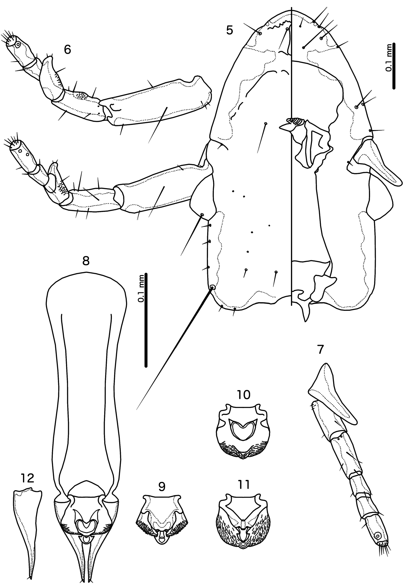
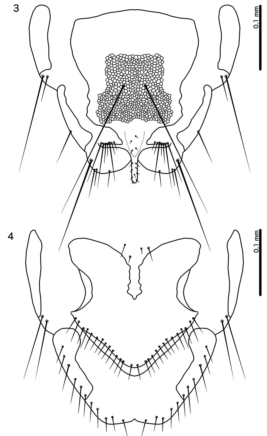
Based on morphological similarities, Calidolipeurus is likely most closely related to Megalipeurus , reflecting the close relationship between the hosts of the two louse genera ( Wang et al. 2013). These two genera share the following characters: coni elongated in both sexes ( Figs 5, 7 View Figs 5–12 ); female subgenital plate with deep median indentation on anterior margin ( Fig. 4 View Figs 3–4 ); female vulval margin convex ( Fig. 4 View Figs 3–4 ); dorsal preantennal suture present ( Fig. 5 View Figs 5–12 ); tergopleurites II–VII medianly separated in both sexes ( Figs 1–2 View Figs 1–2 ); tergopleurites IX +X and XI fused ( Figs 1–2 View Figs 1–2 ).
Calidolipeurus can be separated from Megalipeurus by the following characters: marginal carina interrupted only near as 1 in Megalipeurus , but interrupted near as1 and as 2 in Calidolipeurus ( Fig. 5 View Figs 5–12 ); dorsal preantennal suture transversal in Megalipeurus , but longitudinal in Calidolipeurus ( Fig. 5 View Figs 5–12 ); postmarginal carina extended medianly in Calidolipeurus ( Fig. 5 View Figs 5–12 ), but not in Megalipeurus ; eyes gigantic in Calidolipeurus ( Fig. 5 View Figs 5–12 ), but not in Megalipeurus ; preocular nodus present in Megalipeurus but absent in Calidolipeurus ( Fig. 5 View Figs 5–12 ); stylus reaches beyond distal margin of abdomen in Calidolipeurus ( Fig. 3 View Figs 3–4 ), but not in Megalipeurus ; ss present on male abdominal segments VI–VII in Megalipeurus , but absent in Calidolipeurus ( Fig. 1 View Figs 1–2 ); psps present and of equal size on male tergopleurites IV–VI in Megalipeurus , but present on tergopleurites IV–VII in Calidolipeurus ( Fig. 1 View Figs 1–2 ) with those of tergopleurites VI–VII microsetae; male subgenital plate with lateral extensions following genital opening in Calidolipeurus ( Fig. 3 View Figs 3–4 ), but without such extensions in Megalipeurus ; parameres very broad proximally, with overall shape roughly triangular in Calidolipeurus ( Fig. 12 View Figs 5–12 ), but with slender heads in Megalipeurus ; proximal mesosome with large rounded lobes on each side in Megalipeurus , but without such lobes in Calidolipeurus ( Fig. 9 View Figs 5–12 ); gonopore with lateral hook-shaped elongations in Calidolipeurus ( Fig. 11 View Figs 5–12 ), but roughly rounded in Megalipeurus .
Etymology
Calidolipeurus is constructed from the Latin ‘ calidus ’ for ‘white spot in the forehead’ and the genus name Lipeurus Nitzsch, 1818 , commonly used for long and slender ischnocerans. This refers to the small, elongated dorsal preantennal suture of the type species, which appears white in many specimens in contrast to the otherwise brownish head. Gender: masculine.
Description
Both sexes
Overall body shape elongated, ‘lipeuroid’ ( Figs 1–2 View Figs 1–2 ). Head longer than wide ( Fig. 5 View Figs 5–12 ). Marginal carina widening considerably anteriorly, interrupted laterally near sites of as1 and as2. Frons slightly protruding; marginal carina at frons with serrated posterior margin. Dorsal preantennal suture narrow, median, longitudinal. Ventral carina uninterrupted, indistinct. Head chaetotaxy as in Fig. 5 View Figs 5–12 ; avs3 anterior, near vsms1–2; mds not visible in all examined specimens; s1–3, s5 and s7 present; pos posterior to eye; mts3 only macroseta. Coni elongated. Antennae sexually dimorphic ( Figs 5–7 View Figs 5–12 ). Eyes large. Preocular nodus absent. Marginal temporal carina slender. Dorsal postantennal suture absent.
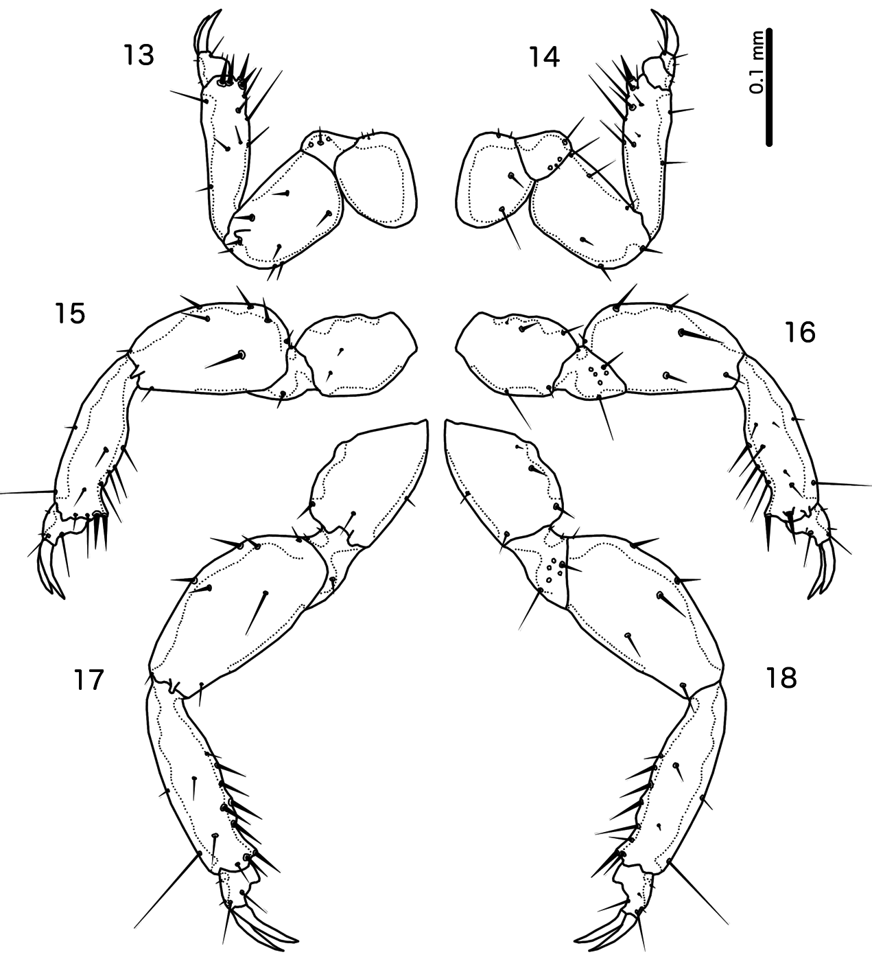
Thoracic and abdominal segments as in Figs 1–2 View Figs 1–2 . Legs as in Figs 13–18 View Figs 13–18 ; similar between sexes but some setae more slender in female than in male. Meso- and metasterna fused. Metepisterna long, with extensive striation. Tergopleurites II–VII medianly separated; tergopleurite VIII medianly continuous in posterior end; tergopleurites IX +X and XI fused. Sternites medianly continuous, reticulated in both sexes. Subgenital plates differ between sexes, formed by fusion of sternites VIII–IX.
Male
Pedicel and flagellomere I with rugose antero-dorsal surface; flagellomere with antero-dorsal claw-like elongation. Thoracic and abdominal chaetotaxy as in Fig. 1 View Figs 1–2 ; ss present on segments II–V; psps present on segments IV–VII, those of VI–VII microsetae. Tergopleurite IX–XI with variable lateral incision reaching apertures of setae. Subgenital plate with extensive reticulation ( Fig. 3 View Figs 3–4 ), distally elongated into protruding stylus; lateral extensions on each side of base of stylus associated with anterior margin of ventral genital opening. Basal apodeme elongated, of roughly equal width, but in some specimens with concave lateral margins ( Fig. 8 View Figs 5–12 ). Mesosome largely unsclerotized, and looks different in specimens with everted or non-everted genitalia. Proximal mesosome flattened to concave, but diffuse and here illustrated approximately ( Figs 8–11 View Figs 5–12 ); on each side sinusoid thickening articulating with parameral heads. Distal mesosome roughly quadratic, with extensive folds and serrations on dorsal and ventral surfaces. Internal sclerite roughly heart-shaped ( Fig. 10 View Figs 5–12 ). Gonopore longer than wide, with lateral hook-shaped in distal end; in anterior end a dark central sclerite may be proximal part of endophallus; one small aperture on each side of presumed endophallus may be microsetae, but setae not visible in examined specimens. In everted genitalia, the gonopore extends beyond distal margin of mesosome, and rugose areas of mesosome contracted ( Figs 8–9 View Figs 5–12 ). Parameres roughly triangular ( Fig. 12 View Figs 5–12 ), median corners of heads almost touching anteriorly; median margin of anterior parameres weakly sclerotized; pst1 not visible; pst2 microsetae near distal end of paramere.
Female
Pedicel and flagellomere I without rugose areas and without claw-like elongation. Head chaetotaxy similar to male, but s7 shorter. Thoracic and abdominal chaetotaxy as in Fig. 2 View Figs 1–2 ; ss present on segments II–V; psps present on segments IV–VII; those on VI–VII microsetae. No lateral incision of tergopleurite IX–XI in examined specimens. Subgenital plate formed by fused sternites VIII–IX ( Fig. 4 View Figs 3–4 ); proximal margin deeply indented medianly. Vulval margin strongly convex; setae situated anterior to margin and do not form distinct sets. Subvulval sclerites absent. Lateral and posterior margins of abdominal segment IX–XI with more or less equally spaced and equal-sized setae.
Host distribution
Only known from the type host of the type species, Rollulus roulroul (Scopoli, 1786) ( Galliformes : Phasianidae ).
Geographical range
Indo-Malayan region, both mainland and the Indonesian archipelago.

No known copyright restrictions apply. See Agosti, D., Egloff, W., 2009. Taxonomic information exchange and copyright: the Plazi approach. BMC Research Notes 2009, 2:53 for further explanation.