
Amrasca Ghauri, 1967
|
publication ID |
https://doi.org/ 10.11646/zootaxa.4353.2.7 |
|
publication LSID |
lsid:zoobank.org:pub:63881055-723D-44A6-A137-006F361512CE |
|
DOI |
https://doi.org/10.5281/zenodo.6021958 |
|
persistent identifier |
https://treatment.plazi.org/id/038087EE-FFA4-FFE6-FF0F-F454A2FFFE4B |
|
treatment provided by |
Plazi |
|
scientific name |
Amrasca Ghauri, 1967 |
| status |
|
Amrasca Ghauri, 1967 View in CoL
Amrasca Ghauri, 1967: 159 View in CoL . Type species: Amrasca splendens Ghauri, 1967 View in CoL by original designation. Sundapteryx Dworakowska, 1970: 708 . Type species: Chlorita biguttula Ishida, 1913 View in CoL by original designation. Synonymized by Dworakowska & Viraktamath, 1975: 530.
Laokayana Dworakowska, 1972: 27 View in CoL . Type species: Empoasca bombaxia Ghauri, 1965 View in CoL by original designation. Synonymized by Dworakowska & Viraktamath, 1975: 530.
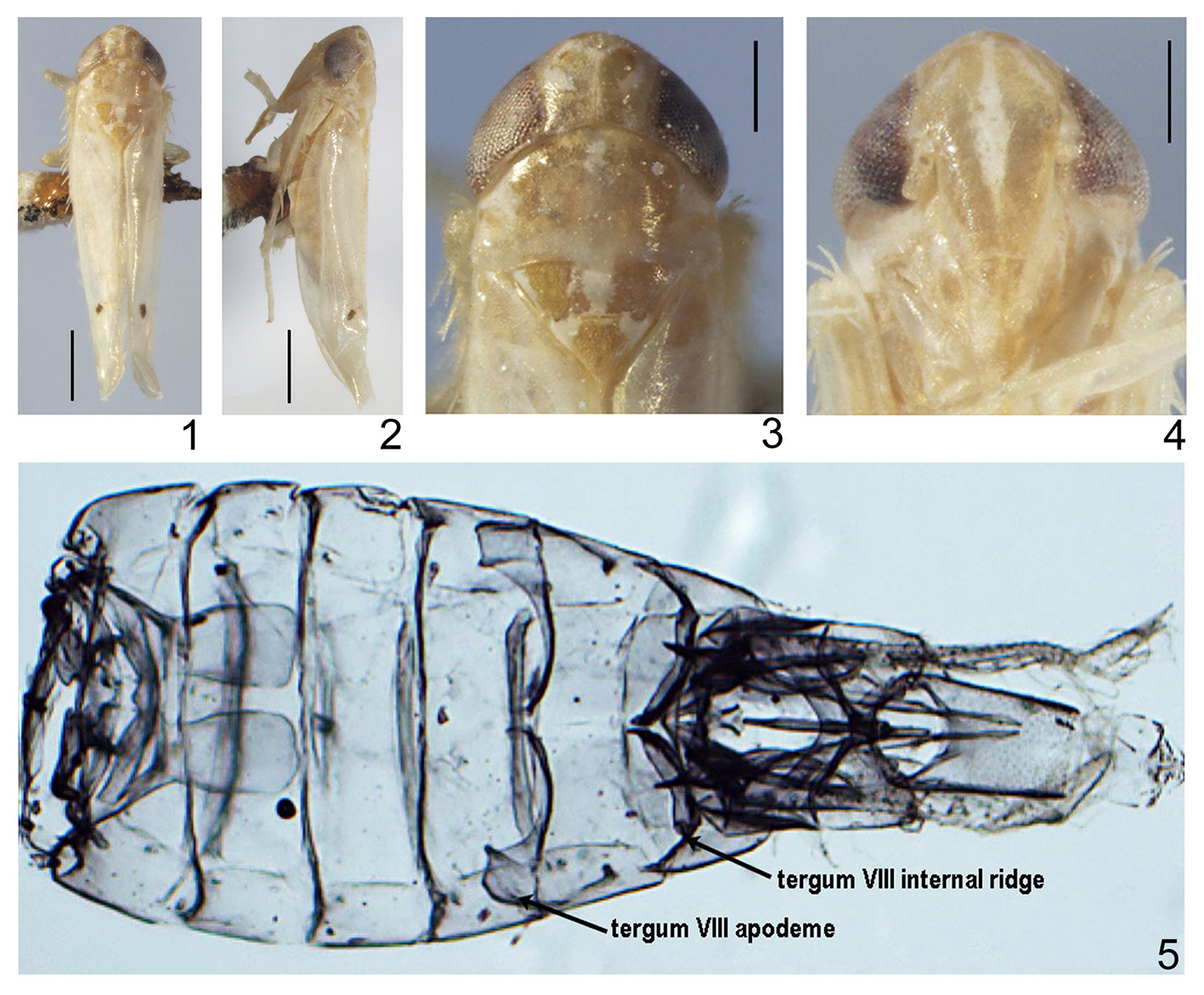
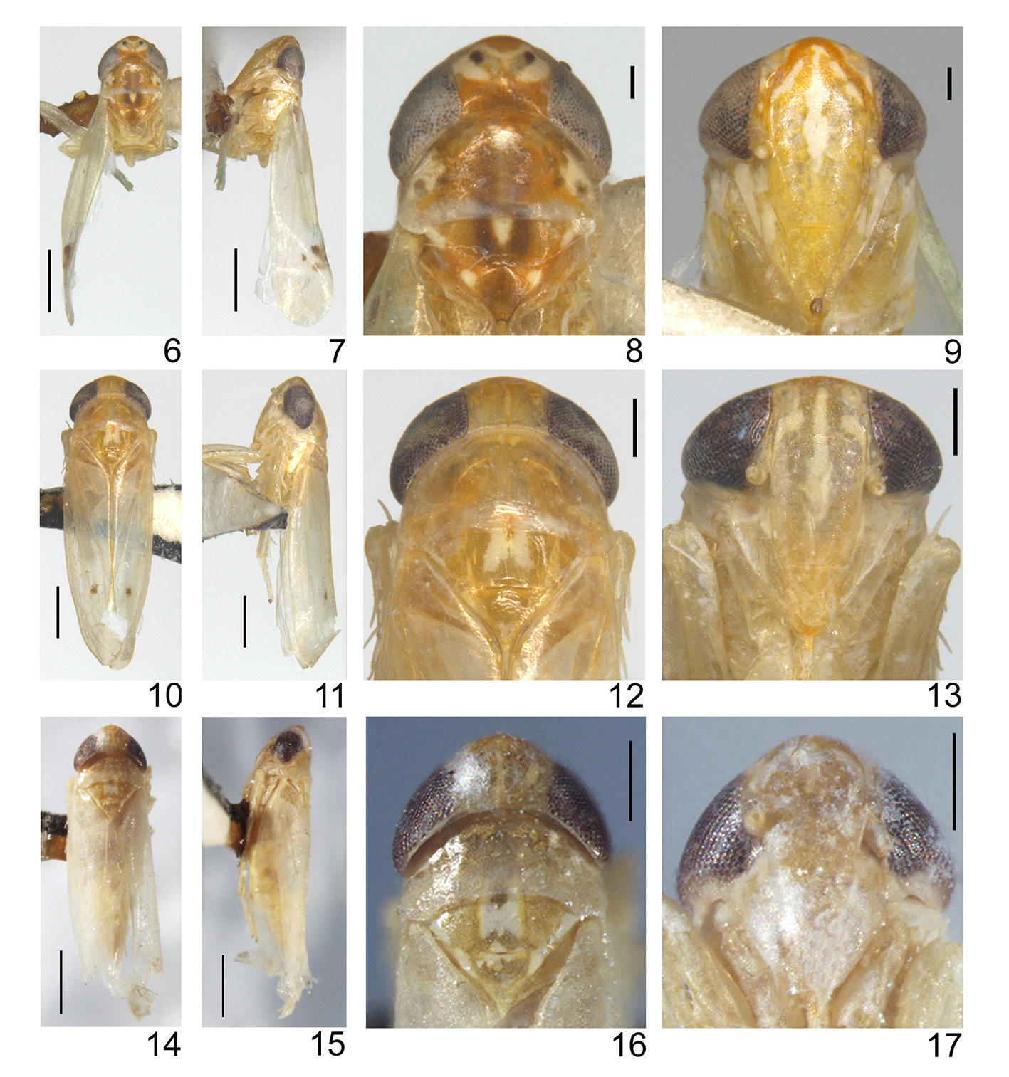
Description. Body delicate. Head including eyes slightly wider than pronotum in dorsal view ( Figs 1, 3 View FIGURES 1–5 , 6, 8, 10, 12, 14, 16 View FIGURES6–17 , 18, 19 View FIGURES18–26 , 37, 39 View FIGURES 37–40 ). Crown produced medially, anterior and posterior margins not parallel, coronal suture not reaching anterior margin ( Figs 1, 3 View FIGURES 1–5 , 6, 8, 10, 12, 14, 16 View FIGURES6–17 , 18, 19 View FIGURES18–26 , 37, 39 View FIGURES 37–40 ). Profile of transition of vertex to face somewhat rounded ( Figs 2 View FIGURES 1–5 , 7, 11, 15 View FIGURES6–17 , 21 View FIGURES18–26 , 38 View FIGURES 37–40 ). Ocelli distinct, located on margin between vertex and frons near eyes ( Figs 3, 4 View FIGURES 1–5 , 8, 9, 12, 13, 16, 17 View FIGURES6–17 , 19, 20 View FIGURES18–26 , 39, 40 View FIGURES 37–40 ). Lateral frontal sutures extended to ocelli but not continuing to midline ( Figs 4 View FIGURES 1–5 , 9, 13, 17 View FIGURES6–17 , 20 View FIGURES18–26 , 40 View FIGURES 37–40 ). Face broad, anteclypeus weakly convex, not swollen ( Figs 4 View FIGURES 1–5 , 9, 13, 17 View FIGURES6–17 , 20 View FIGURES18–26 , 40 View FIGURES 37–40 ). Pronotum moderate to large ( Figs 3 View FIGURES 1–5 , 8, 12, 16 View FIGURES6–17 , 19 View FIGURES18–26 , 39 View FIGURES 37–40 ). Forewing narrow, rounded apically, apical cells occupying nearly one-third of total length; vein RP arising from r cell , MP’ and MP’’+CuA’ dissociated at their bases, both arising from m cell; c and r cells nearly equal in width, both narrower than m and cua cells; 2nd apical cell narrowed at base, broadened apically ( Fig. 22 View FIGURES18–26 ). Hind wing with CuA unbranched ( Fig. 23 View FIGURES18–26 ). Front femur row AV with 1 basal seta distinctly enlarged. Front femur AM1 distinctly enlarged. Middle femur with 1 dorsoapical macroseta. Hind tibia row AV with 4 or 5 preapical macrosetae.
Male basal abdominal apodemes developed, parallel-sided or widely divergent ( Figs 5 View FIGURES 1–5 , 24 View FIGURES18–26 ). Male pygofer with small rigid microsetae scattered over distal portion or restricted to apex of lobe ( Figs 25–29 View FIGURES18–26 View FIGURES 27–36 ); dorsal bridge short ( Figs 25 View FIGURES18–26 , 29 View FIGURES 27–36 ). Ventral appendage present ( Figs 26–28, 30 View FIGURES18–26 View FIGURES 27–36 ). Subgenital plate extended well beyond pygofer side, A and B group setae present or unrecognizable, C group setae sharply pointed near base, reaching or not reaching to apex of the plate, D group setae long and fine ( Figs 26 View FIGURES18–26 , 27, 34 View FIGURES 27–36 ). Paramere broad at base, sharply pointed apically, apophysis bearing prominent dentifer and a few slender setae in apical half ( Fig. 35 View FIGURES 27–36 ). Connective broad anteriorly, strongly narrowed near midlength and tapered to posterior apex, anterior margin straight or weakly concave, without median lobe or with distinct median lobe ( Fig. 33 View FIGURES 27–36 ). Aedeagal shaft tubular, process absent, preatrium developed, dorsoatrium absent ( Figs 31, 32 View FIGURES 27–36 ). Anal tube appendage well developed ( Figs 27, 36 View FIGURES 27–36 ).
Remarks. Among genera in the Empoasca -complex Eastern Hemisphere, Amrasca is most similar to Jacobiasca Dworakowska and Jacobiella Dworakowska in having vein RP arising from r cell , MP’ and MP’’+CuA’ arising from cell m and hind wing vein CuA unbranched, in having the connective not fused with base of aedeagus and in having a well developed ventral pygofer appendage. Amrasca differs from Jacobiasca in having the paramere not strongly curved apically and in lacking a pair of enlarged setae on the pregenital male abdominal sternite, and from Jacobiella in having the subgenital plate not broadened dorsomedially and the anal tube not elongated. Amrasca also differs from both genera in having the male basal abdominal apodemes well developed. Based on its current species composition, Amrasca is somewhat heterogeneous in the form and chaetotaxy of the subgenital plate. Two subgenera have previously been recognized largely based on differences in chaetotaxy: the nominotypical subgenus and A. ( Quartasca ) Dworakowska, which differs from Amrasca (Amrasca) in having the long fine setae of the subgenital plate restricted to the basal half.
Here we recognize a third valid subgenus, A. ( Sundapteryx ) Dworakowska. The latter was originally described as a separate genus by Dworakowska (1970) based on type species Chlorita biguttula Ishida but was subsequently treated as a junior synonym of Amrasca by Dworakowska & Viraktamath (1975). This subgenus is characterized by the presence of macrosetae only in the basal half of the subgenital plate and by the presence of apodemes and other modifications to the male pregenital tergites ( Fig. 5 View FIGURES 1–5 ). Species of the other two currently recognized subgenera have macrosetae extended from the base to or near the apex of the subgenital plate and lack apodemes and other modifications to pregenital abdominal tergites VI-VIII. It may be desirable to subdivide Amrasca further once the genus becomes better known.
Distribution. Oriental and Australian Regions.


No known copyright restrictions apply. See Agosti, D., Egloff, W., 2009. Taxonomic information exchange and copyright: the Plazi approach. BMC Research Notes 2009, 2:53 for further explanation.
|
Kingdom |
|
|
Phylum |
|
|
Class |
|
|
Order |
|
|
Family |
Amrasca Ghauri, 1967
| Xu, Ye, Wang, Yuru, Dietrich, Christopher H., Fletcher, Murray J. & Qin, Daozheng 2017 |
Laokayana
| Dworakowska 1975: 530 |
| Dworakowska 1972: 27 |
Amrasca
| Dworakowska 1975: 530 |
| Dworakowska 1970: 708 |
| Ghauri 1967: 159 |