
Drepanoneura peruviensis ( Fraser 1946 ) Fraser, 1946
|
publication ID |
https://doi.org/ 10.5281/zenodo.183222 |
|
DOI |
https://doi.org/10.5281/zenodo.6231951 |
|
persistent identifier |
https://treatment.plazi.org/id/038087FC-FFC3-4918-FF66-D3D37050F8D8 |
|
treatment provided by |
Plazi |
|
scientific name |
Drepanoneura peruviensis ( Fraser 1946 ) |
| status |
comb. nov. |
Drepanoneura peruviensis ( Fraser 1946) View in CoL comb. nov.
Figures 1 View FIGURE 1 a, 2l–m, 8, 19, 27g – 30g, 36i, 37
Protoneura peruviensis Fraser 1946: 459 –460, figs. 5a–b (description of male, illustration of male S10); — Soukup (1954: 13; listed from Peru); — Rácenis (1959: 472; listed from Peru); — Kimmins (1966: 209; type catalog); — Davies & Tobin (1984: 117; synonymic list); — Steinmann (1997: 453; synonymic list).
Epipleoneura peruviensis Bridges (1994: VII.184; synonymic list); — Tsuda (2000: 12; synonymic list).
Specimens examined. Total 4 ɗ, 2 Ψ. — Holotype ɗ, Peru, Loreto Department, Mishuyacu [near Iquitos] (03°51'S, 73°13'W), 0 2 vii 1930 (BMNH); 1 ɗ, same data as holotype but Río Amazonas, Iquitos, ix 1938, leg. G.G. Klug (RWG); 2 ɗ, same data but i/ iv 1940 (RWG); 2 Ψ, same data but vi 1939 (RWG).
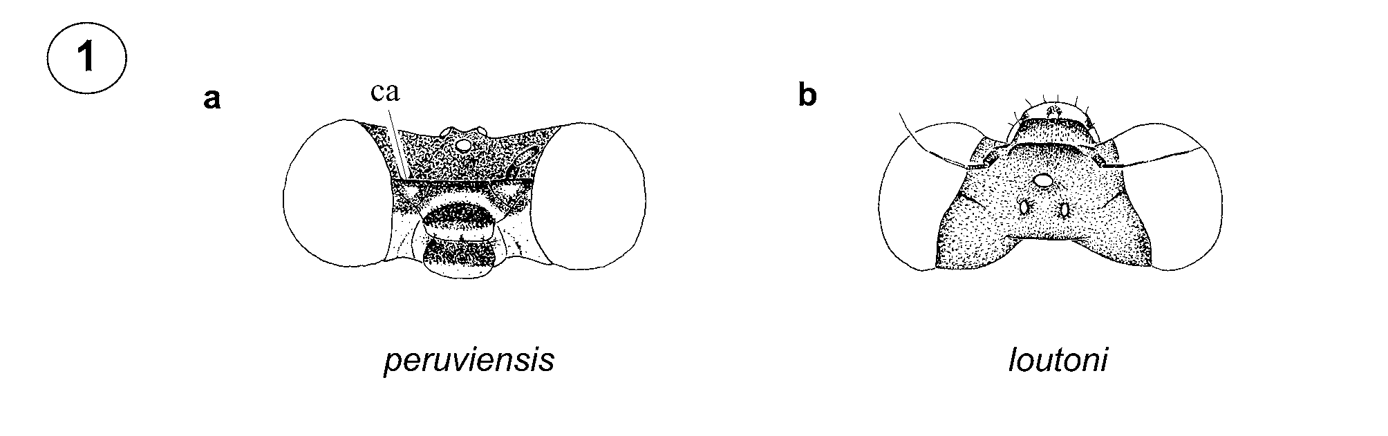
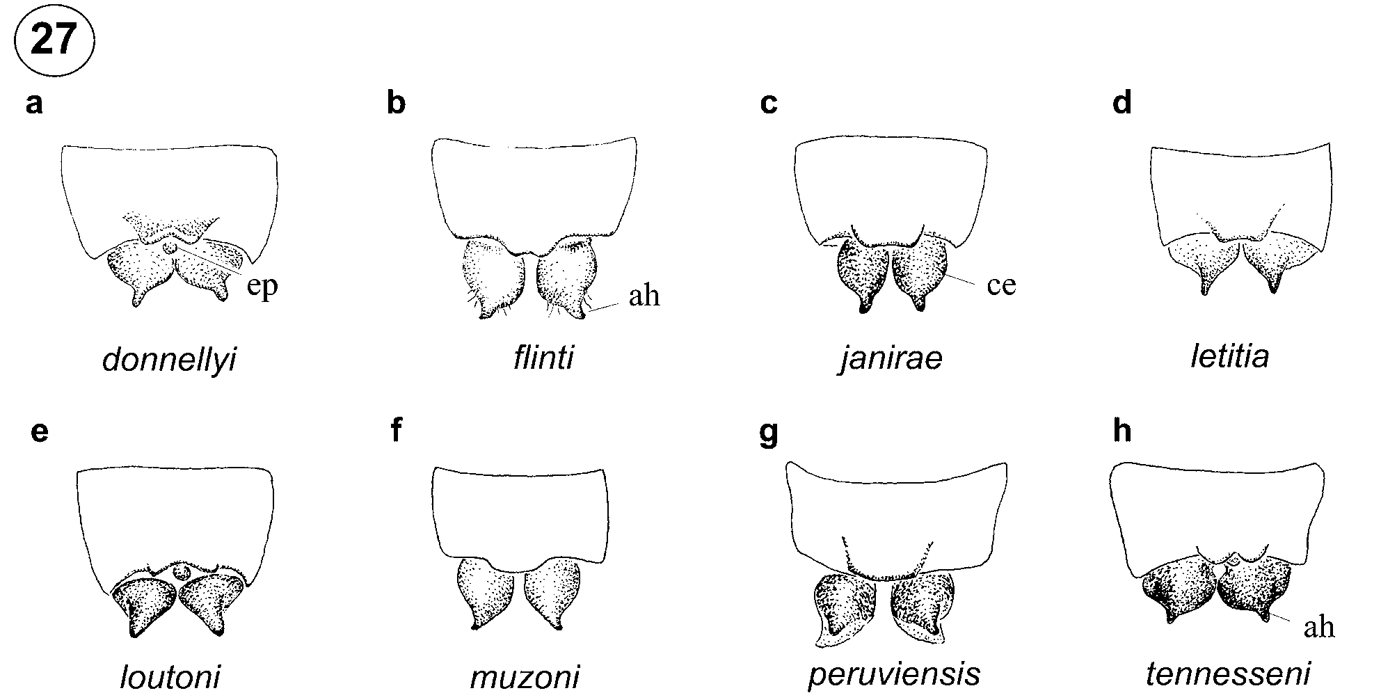
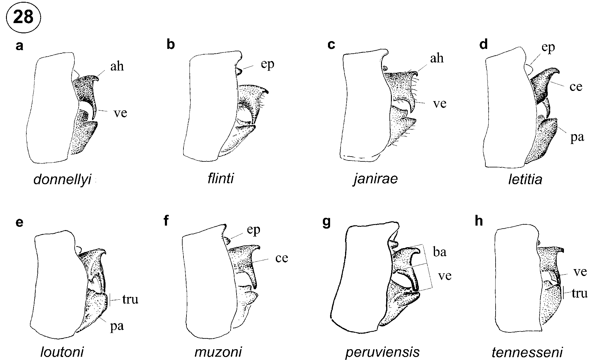
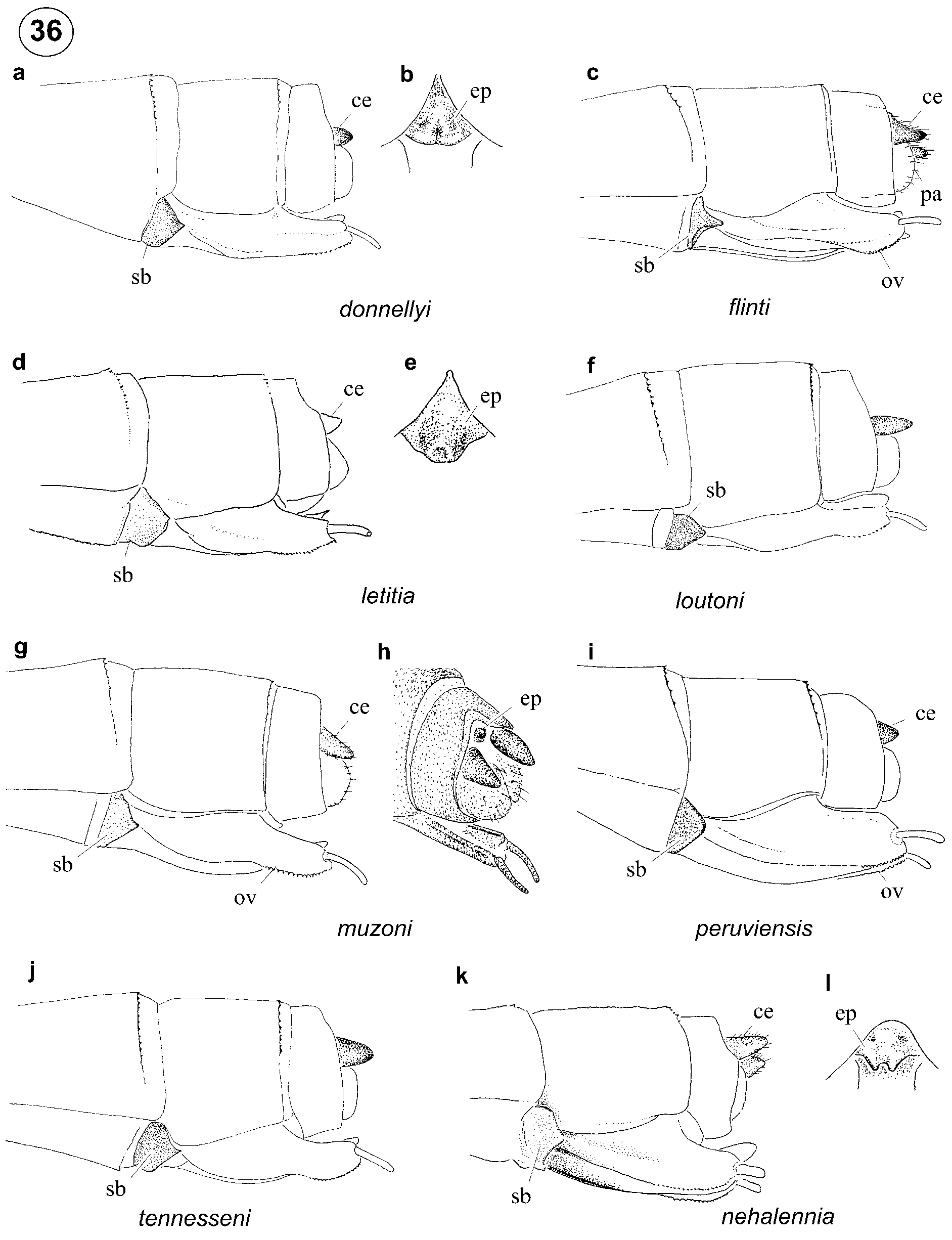
Characterization. Posterior prothoracic lobe smoothly convex in male, with a wide and short semicircular medio-ventral projection in female ( Fig. 8 View FIGURES 3 – 10 ). Pterothoracic dorsum dark to upper 0.7 of metapleural suture in male ( Fig. 2 View FIGURE 2 l), to mid-height of metepisternum and with a black stripe along anterior margin of metapleural suture in female ( Fig. 2 View FIGURE 2 m). Apex of male genital ligula with a deep u-shaped cleft ( Fig. 19 View FIGURES 13 – 20 a) and latero-distal lobes short, broad, and curved medially ( Figs. 19 View FIGURES 13 – 20 b–c). Dorso-posterior margin of male S10 projected posteriorly ( Figs. 27 View FIGURE 27 g– 28g). Ventral branch of male cercus longer than base of cercus, approximately cylindrical ( Fig. 28 View FIGURE 28 g), in posterior view arising at mid-width and convergent to branch of opposite cercus at tip ( Fig. 30 View FIGURE 30 g). Paraproct pointed ( Fig. 28 View FIGURE 28 g). Ventral and dorsal sides of sub-basal plate of female ovipositor linear ( Fig. 36 View FIGURE 36 i).
Diagnosis. Male of D. peruviensis shares the ventral branch of cercus arising at mid-width and convergent to the branch of opposite cercus at tip only with D. flinti and D. muzoni ( Fig. 30 View FIGURE 30 g); it differs from both by having the apex of genital ligula with a deep u-shaped cleft ( Fig. 19 View FIGURES 13 – 20 a), and further from D. muzoni by the ventral branch of male cercus being longer than the base of cercus ( Fig. 28 View FIGURE 28 g) and the absence of pale mesepisternal stripes ( Fig. 2 View FIGURE 2 l). Female is unique by the presence of a medio-ventral semicircular projection on posterior margin of pronotum ( Fig. 8 View FIGURES 3 – 10 ) and by both ventral and dorsal sides of sub-basal plate of ovipositor linear ( Fig. 36 View FIGURE 36 i).
Biology. Adults were collected at streams and rivers.
Distribution. Loreto Department, Peru ( Fig. 37 View FIGURE 37 ).


No known copyright restrictions apply. See Agosti, D., Egloff, W., 2009. Taxonomic information exchange and copyright: the Plazi approach. BMC Research Notes 2009, 2:53 for further explanation.
|
Kingdom |
|
|
Phylum |
|
|
Class |
|
|
Order |
|
|
Family |
|
|
Genus |
Drepanoneura peruviensis ( Fraser 1946 )
| Ellenrieder, Natalia Von & Garrison, Rosser W. 2008 |
Protoneura peruviensis
| Fraser 1946: 459 |