
Epipotoneura machadoi, Ellenrieder, Natalia Von & Garrison, Rosser W., 2008
|
publication ID |
https://doi.org/ 10.5281/zenodo.183222 |
|
DOI |
https://doi.org/10.5281/zenodo.6231962 |
|
persistent identifier |
https://treatment.plazi.org/id/038087FC-FFC5-491F-FF66-D50471F9FB8E |
|
treatment provided by |
Plazi |
|
scientific name |
Epipotoneura machadoi |
| status |
sp. nov. |
Epipotoneura machadoi View in CoL sp. nov.
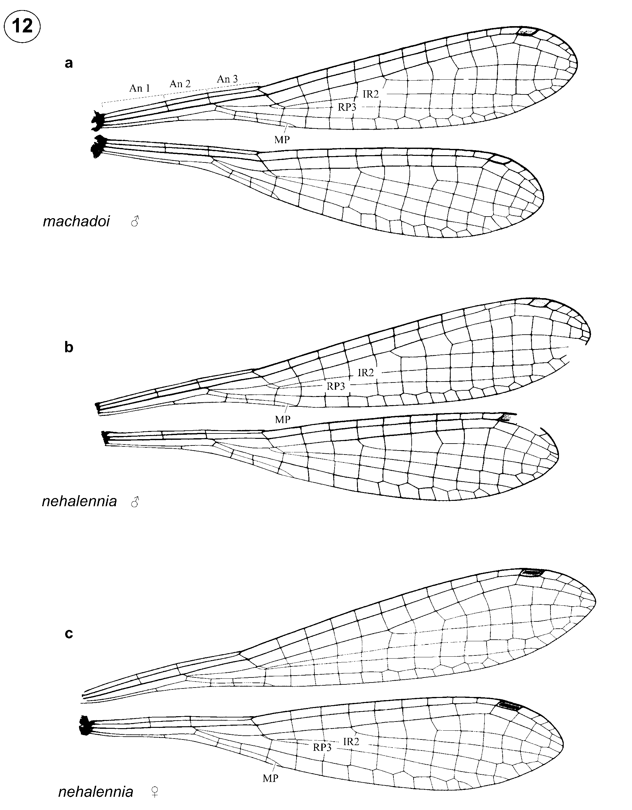
Figures 12 View FIGURE 12 a, 25, 34, 38
Etymology. We name this species machadoi (noun in the genitive case) in honor of our friend and colleague Angelo B.M. Machado, in recognition of his manifold contributions to the knowledge of neotropical Odonata and his continuous assistance in our studies.
Specimens examined. Total 2 ɗ.— Holotype ɗ: Brazil, Pará State, Rio Xingu Camp, ca. 60 km S of Altamira, Igarapé Jabutí, Malaise trap (03°39'S, 52°22'W), 11 x 1986, leg. P. Spangler & O. Flint (USNM). Paratype ɗ, same data as holotype but 15 x 1986 (RWG).
Male holotype. Head. Labium, ventral fourth of labrum, base of mandibles, genae, anteclypeus, and ventral half of antefrons ivory white; remainder of head including rear of head black; dorsum of head with slight green metallic luster.
Thorax. Largely black with metallic green luster; prothorax with rim of anterior lobe and ventral margin of propleuron light yellow; distal tip of mesostigmal plate and metathorax except for upper fourth of metepisternum pale yellow; venter of thorax, coxae, sides of femora pale yellow; posterior surfaces of femora and trochanter-femoral juncture brown, tip of femora edged with black; remainder of legs pale yellow, armature black; 4 spurs on metafemora, 6 on metatibiae. Hw ( Fig. 12 View FIGURE 12 a) 5.1 times as long as wide; 9 Px in Fw, 8 Px in Hw; MP reaching wing margin slightly distal to basal third of first cell posterior to vein descending from subnodus; IR2 arising at vein descending from subnodus; IR2 and RP 3 separated by a short crossvein one cell posterior to their origin; RP 2 beginning closer to Px 4 in Fw and closer to Px 3 in Hw; pt pale brown with yellow marginal hairline, shorter than underlying cell, with costal side slightly longer than posterior side.
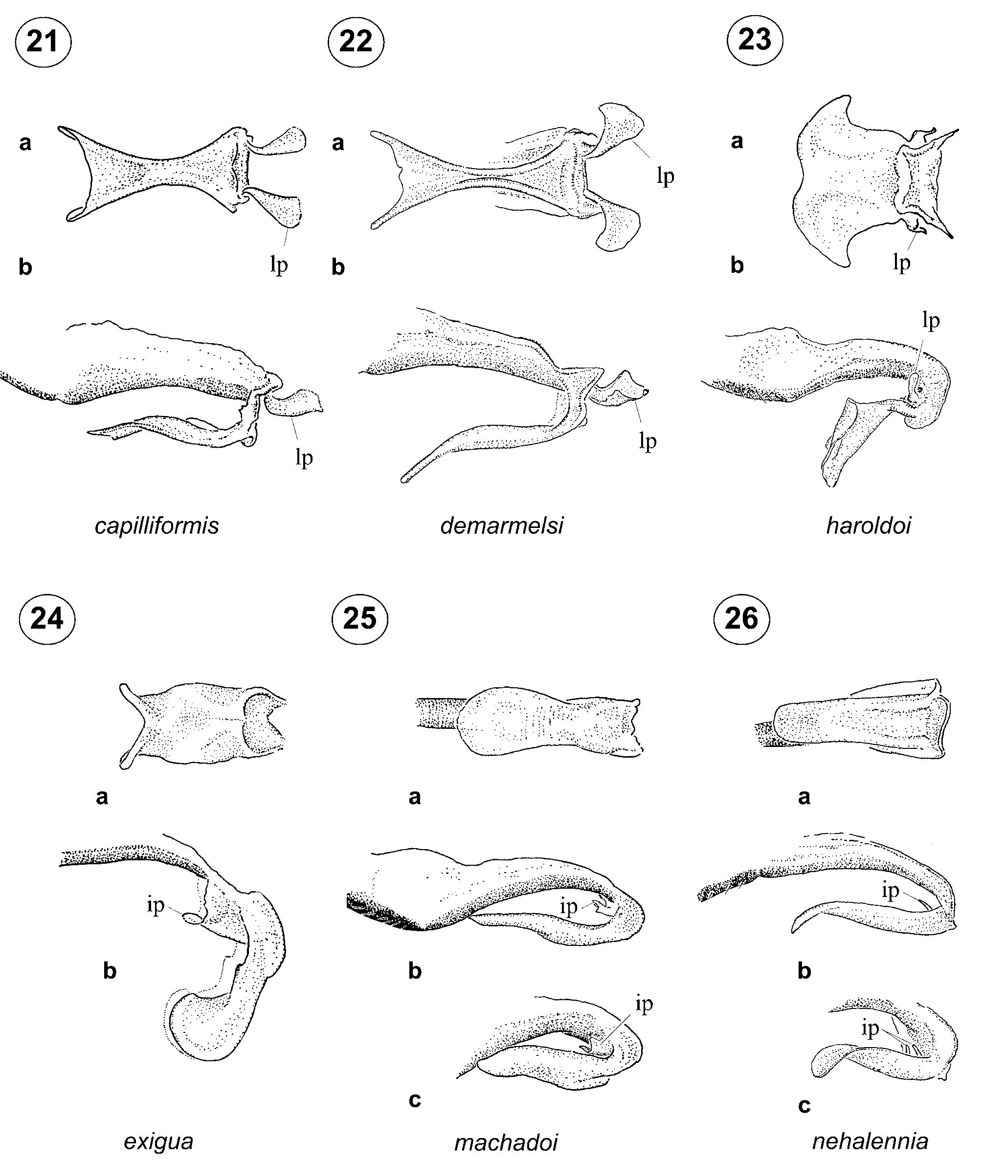
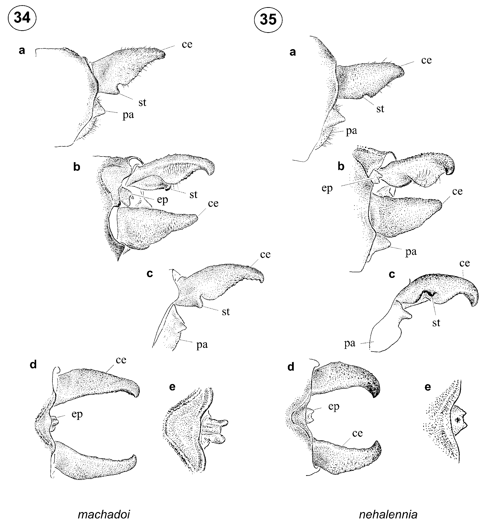
Abdomen. Dorso-laterally black except for narrow yellow bands interrupted medio-dorsally by black on base of S3–7; latero-ventral portion of terga and sterna pale brown to yellow, apical third of S9 pale yellow and connecting with yellow ventrally. Genital ligula with apex entire ( Fig. 25 View FIGURES 21 – 26 a) and a single bifid inner process distal to flexure ( Figs. 25 View FIGURES 21 – 26 b–c). Cercus longer than male S 10 in lateral view, widest at level of sub-basal tooth ( Figs. 34 View FIGURES 34 – 35 a, c), which is prominent and visible in medio-dorsal view ( Fig. 34 View FIGURES 34 – 35 b).
Dimensions. Total length 28.7 mm; abdomen length 24.3 mm; Hw 13.6 mm.
Paratype. Paratype is similar to holotype except for stronger metallic green luster, dorso-lateral yellow on S9 replaced by dull orange and interrupted by black above. Hw is 4.95 times as long as wide; there are 8 Px in Fw, 7 Px in Hw; and MP in Fw reaches wing margin slightly distal to half length of first cell posterior to vein descending from subnodus. Female unknown.
Dimensions. Male (n = 1): total length 27.9 mm; abdomen 23.5 mm; Hw 13.6 mm.
Diagnosis. Male of Epipotoneura machadoi differs from its sibling species E. nehalennia only by morphology of cercus and genital ligula. Cercus in E. machadoi is slightly longer than S10 ( Fig. 34 View FIGURES 34 – 35 a) and widest at base, and there is a single well-developed medially curved sub-basal tooth which extends below ventral margin of cercus (best seen in medio-dorsal, lateral, and ventro-lateral views; st, Figs. 34 View FIGURES 34 – 35 a–c). Cercus in E. nehalennia is sub-equal to S10 ( Fig. 35 View FIGURES 34 – 35 d), comparatively shorter than in E. machadoi ( Fig. 34 View FIGURES 34 – 35 d) and parallelsided at base, and the medially curved sub-basal tooth is hidden in lateral view ( Fig. 35 View FIGURES 34 – 35 a) but it can be seen in latero-ventral view (st, Fig. 35 View FIGURES 34 – 35 c). Genital ligula of E. machadoi has one bifid inner process ( Figs. 25 View FIGURES 21 – 26 b–c), while there are two relatively smaller digit-like inner processes in E. nehalennia ( Figs. 26 View FIGURES 21 – 26 b–c).
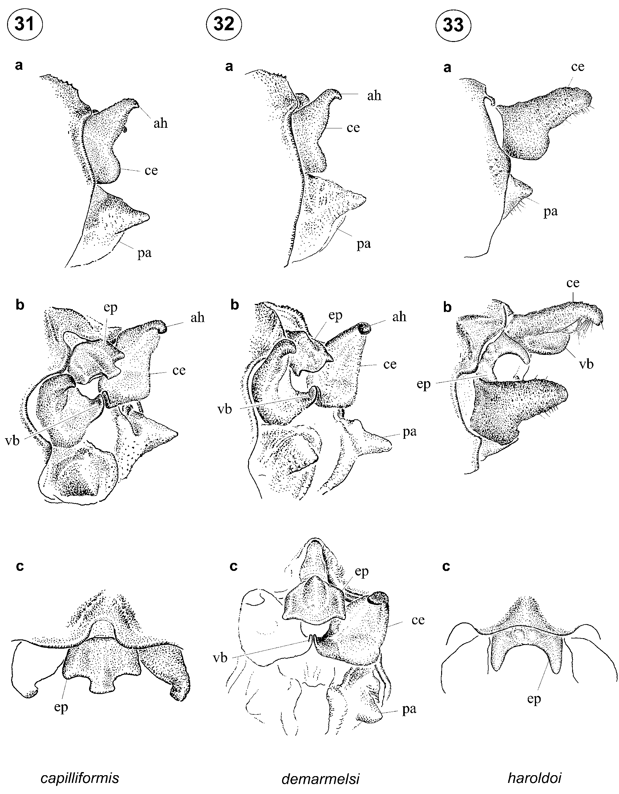
Both species superficially resemble Epipleoneura haroldoi Santos 1964 by caudal appendage morphology ( Figs. 33 View FIGURES 31 – 33 a–b), but in that species the epiproct is large, sclerotized, and apically bifid so that the lateral margins approximate the medial margins of the cerci ( Fig. 33 View FIGURES 31 – 33 c); the epiproct in Epipotoneura is small, not sclerotized, and its sides do not come into contact with the mesal margins of the cerci ( Figs. 34 View FIGURES 34 – 35 d–e–35d–e). The genital ligula of Epipotoneura ( Figs. 25–26 View FIGURES 21 – 26 ) lacks the modified latero-posterior ( Figs. 21-23 View FIGURES 21 – 26 ) pedunculate processes which uniquely characterize Epipleoneura .
Biology. Types were collected at a river.
Distribution. Pará State, Brazil ( Fig. 38 View FIGURE 38 ).

No known copyright restrictions apply. See Agosti, D., Egloff, W., 2009. Taxonomic information exchange and copyright: the Plazi approach. BMC Research Notes 2009, 2:53 for further explanation.