
Drepanoneura, Ellenrieder, Natalia Von & Garrison, Rosser W., 2008
|
publication ID |
https://doi.org/ 10.5281/zenodo.183222 |
|
DOI |
https://doi.org/10.5281/zenodo.6231932 |
|
persistent identifier |
https://treatment.plazi.org/id/038087FC-FFD8-4907-FF66-D1DE728BFE35 |
|
treatment provided by |
Plazi |
|
scientific name |
Drepanoneura |
| status |
gen. nov. |
Drepanoneura View in CoL gen. nov.
Figures 1 View FIGURE 1 , 2 View FIGURE 2 , 3–9 View FIGURES 3 – 10 , 11 View FIGURE 11 a–b, 13–20, 27–30, 36a–j, 37
Type species: Drepanoneura loutoni sp. nov. by present designation.
Etymology. Latinized from Greek drepanon meaning 'sickle' — referring to the shape of male cerci — and Greek neura, a common suffix for protoneurid genera.
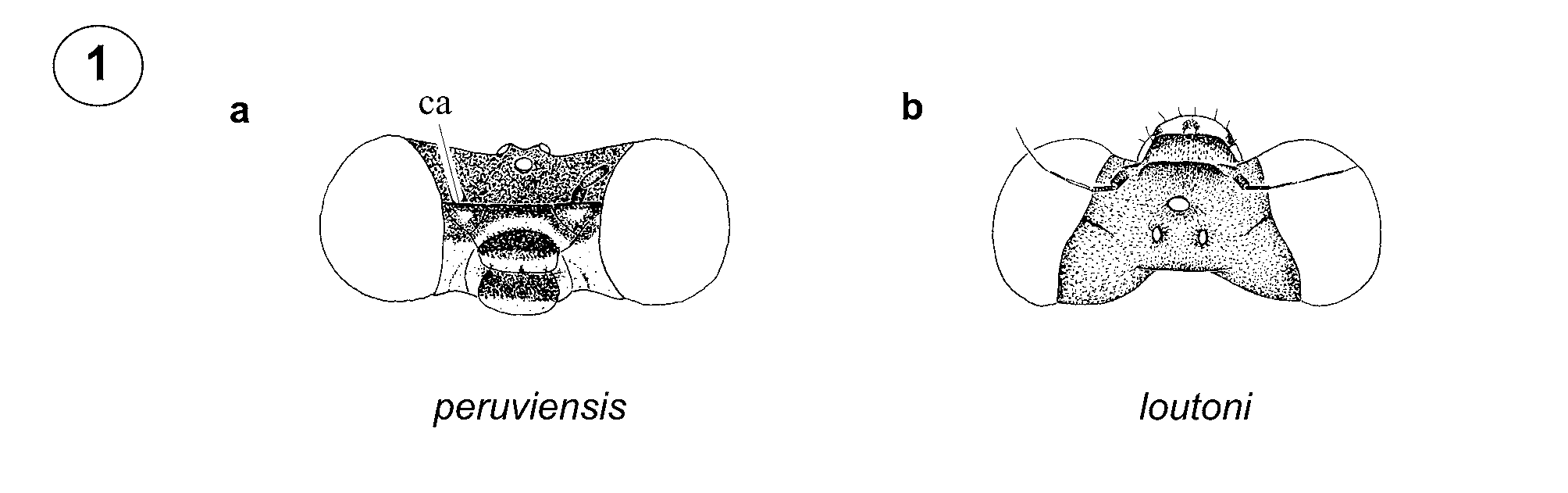
Generic characterization. Head. Frons angulate in profile, with carina continued on dorsal surface of antennifer (ca, Fig. 1 View FIGURE 1 a); labium, ventral third of labrum, anteclypeus, and triangular spot on anterior surface of antennifer pale yellow; base of mandibles, genae, and ventral half of antefrons pale blue ( Fig. 1 View FIGURE 1 a), remainder of head black ( Fig. 1 View FIGURE 1 b).
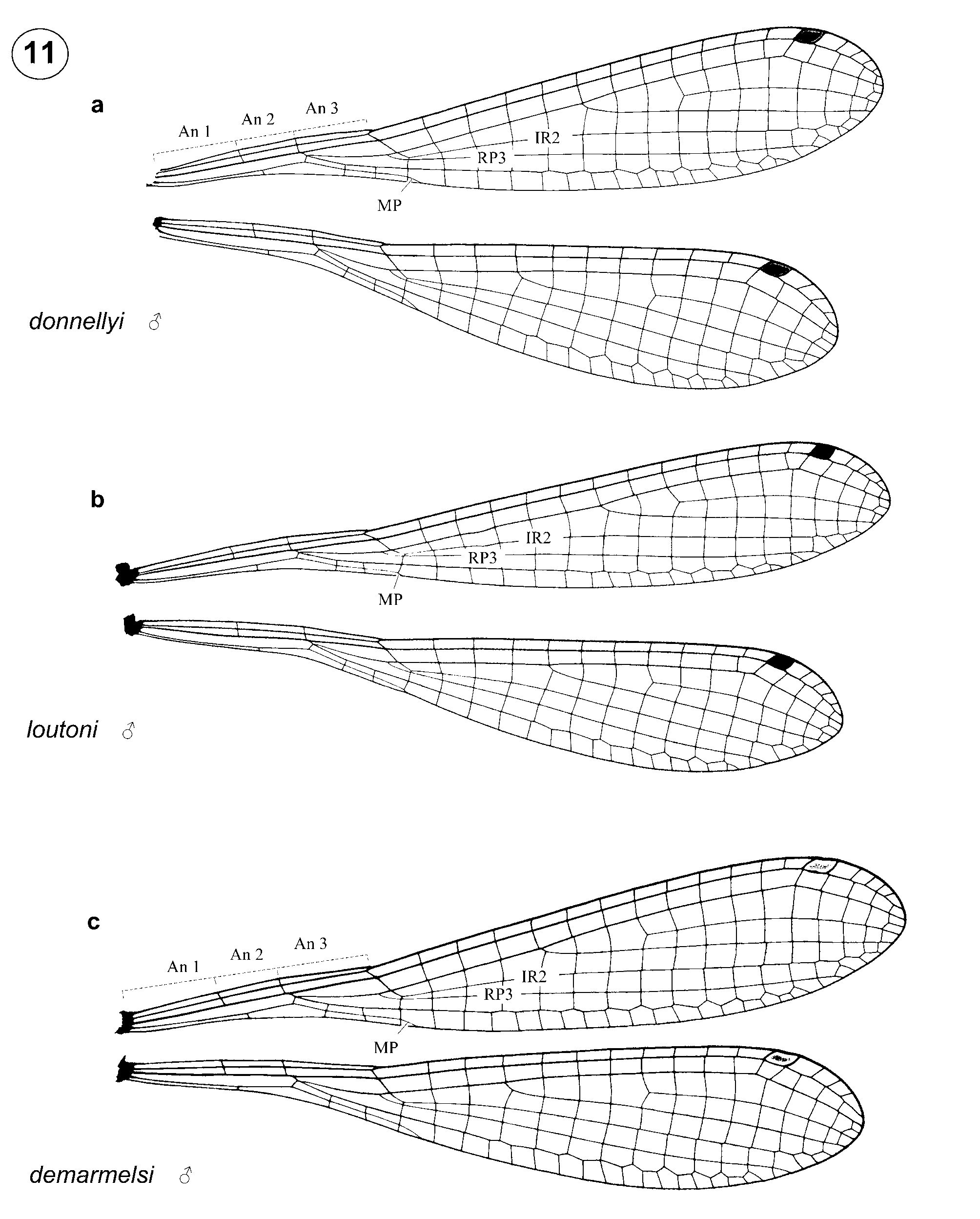
Thorax. Pterothorax black with metallic copper, greenish or bluish reflections; part of metepisternum, metepimeron, and venter pale yellow ( Fig. 2 View FIGURE 2 ). Mesepisternum with a narrow, yellow humeral stripe ( Figs. 2 View FIGURE 2 b, e, g–k), which can become obscured in mature specimens of some species. Legs relatively long and thin, with hind femur reaching or surpassing anterior margin of S1 ( Fig. 2 View FIGURE 2 ); spurs on femora and tibiae shorter than twice intervening spaces ( Fig. 2 View FIGURE 2 ); pretarsal claws each with well developed supplementary tooth. Postero-lateral margin of prothorax with moderate lobes, not longer than wide, and posterior margin of pronotum smoothly rounded in males. In females interspecifically variable, from smoothly curved ( Fig. 5 View FIGURES 3 – 10 ) or linear ( Fig. 4 View FIGURES 3 – 10 ), to medially cleft ( Fig. 7 View FIGURES 3 – 10 ) or trilobed ( Figs. 6, 8–9 View FIGURES 3 – 10 ), lacking projections along posterior margin ( Figs. 4–5 View FIGURES 3 – 10 ), or with one to five ( Figs. 3, 6-9 View FIGURES 3 – 10 ). Mesostigmal plates in both sexes triangular and flat, with transverse length shorter than width of medial disc ( Figs. 3 View FIGURES 3 – 10 c–9c). Wings ( Fig. 11 View FIGURE 11 a–b) one sixth or less as wide as long; antenodal space 1 shorter than twice the length of 2, and slightly shorter than or as long as 3; CuA and CuP&AA completely fused to wing margin; MP reaching wing margin at vein descending from subnodus or within the basal half of first cell posterior to it; IR2 arising at vein descending from subnodus or slightly distal to it (intra- and interspecifically variable); IR2 and RP 3 separated by a short crossvein or joined one cell posterior to their origin (intra- and interspecifically variable); divergence of RP-RA (arculus) distal to Ax 2; RP 2 in Fw beginning closer to Px 4–7 (most frequently at 5) and in Hw closer to Px 3–4 (most frequently at 3); pt shorter than or as long as underlying cell, with its costal side shorter than or as long as its posterior side.
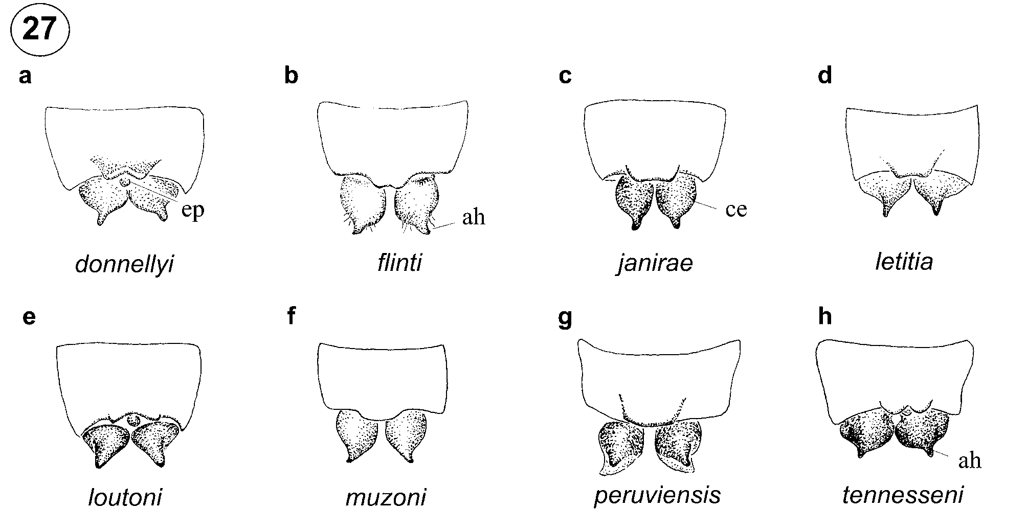
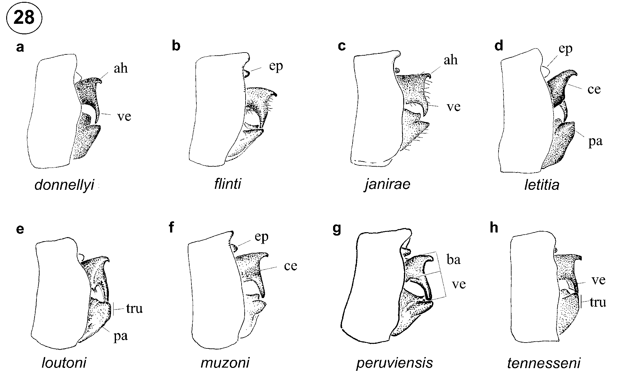
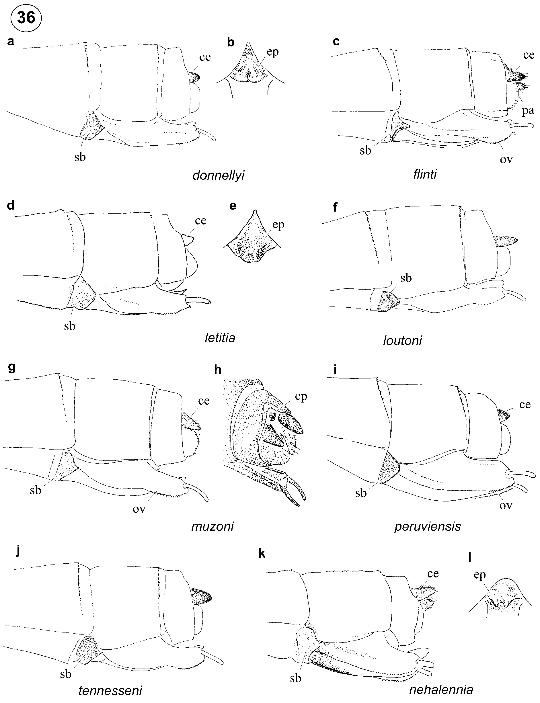
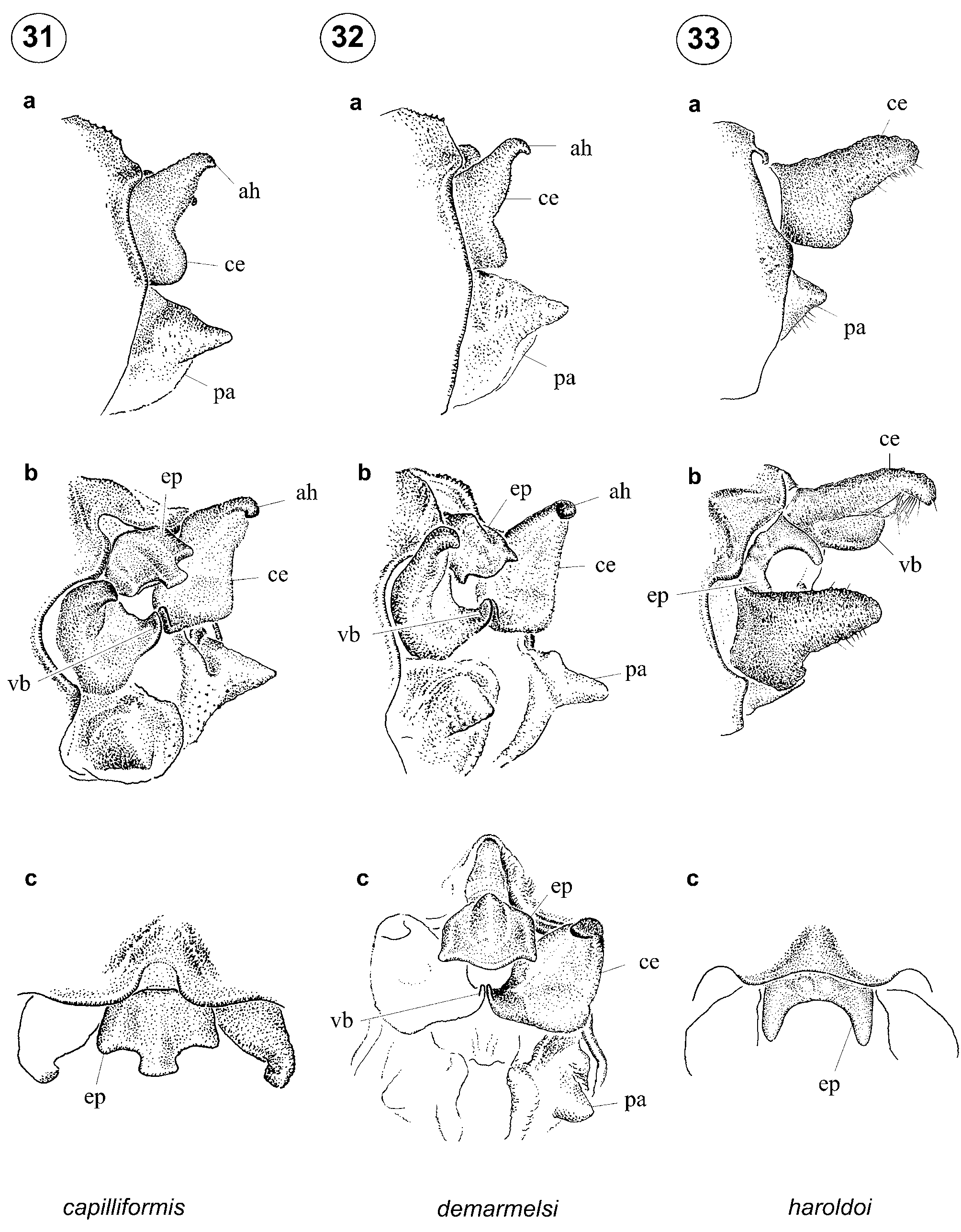
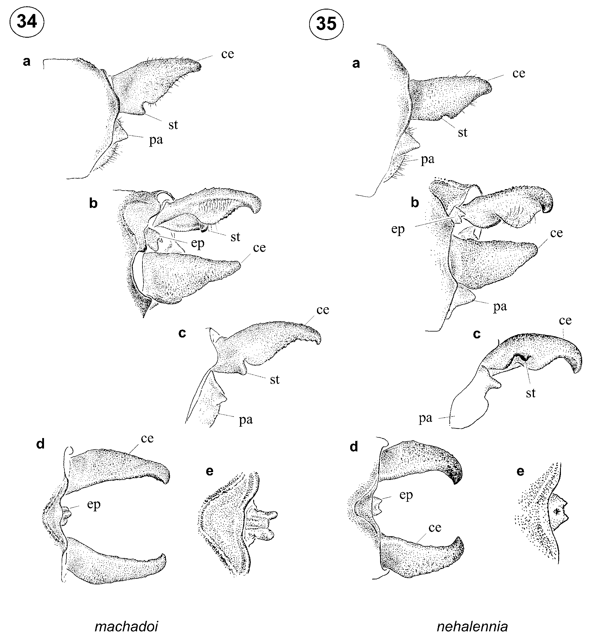
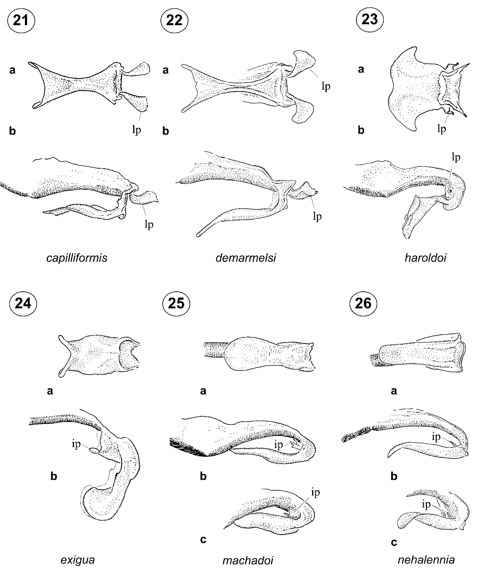
Abdomen. Abdomen dorso-laterally entirely or almost entirely black with metallic reflections, except for narrow distal yellow rings on S 3–6 in some species; latero-ventral portion of terga and sterna pale brown to yellow. Genital ligula lacking an inner fold basal to flexure, with a short medial membranous triangular inner process distal to flexure (ip, Figs. 13 View FIGURES 13 – 20 b–16b, 20b); apex entire ( Fig. 13 View FIGURES 13 – 20 a) or with a v- ( Figs. 14 View FIGURES 13 – 20 a–16a, 18a) or ushaped cleft ( Figs. 17 View FIGURES 13 – 20 a, 19a–20a), and with latero-distal corners projected into short (ld, Figs. 14 View FIGURES 13 – 20 a, c–16a, c, 18a, c–19a, c) to long (ld, Figs. 13 View FIGURES 13 – 20 a, c, 17a, c, 20a, c) lateral lobes curved medially ( Figs. 14 View FIGURES 13 – 20 c–20c) or externally ( Fig. 13 View FIGURES 13 – 20 c). Postero-dorsal margin of male S10 projected posteriorly ( Figs. 28 View FIGURE 28 b–d, f–g) or not projected ( Figs. 28 View FIGURE 28 a, e, h). Male cercus in lateral view shorter than S10, with a pointed ventro-apical process perpendicular to main axis, slightly shorter to slightly longer than base of cercus, and with a short dorso-apical hook (ah, Figs. 27–30 View FIGURE 27 View FIGURE 28 View FIGURE 29 View FIGURE 30 ). Male paraproct bilobed in posterior view, subequal to cercus in length ( Fig. 28 View FIGURE 28 ). Epiproct much smaller than cercus, rounded and undifferentiated in both sexes (ep, Figs. 27–30 View FIGURE 27 View FIGURE 28 View FIGURE 29 View FIGURE 30 , 36 View FIGURE 36 b, e, h). Female cerci conical, shorter than S10; outer valves of ovipositor with a single row of teeth along distal fourth ( Figs. 36 View FIGURE 36 a, c–d, f–g, i–j); sub-basal plate of ovipositor meeting its counterpart ventrally, with dorsal side linear ( Fig. 36 View FIGURE 36 i) to concave ( Figs. 36 View FIGURE 36 a, c–d, g, j); tip of ovipositor (excluding stylus) extending beyond posterodorsal margin of S10 but not surpassing tip of cerci ( Figs. 36 View FIGURE 36 a, c–d, f–g, j). Larva unknown.
Diagnosis. Among neotropical Protoneuridae , Drepanoneura differs from Lamproneura De Marmels 2003 , Idioneura Selys 1860 , Junix Rácenis 1968 , Neoneura Selys 1860 , Peristicta Hagen in Selys 1860, and Proneura Selys 1889 by the complete fusion of CuA and CuP&AA to wing margin (absence of anal vein). Among genera which share this state ( Forcepsioneura Lencioni 1999 , Microneura Hagen in Selys 1886, Phasmoneura Williamson 1916 , Psaironeura Williamson 1915 , and Roppaneura Santos 1966 ), both sexes of Drepanoneura differ by the carinated antennifer (ca, Fig. 1 View FIGURE 1 a), which is cylindrical in the five genera mentioned. Drepanoneura differs from Protoneura by antenodal space 1 being shorter than twice the length of 2 ( Figs. 11 View FIGURE 11 a–b); from Epipleoneura and Epipotoneura by the small unmodified button-shaped epiproct ( Figs. 27–30 View FIGURE 27 View FIGURE 28 View FIGURE 29 View FIGURE 30 , 36 View FIGURE 36 b, e, h), which in Epipleoneura is always enlarged and longer than wide ( Figs. 31 View FIGURES 31 – 33 b–c–33b–c) and in Epipotoneura is small but bifid ( Figs. 34 View FIGURES 34 – 35 b, d–e–35b, d–e, 36l). Additionally, male cercus morphology (approaching an inverted 'L', with a ventro-apical process perpendicular to main axis and subequal in length to base of cercus, Figs. 27–30 View FIGURE 27 View FIGURE 28 View FIGURE 29 View FIGURE 30 ) is unique for Drepanoneura . In Epipleoneura there is usually a ventro-basal swelling or branch ( Figs. 31 View FIGURES 31 – 33 b–c–33b–c) and in Epipotoneura there are no apical or basal ventral branches, and there is instead a small sub-basal tooth (st in Figs. 34 View FIGURES 34 – 35 a–c–35a, c). The absence of an inner fold basal to flexure in male genital ligula of Drepanoneura ( Figs. 13 View FIGURES 13 – 20 b–c–20b–c) is shared only with Epipleoneura ( Figs. 21 View FIGURES 21 – 26 b– 23b), Epipotoneura ( Figs. 25 View FIGURES 21 – 26 b–26b) and Phasmoneura ( Fig. 24 View FIGURES 21 – 26 b), and the presence of an inner membranous process distal to flexure (ip in Figs. 13–20 View FIGURES 13 – 20 ) is shared only with Epipotoneura ( Figs. 25 View FIGURES 21 – 26 b–c–26b–c) and Phasmoneura ( Fig. 24 View FIGURES 21 – 26 b). However, the morphology of the inner processes in these three genera is different, consisting of a single small medial narrow process in Drepanoneura ( Figs. 13 View FIGURES 13 – 20 b–16b, 20b), a medial process bifid at tip to completely split into two narrow small processes in Epipotoneura ( Figs. 25 View FIGURES 21 – 26 b–26b), and a large, ushaped wide fold medially attached to the distal segment of genital ligula in Phasmoneura ( Fig. 24 View FIGURES 21 – 26 b). Epipleoneura further differs from Drepanoneura ( Figs. 13–20 View FIGURES 13 – 20 ) and Epipotoneura ( Figs. 25–26 View FIGURES 21 – 26 ) by the presence of a pair of latero-posterior membranous processes (lp, Figs. 21–23 View FIGURES 21 – 26 ).
The key to males of neotropical genera of Protoneuridae provided by Pessacq (2008) can be modified to include Drepanoneura as follows:
8. Cerci usually shorter than S10 (i.e. Figs. 28 View FIGURE 28 a–h).........................................................................................9 8'. Cerci and/or paraprocts usually longer than S10..................................10 [to Microneura and Protoneura ] 9. Epiproct enlarged, sclerotized, longer than wide, and adjacent to mesal margin of cerci; cercus with a ventro-basal branch (i.e. Figs. 31 View FIGURES 31 – 33 b–33b); genital ligula lacking inner processes on distal segment and with a pair of latero-posterior outer processes at level of flexure (i.e. lp, Figs. 21–23 View FIGURES 21 – 26 ).................... Epipleoneura 9'. Epiproct small, rounded, not forming a sclerotized plate, and not adjacent to mesal margin of cerci ( Fig. 30 View FIGURE 30 ); cercus with a ventro-apical process perpendicular to main axis and subequal in length to base of cercus ( Figs. 28–30 View FIGURE 28 View FIGURE 29 View FIGURE 30 ); genital ligula with a small medial narrow inner process distal to flexure (ip, Figs. 13– 20 View FIGURES 13 – 20 ) and lacking outer latero-posterior processes at level of flexure..................................... Drepanoneura Distribution. Members of this genus occur along rivers and streams within forests and occupy largely allopatric distributions from Panama south through foothills of the Andean cordillera in Colombia, Ecuador, and Peru. Three species, D. flinti , D. peruviensis , and D. janirae , occur in the Amazonian regions of Colombia, Peru, SW Brazil, and NW Bolivia; D. muzoni is sympatric with D. tennesseni in Ecuador and with D. loutoni in Peru ( Fig. 37 View FIGURE 37 ). Due to their somber coloration and cryptic appearance in nature, we suspect that more species will be discovered within intervening areas.
Species included. Drepanoneura donnellyi sp. nov.; D. flinti sp. nov.; D. janirae sp. nov.; D. letitia ( Donnelly 1992) comb. nov.; D. loutoni sp. nov.; D. muzoni sp. nov.; D. peruviensis ( Fraser 1946) comb. nov.; D. tennesseni sp. nov.


No known copyright restrictions apply. See Agosti, D., Egloff, W., 2009. Taxonomic information exchange and copyright: the Plazi approach. BMC Research Notes 2009, 2:53 for further explanation.