
Atractides (Atractides) primoryensis, Tuzovskij, Petr V., 2011
|
publication ID |
https://doi.org/ 10.5281/zenodo.206972 |
|
DOI |
https://doi.org/10.5281/zenodo.6188382 |
|
persistent identifier |
https://treatment.plazi.org/id/03812A52-FFC0-7952-F3B1-FDB97B20E37E |
|
treatment provided by |
Plazi |
|
scientific name |
Atractides (Atractides) primoryensis |
| status |
sp. nov. |
Atractides (Atractides) primoryensis sp. n.
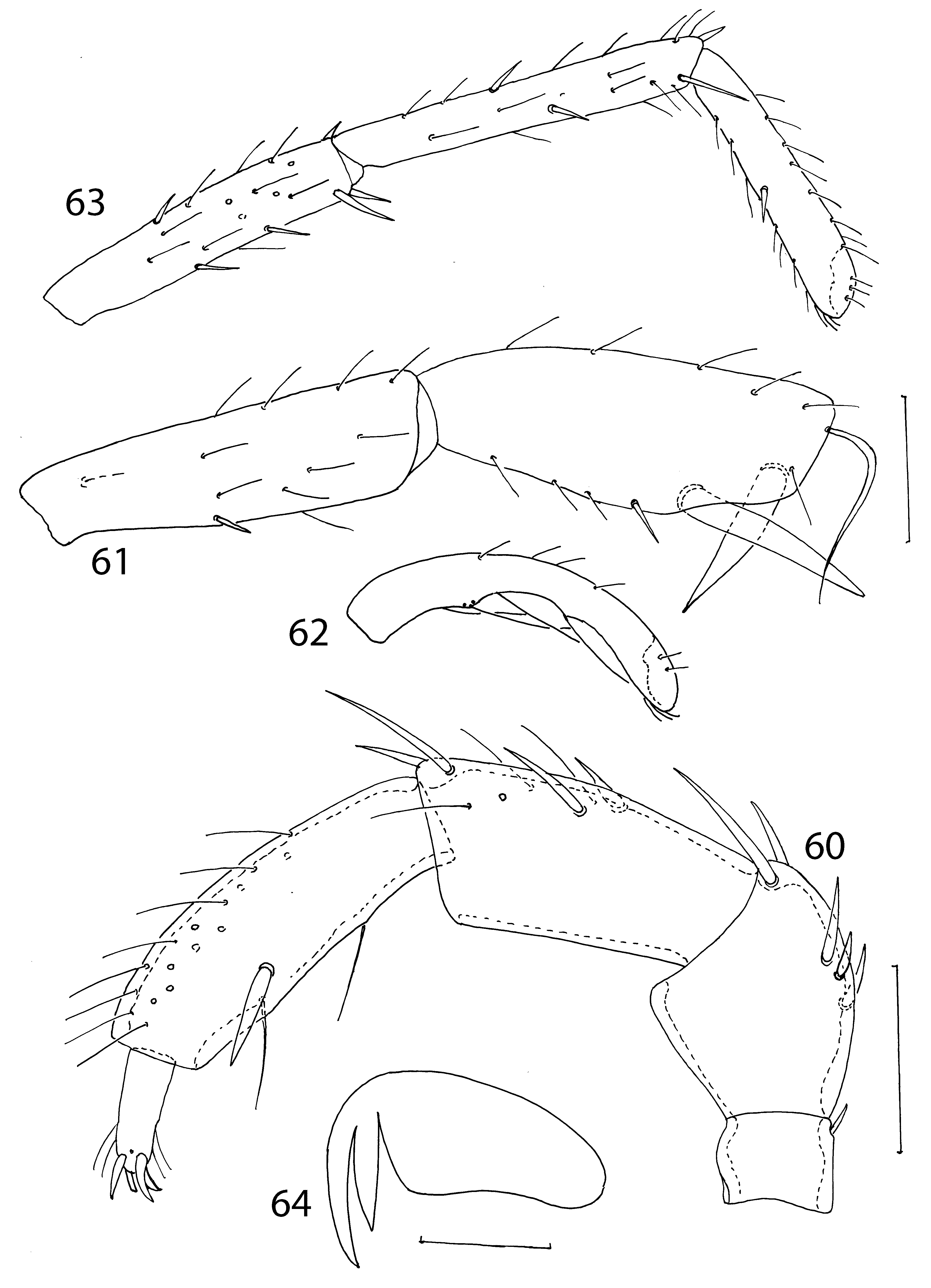
( Figs 52–64 View FIGURES 52 – 59 View FIGURES 60 – 64 )
Material examined. Holotype female, Russia, Primorie Territory, Octyabrsky District, Razdolnaya River, 4. 06. 1990, coll. T. S. Vshivkova, slide 6308. The holotype is deposited in the collection of Institute for Biology of Inland Waters (Borok, Russia).
Diagnosis. Setae Fch thick, stick–shaped with parallel lateral margins; setae Oe, Hi and He slightly longer but considerably thinner than Fch; the tibia of leg I well thickened in distal half, with not parallel dorsal and ventral margins, both the sword setae (S1 and S2) wide in proximal half and not narrowed near their bases; tarsus of leg I moderately curved, ratio length tibia/length tarsus 1.32; genital plates longer than gonopore, all genital acetabula approximately subequal in length, anterior and posterior genital sclerites approximately equal in width.
Description. Female. Color yellow in preservative liquid. Body flat oval, integument smooth with very fine strips. Number and position of idiosomal setae typical for representatives of the genus Atractides . Setae Fch ( Fig. 52 View FIGURES 52 – 59 ) thick, stick–shaped with more or less rounded tips; setae Oe, Hi and He ( Fig. 53 View FIGURES 52 – 59 ) slightly longer but considerably thinner than Fch; trichobothria Fp and Oi thin without glandularia ( Fig. 54 View FIGURES 52 – 59 ), other dorsal idiosomal setae hair– like and associated with glandularia short ( Fig. 55 View FIGURES 52 – 59 ). Coxal plates of legs I–IV covering about half of ventral surface. Median suture line between coxae I clearly developed; median sub–dermal process short, apodemes of anterior coxal plates rather long, slightly curved and forming right angle ( Fig. 56 View FIGURES 52 – 59 ). Posteromedial margin of coxal plates I wide and convex. Setae Hv located in lateral portion of coxal plates II. Coxal plate IV trapezoidal, its medial margins slightly concave. Glandularia Pe situated at anterior margins of coxae IV. Setae Pi without glandularia, their bases separated from setae Ci, excretory pore unsclerotized ( Fig. 57 View FIGURES 52 – 59 ). Genital plates ( Fig. 58 View FIGURES 52 – 59 ) elongate (ratio length/width 2.6–2.8), with 3 acetabula and 12 thin setae each; genital acetabula arranged in an arc, all acetabula approximately subequal in length, but posterior pair of acetabula wider than anterior and median acetabula. Genital plates longer than gonopore, anterior and posterior genital sclerites approximately equal in width. Basal segment of chelicera ( Fig. 59 View FIGURES 52 – 59 ) large with straight dorsal margin, cheliceral stylet crescent and relatively short, ratio length of basal segment of chelicera/length cheliceral stylet 2.4. Cheliceral stylet with a few small teeth on concave side.
Pedipalps ( Fig. 60 View FIGURES 60 – 64 ) slender; trochanter short with single dorsodistal seta; femur with straight ventral margin and rounded ventrodistal edge, with five dorsal unequal setae; genu considerably longer than femur with straight ventral margin, with two short proximal and two long stout dorsodistal unequal setae and several hair-like setae. Pedipalpal tibia slightly longer than genu, with a few thin dorsal setae; bases of two ventral setae divide tibia into three sectors approximately as 2–3–2; sword seta pointed, shorter than distance between ventral setae and placed near ventrodistal seta.
Legs without swimming setae. Genu of leg I ( Fig. 61 View FIGURES 60 – 64 ) slightly thickened distally with a few thin setae and one short ventral spine near middle of segment. Tibia of leg I thickened in distal half with maximum thickness near proximal sword seta; setae S1 and S2 separated by rather greater interspace, both these setae approximately equal in width but S1 longer than S2, S2 pointed, S1 with rounded tip; curved seta moderately developed. In addition, tibia I with short ventral spine near proximal sword seta and with a few thin setae. Tarsus of leg I ( Fig. 62 View FIGURES 60 – 64 ) moderately equally curved dorsoventrally, thickened basally and distally with minimum thickness near middle of segment. Posterior legs, especially legs IV ( Fig. 63 View FIGURES 60 – 64 ), slender; genu and tibia with a few thin setae and several spines, tarsus with a few thin setae and one ventral spine near middle of segment. Claws of legs I lesser than claws II–IV. Claws all legs with two denticles, external denticle longer than internal denticle; claw lamella well developed with slightly concave ventral margin ( Fig. 64 View FIGURES 60 – 64 ).
Measurements, n=1. Length of body 770; length of setae Fch 72, length of setae Oe, Hi and He 78–95; diameter or length of sclerite bearing setae Fch 24, diameter of sclerite bearing setae Le 21, diameter or length of sclerite bearing other idiosomal setae Fch 18; length of coxae I+II 210, width 370; length of coxae III+IV 230, width 180; lengths of genital plate 95–100, width 35; length/width of genital acetabula (ac. 1–3): 33–36/27–30, 36–42/27–30, 36–37/30–32; length of capitulum 125, length of basal segment of chelicera 155, length of cheliceral stylet 65; lengths of pedipalpal segments (P–1–5): 30, 66–70, 90–95, 95–102, 30; length/width of sword seta on P– 4 24/9; lengths of leg segments: I–Leg. 1–6—48, 95, 145, 215, 205, 155; II–Leg. 1–6: 55, 78, 108, 150, 150, 145; III– Leg.1–6: 60, 65, 115, 160, 175, 150; IV–Leg. 1–6—125, 110, 180, 235, 260, 200; tibia of leg I: length of seta S1 100, width 15; length of seta S2 78, width 16; distance between setae S1 and S2 35.
Differential diagnosis. The present species is similar to Atractides montanus (Halbert, 1911) , from which it is easily distinguishable by the structure of the tibia and tarsus of leg I, small genital acetabula and arranged of anterior coxal apodemes. The female of A. montanus characterized by follows features: the tibia of leg I with nearly parallel dorsal and ventral margins, both the sword setae (S1, S2) slightly narrowed near their base, interspace between setae S1 and S2 26 µm; tarsus of leg I strongly curved, ratio length tibia/length tarsus 1.43; posterior acetabula considerably larger than anterior and median acetabula, length of acetabula (ac. 1–3): 39, 43, 61 µm, anterior genital sclerite considerably wider than posterior sclerite ( Gerecke 2003). In contrast, in the female A. primoryensis the tibia of leg I without parallel dorsal and ventral margins, both the sword setae (S1, S2) wide and not narrowed near their bases, interspace between setae S1 and S2 35 µm; the tarsus of leg I moderately curved, the ratio length tibia/length tarsus 1.32; all acetabula approximately subequal in length, length of acetabula (ac. 1–3): 33–36, 38–42, 33–37 µm, anterior and posterior genital sclerites equal in width.
Etymology. The species epithet primoryensis is derived from the name of the Province where it was collected (Primory).
Habitat. Running waters.
Distribution. Asia ( Russia, Far East, Primory Territory).

No known copyright restrictions apply. See Agosti, D., Egloff, W., 2009. Taxonomic information exchange and copyright: the Plazi approach. BMC Research Notes 2009, 2:53 for further explanation.