
Crocodylus undetermined
|
publication ID |
https://doi.org/ 10.1111/j.1096-3642.2007.00248.x |
|
persistent identifier |
https://treatment.plazi.org/id/0381631A-9774-517D-FEE3-5A4EF0C5F920 |
|
treatment provided by |
Felipe |
|
scientific name |
Crocodylus undetermined |
| status |
|
Locality
All crocodylian specimens were found in karstic fissure fillings exposed by quarrying activities in the Gargano pedemountain belt (41.8°N, 15.4°E, Apulia, south-eastern Italy). The name or the code of the karstic fissures are given in parentheses in the ‘ Referred material’ section below GoogleMaps .
nino 1973); DSTF GH1 (F9) ]; 1 maxilla ( BSP 2004 I 1 ]; 1 frontal + prefrontal [ RGM 455203 View Materials (San Giovannino 1973)]; 1 jugal [ RGM 455202 View Materials (San Giovannino 1973)]; 1 skull fragment [ RGM 455200 View Materials (San Giovannino 1973)]; 1 lower jaw [ RGM 335893 View Materials (San Giovannino 1973)]; 6 isolated teeth [ RGM 453783 View Materials (Fina D), RGM 454280 View Materials (Pizzicoli 2), RGM 454950-1 View Materials (San Giovannino 1969), RGM 455205-6 View Materials (San Giovannino 1973)]; 2 coracoids [ RGM 455204 View Materials (San Giovannino 1973), RGM 455327 View Materials (Pepo N)]; 1 scapula [ RGM 455328 View Materials (Pepo N)]; 1 humerus [ RGM 455329 View Materials (Pepo N)]; 1 ulna [ RGM 455336 View Materials (Pepo N)]; 1 phalanx [ RGM 455337 View Materials (Pepo N)]; 6 vertebrae [ RGM 215348 View Materials (San Giovannino 1971); RGM 453472 View Materials (Gervasio 1975), RGM 455330-2 View Materials (Pepo N); DSTF GH2 (F40)]; 3 ribs [ RGM 455333-5 View Materials (Pepo N)]; 19 osteoderms [ RGM 335894-5 View Materials (Pepo N), RGM 451432-3 View Materials (Chiro 12 Penalba), RGM 453781-2 View Materials (Fina D), RGM 454946-9 View Materials (San Giovannino 1969), RGM 455320-6 View Materials (Pepo N); DSTF GH3-4 (F9)] .
Preservation
The fossil materials come from karst fissure fillings and they are therefore represented by completely isolated skeletal remains the surface of which is usually perfectly clean and readable, only rarely being covered by thin concretionary layer. Although sometimes fragmentary, the remains are well preserved in three dimensions, and do not show any signs of deformation.
Description
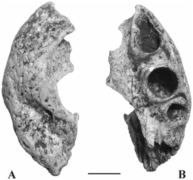
Premaxilla ( Fig. 1 View Figure 1 ): The best preserved premaxilla (DSTF GH1) is a nearly complete left element 53 mm Horizon
Reddish, massive or crudely stratified silty-sandy clays ( Abbazzi et al., 1996) yielding the so-called ‘ Microtia fauna’, which is biochronologically dated at between 5 and 6 Myr (Upper Messinian to lowermost Pliocene; Abbazzi et al., 1996; Rook, Abbazzi & Engesser, 1999).
Referred material
The material described here belongs to the following institutions: Naturalis, Nationaal Natuurhistorisch Museum (Leiden, The Netherlands; RGM), Dipartimento di Scienze della Terra dell’Università degli Studi di Firenze ( Italy; DSTF), and Universitätsinstitut und Staatssammlung für Paläontologie und historische Geologie, München ( Germany; BSP). It consists of: 2 premaxillae [RGM-455201 (San Giovan- long. It lacks the antero-medial region and shows the last four alveoli (the first two that are preserved are confluent). No teeth are preserved. The third preserved alveolus is the largest and is separated from the contiguous alveoli by deep mesial occlusal pits. A lateral notch at the level of the suture with the maxilla is present. Nasals contacted the premaxillae but it is not possible to establish if they bisected the naris. The postero-lateral margin of the naris is raised above the premaxilla surface and delimited by a shallow but evident ‘groove’; this condition is, however, different from that shown by Diplocynodon (presence of a deep notch).
A second premaxilla (again a left element; RGM 455201) is smaller and rather damaged (length of the fragment: 36 mm). It partly preserves the dorsal rim of the external naris. In lateral view, the premaxilla is ventrally festooned. The last four alveoli are preserved; they do not retain teeth. As in the previous case, the third preserved alveolus is the largest, and the first two seem to be confluent (but the interalveolar space is not completely preserved here); deep occlusion pits are mesially (but close to the lateral edge) developed in the (preserved) second and third interalveolar spaces.
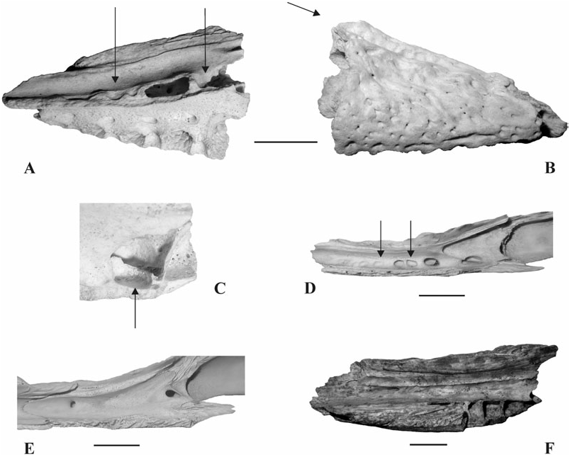
Maxilla ( Fig. 2 View Figure 2 ): The total length of the maxilla fragment (BSP 2004 I 1; Fig. 2A, B View Figure 2 ) is 37.5 mm. Teeth are not preserved. The alveoli, five in number (the last one is not complete), show an increasing size in backward direction. The dorsal, lateral and ventral surfaces of the fragment are intact; the palatal lamina is broken off nearly at its base. Each interalveolar space shows a lateral depression but the third and fourth show a true pit. The latero-ventral margin of the maxilla is not festooned. The medial edge of the maxilla, corresponding to the suture with the nasal, is elevated in a marked sagittal ‘ridge’, corresponding to the last preserved alveoli, medially delimited by a deep para-sagittal groove (see arrow in Fig. 2B View Figure 2 ). In the caviconchal recess, close to the palatal lamina, three depressions are visible between the first interalveolar space and the third alveolus. At the level of the fifth alveolus, fragments of the anterior and ventral wall of a blind pocket (cecal recess) are clearly preserved ( Fig. 2C View Figure 2 ).
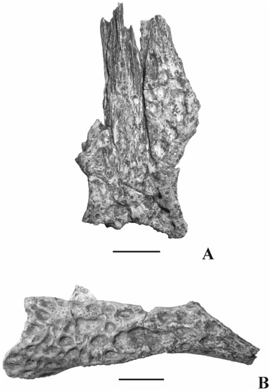
Frontal + prefrontals ( Fig. 3A View Figure 3 ): The material catalogued as RGM 455203 is represented by the anterior frontal process still attached with the prefrontals, of which that on the right is nearly completely preserved and that on the left shows only a proximal fragment. The long frontal process dorsally shows a wide suture area for a firm link with nasals (not preserved). Lateral edges of the frontal and posterior edges of prefrontal are distinctly raised and rounded and constitute the medial rims of the orbits. The dorsal surface of all these elements is irregularly ornate with roundish pits; no crest is present on the frontal surface between the orbits. The minimum interorbital distance is 21 mm. The total length of the fragment is 60 mm.
Jugal ( Fig. 3B View Figure 3 ): Left jugal RGM 455202 completely lacks the region anterior to the postorbital bar; the last one is broken off nearly at the base. The posterior region medially shows an extensive area for the suture with the quadratojugal while the ‘root’ of the postorbital bar is characterized by an evident suture area for the junction with the ascending process of the ectopterygoid. The postero-dorsal margin of the jugal constitutes the lateral rim of the left infratemporal fenestra. On the medial surface of the element, the medial jugal foramen is rather large. The lateral jugal surface is sculpted by deep variably sized and shaped pits. Its total length is 55 mm.
Skull fragment ( Fig. 4 View Figure 4 ): RGM 455200 represents the postero-left region of the skull and preserves quadrate, exoccipital, squamosal and a posterior fragment of the postorbital. The dorsal surface of the squamosal and postorbital is approximately flat and ornate with several pits; the lateral edge to these elements represents the lateral edge of the skull table and does not seem to be convex; their medial edge, devoid of any particular ridge, represents the lateral rim of the left supratemporal fenestra (which does not seem to have markedly overhanging rims; the squamosal overhangs only slightly at the posterior rim of the fenestra). Although the area is not perfectly preserved, it seems that the dorsal and ventral rims of the squamosal groove for the external ear valve musculature are approximately parallel. The squamosal prongs are rather elongated.
The quadrate is relatively well preserved; its lateral edge is free and is represented by the suture surface with the missing quadratojugal; it clearly shows ( Fig. 4B View Figure 4 ) a foramen aerum placed close to the mediodorsal angle. Even if this region has been partly damaged and is still partly covered by concretionary material, SEM analysis ( Fig. 4C View Figure 4 ) revealed the presence of a true foramen. Taking into consideration that its medio-dorsal surface has been abraded, the medial hemicondyle is considered to be tall (characters 112 and 113). It is not as tall as in modern comparative material of Crocodylus but not as small as in the Diplocynodon ratelii specimen used for comparison (Naturhistorisches Museum Basel, Switzerland; NHMB-MA 2275; and all the specimens from Saint Gérand-le-Puy stored in the collections of Museum national d’Histoire naturelle, Paris).
The exoccipital is fairly complete: only a small area of its lateral expansion dorsally delimiting the wide cranio-quadrate passage is missing; the medial edge shows a sector, free of suture surfaces or possible breakages, corresponding to the left rim of the foramen magnum; four foramina open ventro-laterally to the foramen magnum: the most evident is the foramen vagi, then, ventral to this, opens the posterior carotid foramen; two other small foramina filled with matrix are placed medially to the foramen vagi and could represent the foramina for the twelfth cranial nerve (but the smallest of them is possibly a bifurcation of the foramen vagi).
The dorso-medial margin of the exoccipital shows the suture with supraoccipital. The area corresponding to the cranio-quadrate passage is highly incomplete.
The length of the fragment is 73 mm, its maximum width is 45 mm; condyle width is nearly 23 mm.
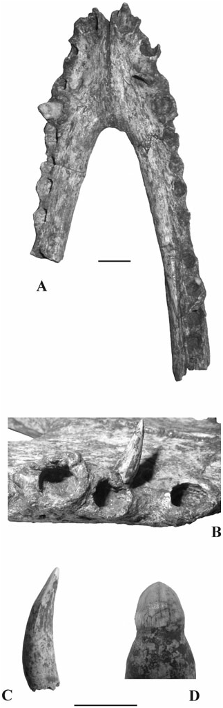
Lower jaw ( Fig. 5A, B View Figure 5 ): The fragmentary lower jaw (RGM 335893) is represented by incomplete dentaries sutured at the level of the symphysis. The right dentary is 128 mm long and preserves 12 alveoli (plus a part of the thirteenth), while the left one is only
89.4 mm long and shows eight alveoli (plus part of the ninth). Altogether, only four teeth are preserved (for a description see ‘Dentition’ below): the tooth corresponding to the third right alveolus is completely out of the alveolus and it is probably fixed by the matrix on the dorsal surface of the dentary ( Fig. 5B View Figure 5 ). In dorsal view, the symphysis nearly reaches the anterior bor-
der of the fifth alveolus. Several alveoli show margins distinctly ‘protruded’ outward (particularly developed sal view; the lateral concavities represent the rims of the orbits. B, left jugal (RGM 455202) in lateral view. Scale bar equals 10 mm.
tinctly at medio-dorsal angle. A, dorsal view of the left postero-lateral skull fragment preserving quadrate, exoccipital, squamosal and postorbital (RGM 455200). B, detail of the quadrate showing the position of the foramen aerum;
SEM image of the foramen aerum opening on the quadrate.
Scale bar equals 10 mm.
in alveoli 1, 2 and 4) that confer to the dentaries a slightly festooned appearance in dorsal and lateral views. In lateral view, the dentary shows an anterior dorsal convexity centred on the fourth alveolus and a posterior one centred on the tenth interalveolar space.
The measurements of the medio-lateral diameter, mesio-distal diameter and interalveolar length are reported below (mm) for each alveolus of the right dentary (owing to the morphology and preservation of the material, all measurements are somewhat imprecise but ‘*’ indicates a genuine approximation): 6.8, 6.7, 5.5; 6.6, 6.3, 6.8; 4.7, 6.2, 2.9; 6.6, 6.5, 4.4*; 4.2, 5.9*, 5.1; 5.4, 5.3, 2.9; 5.1, 5.5, 4.5; 4.6, 5.6, 9.3; 4.9, 5.7, 3.1*; 5.7, 6.6*, 3.9; 5.4, 6.3*, 2.4; 5.0, 7.6*, 2.9; and for the left dentary: 7.4, 7.0, 6.5; 4.7, 5.7, 6.7; 5.2, 5.5, 3.0; 6.7, 6.3, 3.5*; 4.6, 5.8, 4.2; 5.1, 5.2, 2.7; 5.2, 6.0, 4.9; 4.8, 5.9, 9.1.
The third a fourth alveoli are not confluent and the fourth is slightly larger than the previous one ( Fig. 5B View Figure 5 ).
On the dorsal surface of the dentaries, no occlusal pits are visible but several small depressions, filled by matrix and probably hosting a foramen at the bottom, are aligned medially to the tooth row in the anterior region and fuse together forming a groove in the posterior one.
The imprint of the splenials allows us to exclude their participation in the symphysis; their anterior tip passes ventrally to the Meckelian groove (which nearly reaches the symphysis).
The external surface of the dentaries is not particularly ornate: the pits become more frequent toward the ventral surface where they are grouped into longitudinal grooves.
Dentition ( Fig. 5C, D View Figure 5 ): Ten teeth are preserved: four associated with the lower jaw and six isolated. They show a crown usually furnished by two non-denticulated mesio-distal keels corresponding to the maximal diameter and usually separating the crown surface into a labial surface that is more developed than the lingual one. The crown surface is sometimes ornate by secondary small but evident ridges longitudinally developed that do not reach the crown base (as in RGM 454950) or by an unordered but homogeneous pattern of microreliefs (as in RGM 454280). Crowns are variably shaped (from acutely conical to nearly blunt) and sized (from 12.7 to 7.2 mm). Slender and pointed teeth such as RGM 454950 are probably anterior teeth whereas more massive teeth such as RGM 454280 probably represent the maxillary or dentary posterior region. Few teeth still preserve the root.
The dentition pattern can be defined as not homodont given that the teeth variably sized and shaped. Nothing can be said regarding the maxillary dentition but the dentary shows that the largest alveolus is the first, followed by the fourth; there is no sign of confluent third and fourth alveoli but, even if the fourth is larger that the third, they are of rather similar size. The eighth interalveolar space is by far the largest in both dentaries. Based on the presence of a notch at the boundary between premaxilla and maxilla, it seems likely that the fourth dentary tooth was occluded in a lateral notch. Nothing can be directly said about the occlusal pattern as there are no occlusal pits on the dentary.
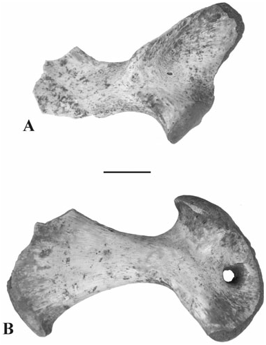
Scapula ( Fig. 6A View Figure 6 ): The right scapula (RGM 455328) lacks part of its dorsal blade but clearly preserves the proximal sector. The inferior area of its anterior edge is elevated into a high and thin deltoid crest separated from the glenoid area by a wide and deep lateral con-
cavity. The scapulocoracoid facet anterior to the glenoid fossa markedly tapers anteriorly.
Coracoids ( Fig. 6C View Figure 6 ): Neither of the coracoids shows any sign of fusion with the scapula. The best preserved specimen is a right element (RGM 455327), 57 mm long, but lacking the postero-ventral tip. RGM 455204 is the result of restoration of several fragments: it is a right element that is slightly damaged, mainly in its dorsal sector where the coracoid foramen is not entirely surrounded by bone; it is similar in size to that of the previously described coracoid.
Humerus: The only preserved humerus (RGM 455329) is a fragment of a right element, 78 mm long, which lacks the proximal epiphysis; only the distal part of a relatively robust deltopectoral crest is therefore preserved.
Ulna: The fragmentary right ulna (RGM 455336) is 54 mm long and lacks its distal epiphysis. The proximal epiphysis shows a rounded olecranon process. It seems likely that this ulna, the right coracoid RGM 455327, the right scapula RGM 455328 and the right humerus RGM 455329 could have belonged to a single specimen as they come from the same locality (Pepo N) and show matching size and preservation.
Phalanx: The only available phalanx (RGM 455337) is perfectly preserved, 10 mm long, and is rather stout in general appearance.
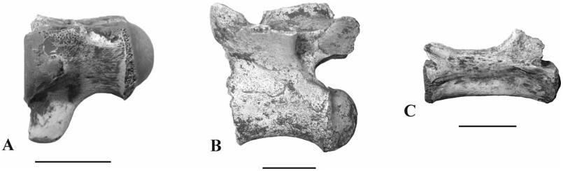
Vertebrae ( Fig. 7 View Figure 7 ): All the vertebrae identified show a procoelous centrum. Two vertebrae are represented by their centra only as they were separated from their neural arch along the neurocentral suture: centrum RGM 453472 (total length 16 mm; Fig. 7A View Figure 7 ) represents a cervical vertebra as it shows evident parapophyses laterally to the condyle and a robust and long hypapophysis that is ventrally (and slightly anteriorly) directed; centrum RGM 455330 is larger (total length approximately 23 mm) and is probably one of the first dorsal vertebrae given that it shows no trace of parapophyses but a suggestion of hypapophysis; a left prezygapophysis and transverse process RGM 455331 probably belong to the centrum previously described.
DSTF GH2 ( Fig. 7B View Figure 7 ) probably represents one of the last dorsal or a lumbar vertebra: its centrum is 23.2 mm long, strongly convex ventrally and it is sutured with the neural arch, which preserves prezygapophyses and postzygapophyses (the distance between the anterior edge of prezygapophyses and the posterior one of postzygapophyses is 26.1 mm) but only a proximal fragment of transverse processes and neural spine.
The caudal vertebra RGM 215348 ( Fig. 7C View Figure 7 ) has a centrum 23 mm long, cotyle and condyle are weakly developed, the neurocentral suture is closed, transverse processes are absent while neural spine and postzygapophyses are broken off.
Vertebra RGM 455332 is still embedded in its matrix: it seems to be fractured and is probably not complete.
Ribs: Three ribs are preserved. Two come from the cervical region as they comprise a longitudinal shaft and two processes joining the shaft almost perpendicularly; RGM 455335 is a right rib and is the best preserved: the shaft is 21 mm long (but it is not complete) and the capitular articular surface (lower) is larger than the tubercular one (upper); it may be one of the first cervical ribs. RGM 455334 is similar in general shape, but the shaft is more elongate and entirely preserved (28 mm), and the capitulum is broken off at its base; it is a left cervical rib. RGM 455333 is fragmentary and preserves only a long capitulum and part of the shaft; it comes from a posterior area on the right side and could be the last cervical rib or one of the first dorsal ribs. These tree ribs come from the same fissure (Pepo N) and could have belonged to the same individual.
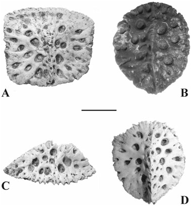
Osteoderms ( Fig. 8 View Figure 8 ): These elements represent nearly half of the crocodylian fossil remains from the Gargano area. Their shape varies from rectangular to oval and their length from 31.8 to 14.7 mm. They are invariably characterized by a nearly flat ventral surface and a longitudinal (or nearly longitudinal) keel that in some cases is so developed that the element is triangular in cross-section (suggesting that it could be a lateral osteoderm – not caudal given the large size; RGM 454949; Fig. 8C View Figure 8 ). The rectangular osteoderms have a small anterior smooth surface; most of the osteoderms have smooth edges although some (those that are triangular in cross-section) have spiny edges. The external surface is ornate with deep roundish pits that are relatively large.
On the ventral surface it is occasionally (as in RGM 453781) possible to perceive the criss-crossed pattern. There is no evidence of paired and keel-less ventral osteoderms.
Fragments RGM 451432–33 have been considered as osteoderms because of the pits that ornate the external surface but they could also represent skull fragments.

No known copyright restrictions apply. See Agosti, D., Egloff, W., 2009. Taxonomic information exchange and copyright: the Plazi approach. BMC Research Notes 2009, 2:53 for further explanation.
|
Kingdom |
|
|
Phylum |
|
|
Class |
|
|
Order |
|
|
Family |
|
|
Genus |