
Pachytomella Reuter, 1890
|
publication ID |
https://doi.org/10.5281/zenodo.4503680 |
|
DOI |
https://doi.org/10.5281/zenodo.4604771 |
|
persistent identifier |
https://treatment.plazi.org/id/038287C7-FF97-9370-068F-FE141B640805 |
|
treatment provided by |
Felipe |
|
scientific name |
Pachytomella Reuter, 1890 |
| status |
|
Pachytomella Reuter, 1890 View in CoL View at ENA
Pachytoma A. Costa, 1842: 288 (junior homonym of Pachytoma Swainson, 1840 , Mollusca). Type species by subsequent designation ( KIRKALDY 1906: 131): Pachytoma minor A. Costa, 1842 View in CoL (= Phytocoris passerinii A. Costa, 1842 View in CoL ).
Pachytomella Reuter, 1890: 253 View in CoL (new name for Pachytoma A. Costa, 1842 ).
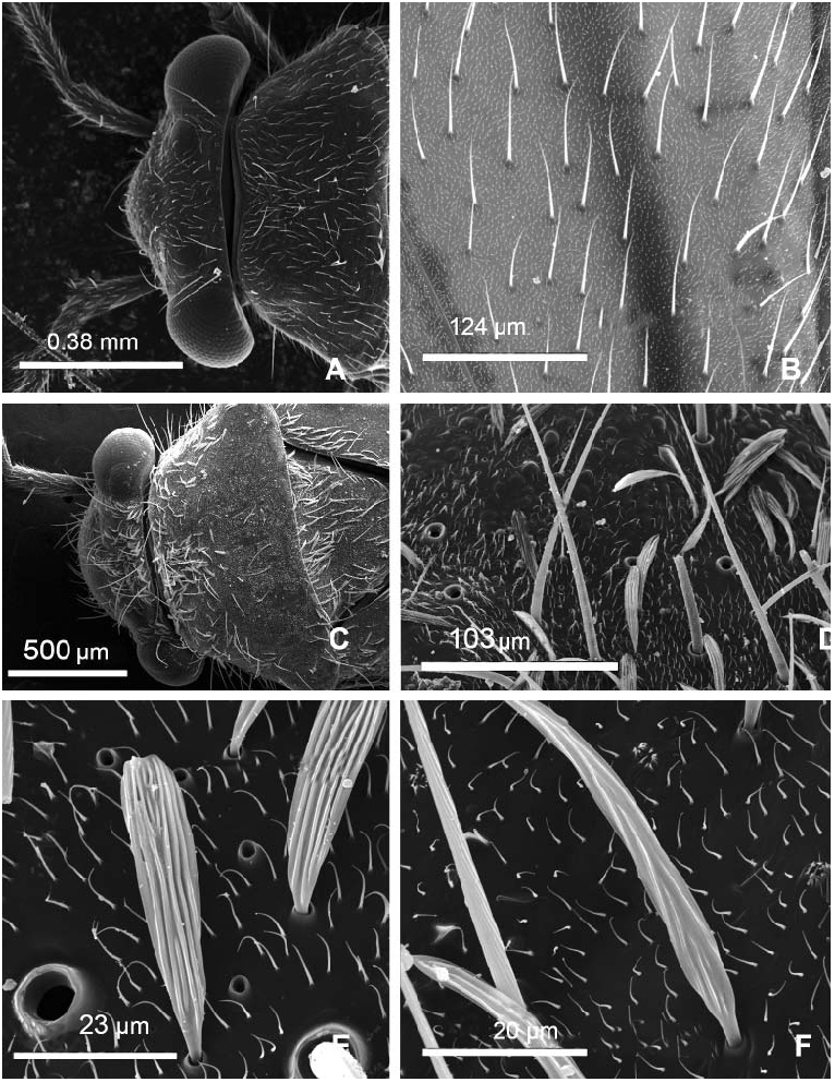
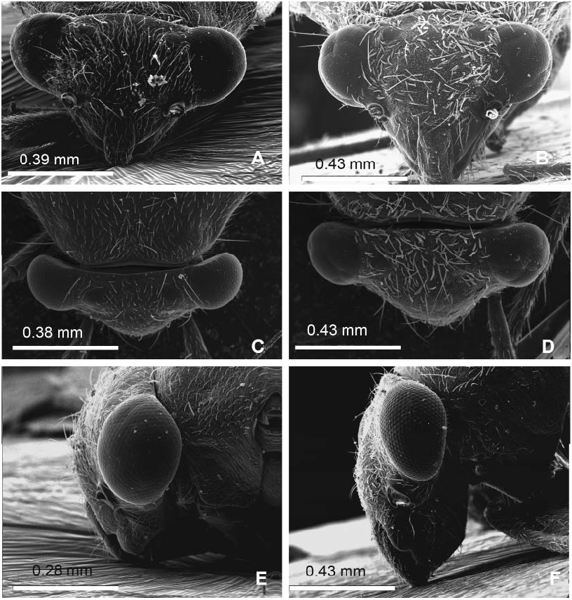
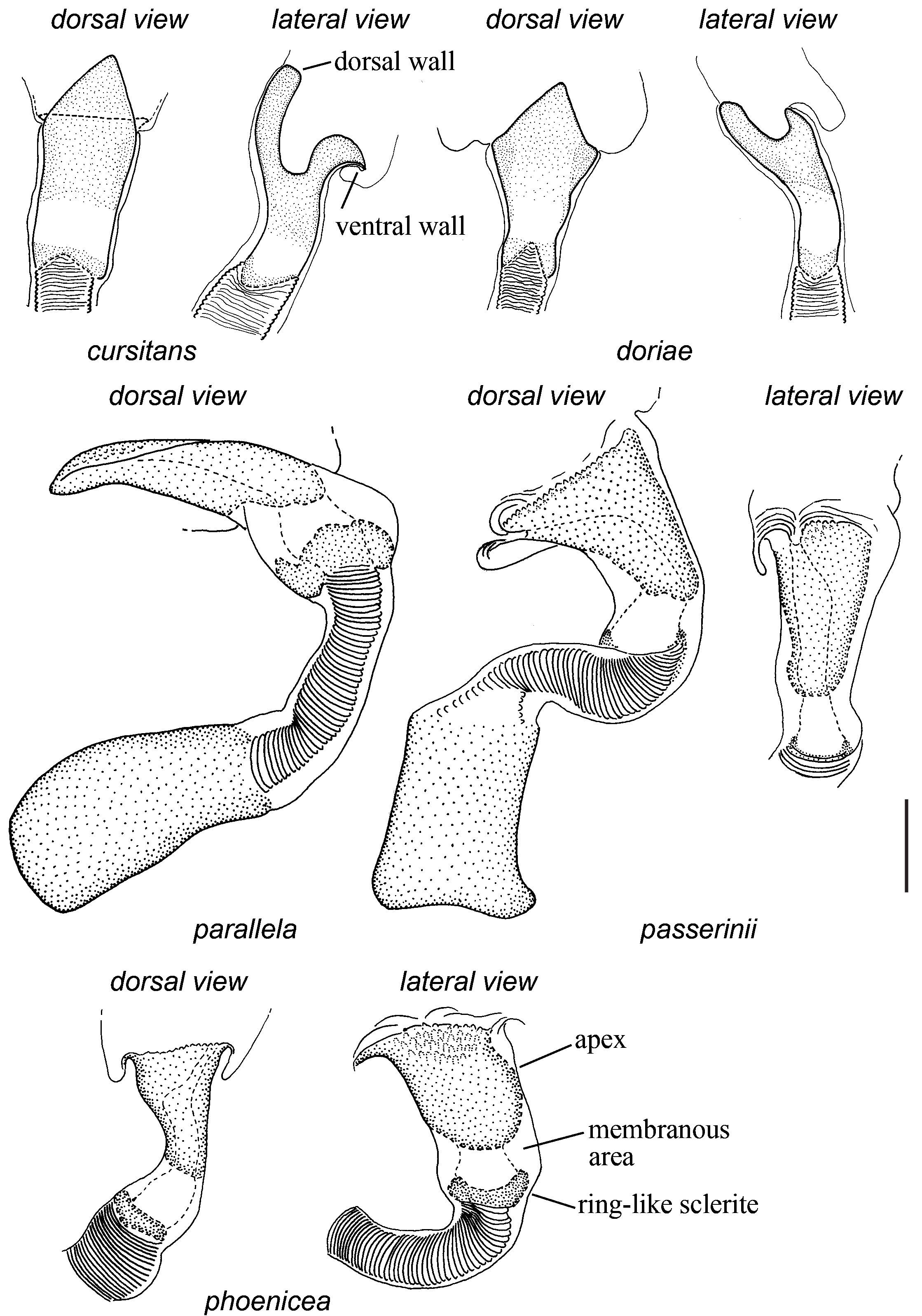
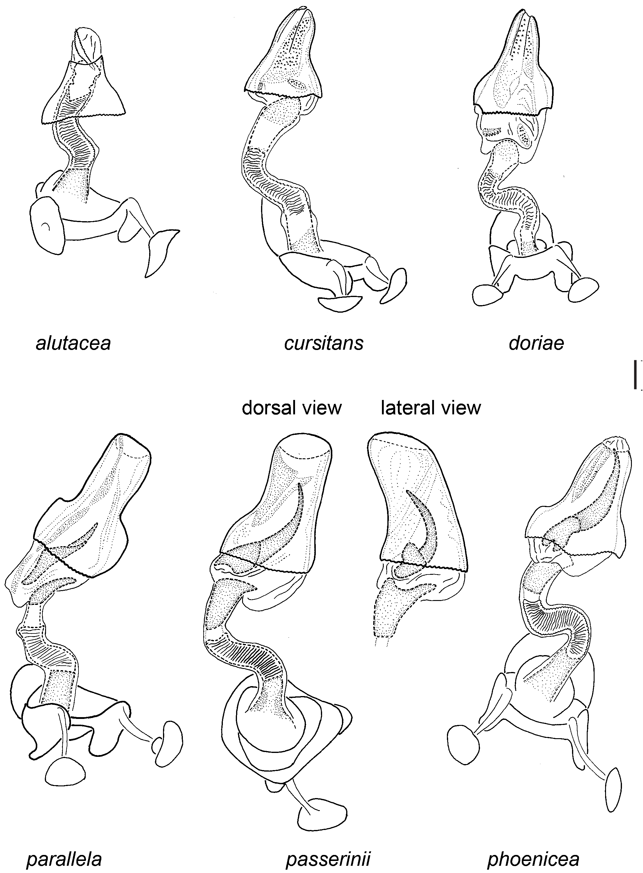
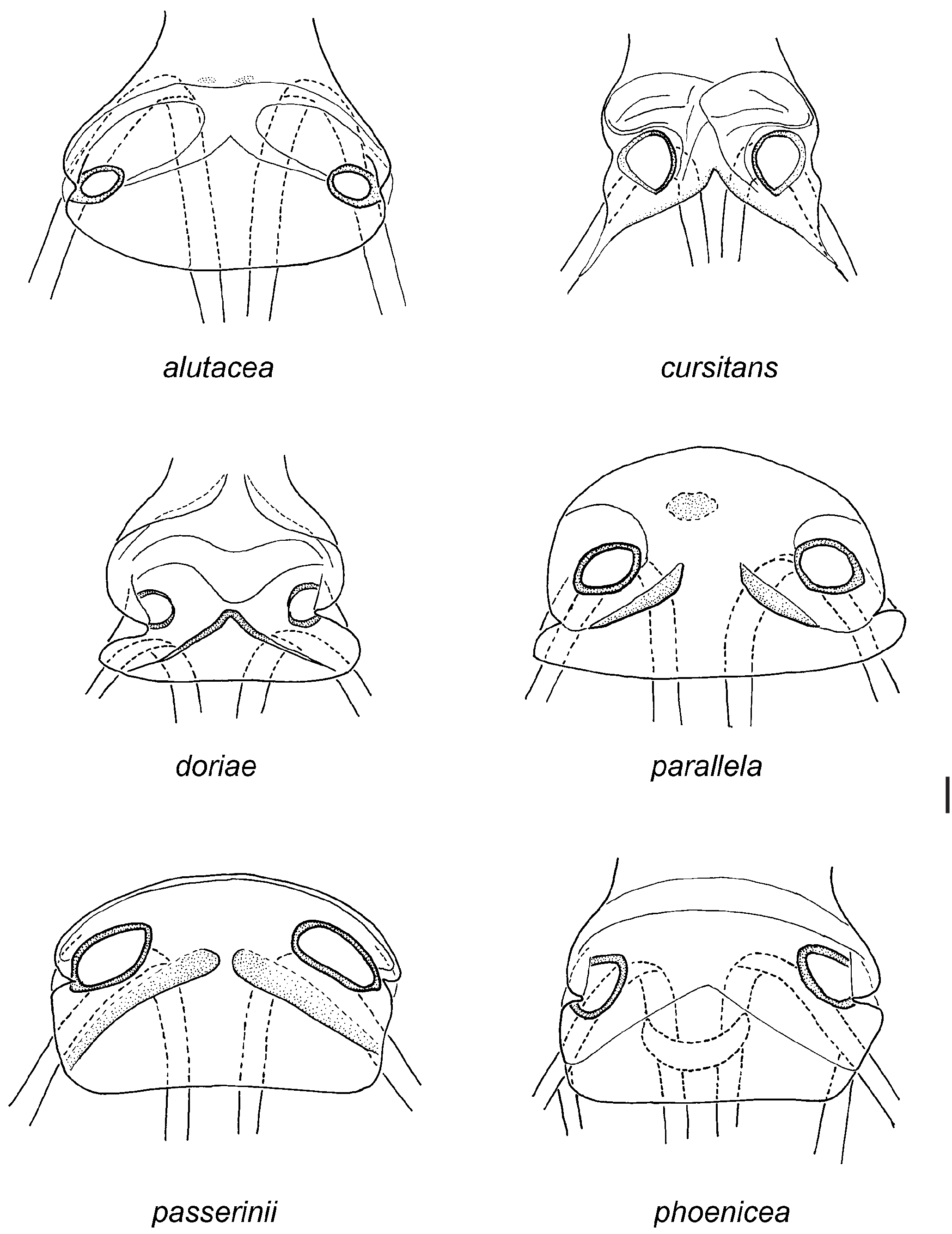
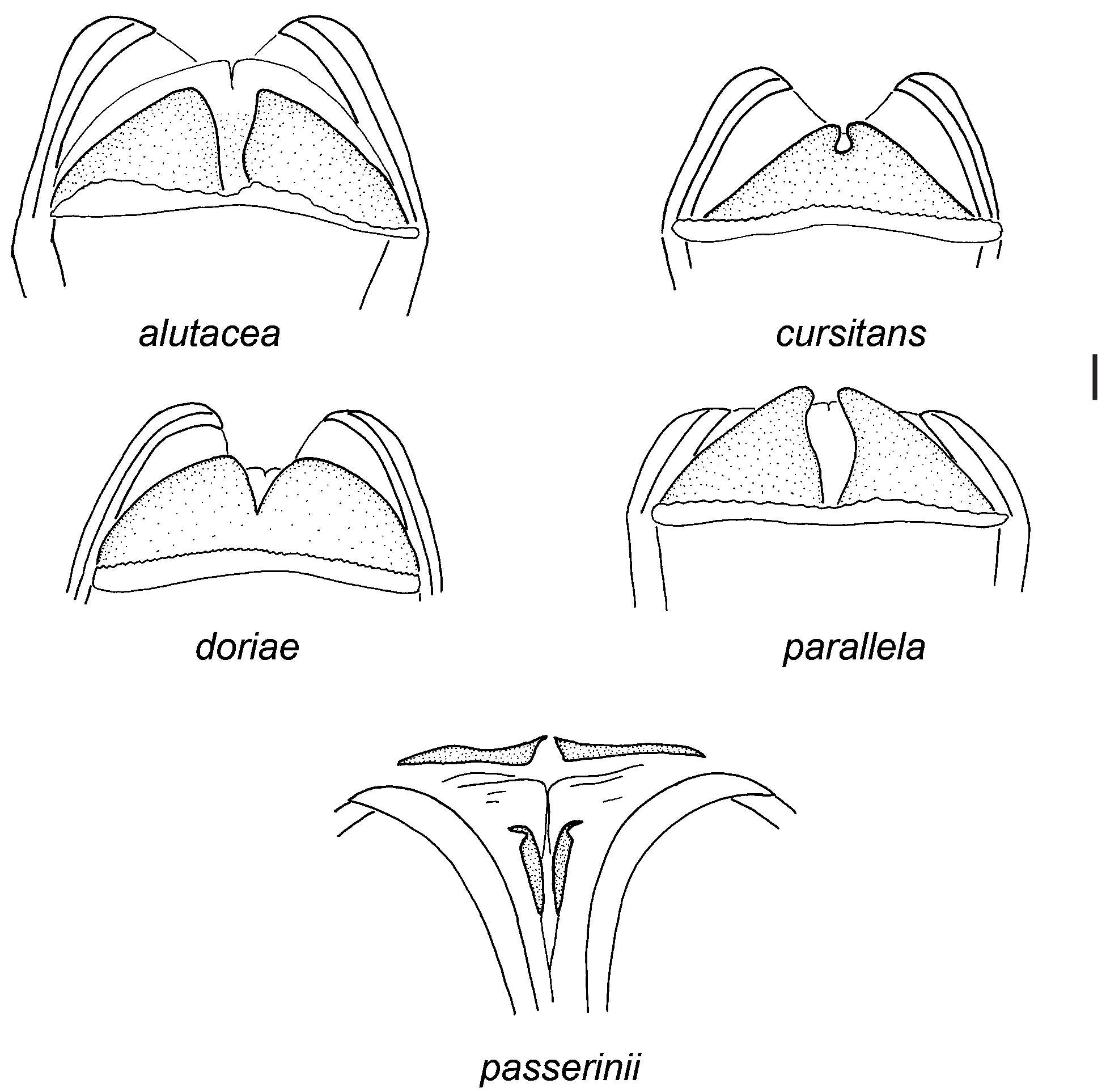
Redescription. Male: Macropterous, rarely brachypterous; small, elongate, total length 1.4–4.0 mm. COLOURATION ( Fig. 1 View Fig ): Body dark brown to black, sometimes with yellowish or pale brown areas; head dark brown to black, sometimes with pale brown or yellowish roundish spot near inner margin of each eye or with single spot between eyes; maxillary plate rarely with yellowish stripe below antennal fossa; labium of same colour as or somewhat paler than head; eye often dark brown to black sometimes with yellowish margins, rarely yellowish with darker spots or uniformly yellowish; colouration of appendages ranging from yellowish to uniformly black; hemelytron usually uniformly dark brown to black, sometimes dirty yellowish or pale brown with dark brown outer margin of corium and inner margin of clavus; membrane uniformly pale brown or yellowish. SURFACE AND VESTITURE: Dorsum smooth or slightly rugose, matt or shining, pronotum and scutellum rugose, hemelytron smooth; entire body clothed with semi-adpressed short simple setae, setae somewhat longer and darker on appendages and abdomen, head and pronotum with few bristle-like setae ( Figs. 9A, B View Fig ); antennal segment I with a pair of spinelike mesial setae; tibial spines dark brown. STRUCTURE: Head: Distinctly transverse ( Figs. 8A, C View Fig ), wider than or as wide as basal width of pronotum (9A); head only slightly protruding ventrally below inferior margins of eyes, diameter of eye wider than distance between inferior margin of eye and apex of clypeus ( Figs. 8A, E View Fig ); eye oval from lateral view and semi-circular in anterior view, almost not protruding ( Figs. 8A, C View Fig ), located at small distance from anterior pronotal margin or in contact with anterior margin of pronotum ( Fig. 9A View Fig ); frons convex; vertex smooth, without depression along hind margin ( Figs. 8A, C, E View Fig ); mandibular and maxillary plates sub-quandrangular, antennal fossa placed lower than and at a small distance from inferior margin of eye ( Fig. 8A View Fig ); antennal segment I somewhat longer than diameter of eye and distinctly shorter than width of vertex; antennal segment I as thick as antennal segment II or slightly thicker, segment II cylindrical, somewhat incrassate, distinctly longer than width of vertex; antennal segments III and IV thin, combined length of these segments shorter than or as long as segment II; labium reaching middle coxa. Thorax: Pronotum trapeziform with almost straight sides, posterior margin straight or slightly concave, posterior angles rounded, calli more or less distinct, flattened; evaporatorium of metathoracic scent gland ellipsoidal, with posterior angle distinctly elongated and extended backwards; scutellum almost flat; hind femur of typical shape, not swollen or somewhat swollen; tarsal segment II approximately 2× as long as segment I, tarsal segments II and III almost equal in length; in brachypterous specimens hemelytron with rounded posterior margin, cuneus and clavus indistinct, claval comissure shorter than inner margin of clavus. GENITALIA: Genital segment trapeziform, parameres of typical Halticini shape: right paramere spoon-shaped, with oval body and apex pointed or truncate; left paramere L-shaped, with hooked apex, sometimes with an additional process directed upward ( Fig. 5 View Fig ). Aedeagus: theca membranous, with sclerotised dorsal wall sometimes bearing apical depression; middle part of ductus seminis membranous, with ribs; basal part of ductus seminis entirely sclerotised or membranous distally; distal portion of ductus seminis strongly sclerotised, divided in two parts separated by membranous area, flattened dorsoventrally, bowl-shaped or rarely elongated, its dorsal wall as long as or slightly longer than ventral wall, with secondary gonopore slitlike; sculpture around secondary gonopore absent or hardly visible ( Fig. 2 View Fig ); endosoma voluminous with one or two sclerotised spicules, sometimes also with denticulate areas ( Fig. 4 View Fig ).
Female: Brachypterous; broadly-oval, small, total size 1.7–2.9 mm. COLOURATION ( Fig. 1 View Fig ): Similar to that of male, but hemelytron often dark brown to black, rarely uniformly dirty yellowish with dark brown inner margin. SURFACE AND VESTITURE: Similar to male, hemelytron sometimes rugose or with indistinct punctation. STRUCTURE: Similar to male, but more oval and broader than male; head broader than in male, eye not protruding and in contact with anterior pronotal margin; hemelytra with posterior margin truncate or slightly rounded, clavus indistinct or separated by shallow suture, claval commissure as long as inner margin of clavus. GENITALIA: Dorsal labiate plate with well-developed sclerotised rings of differing shapes and often more or less distinctly sclerotised areas near the rings ( Fig. 6 View Fig ); posterior wall of bursa copulatrix with trapeziform sclerite or with a pair of triangular sclerites at sides ( Fig. 7 View Fig ); vulva surrounded by a pair of straight sclerites ( Fig. 7 View Fig ).
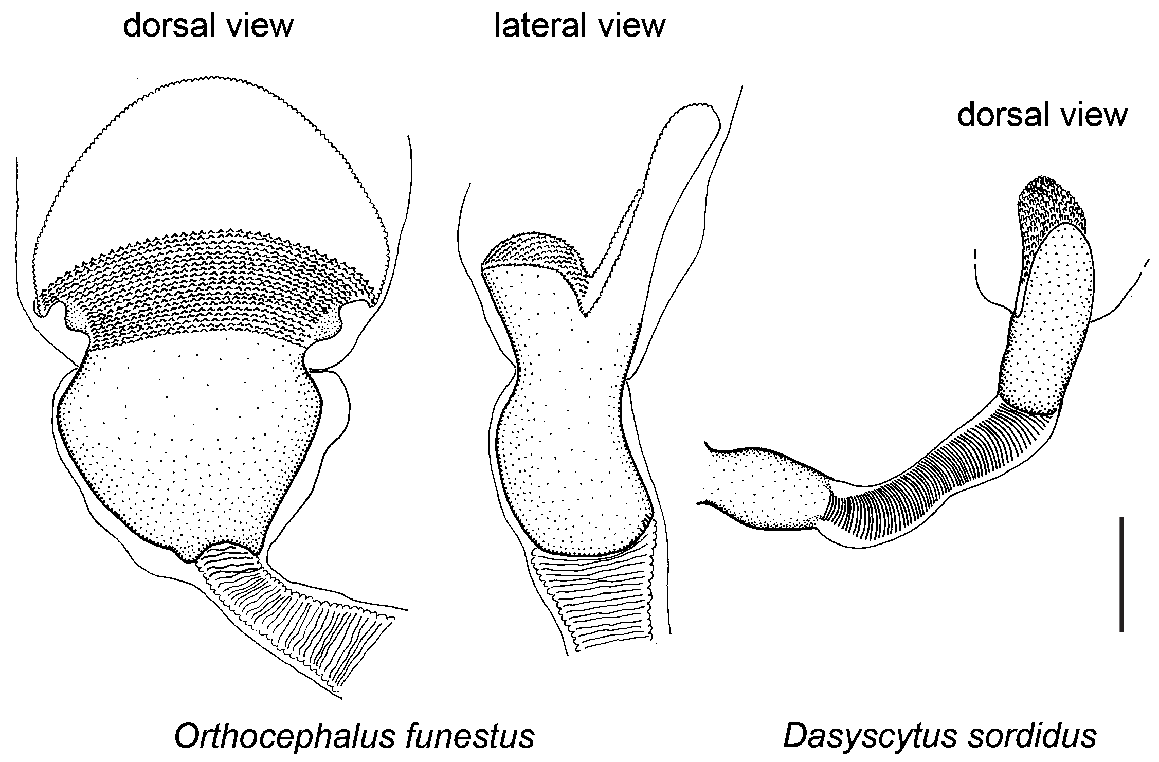
Differential diagnosis. Recognized by the following combination of characters: body relatively small, males 1.4–4.0 mm long and females 1.7–2.9 mm long, gracile, often dark coloured, sometimes with pale brown or yellowish appendages and rarely with pale inner part of corium ( Fig. 1 View Fig ); dorsum smooth, sometimes rugose, hemelytron rarely with indistinct punctation; head distinctly transverse, slightly protruding ventrally below ventral margins of eyes, diameter of eye wider than distance between eye and apex of clypeus ( Figs. 8A, E View Fig ); head as wide as or wider than posterior margin of pronotum ( Fig. 9A View Fig ); vertex smooth without depression along posterior margin of head ( Figs. 8A, C, E View Fig ); eye bulging and slightly protruding ( Fig. 8A View Fig ), at small distance from or in contact with anterior margin of pronotum ( Fig. 9A View Fig ); antennal segment I short, always shorter than width of vertex, with mesial spine-like setae; dorsum covered only with adpressed, short, simple setae ( Fig. 9A, B View Fig ), without any flattened, scalelike setae, with bristlelike setae occurring only on head and anterior angles of pronotum; hemelytral membrane without setae; hind tarsal segment II at least twice as long as segment I, and longer than segment III; endosomal membrane of aedeagus with one or two spicules, sometimes with denticulate areas basal to spicules ( Fig. 4 View Fig ); ductus seminis short, with strongly sclerotised basal portion and membranous middle portion bearing sclerotised ribs ( Fig. 2 View Fig ); distal portion of ductus seminis with ring-like sclerite distally followed by membranous area and strongly sclerotised, distinctly bowl-shaped (elongated in P. parallela ), dorso-ventrally flattened apex ( Fig. 2 View Fig ); dorsal and ventral walls of apical portion of ductus seminis equally sclerotised, often equal in length, sometimes with ventral wall somewhat longer but not leaf shaped or broadened; secondary gonopore slit-like, with indistinct sculpture ( Fig. 2 View Fig ). Pachytomella is close to the genera Dasyscytus Fieber, 1864 , Piezocranum Horváth, 1877 , and Orthocephalus Fieber, 1858 . Representatives of all these genera have more or less equal body proportions, smooth, slightly rugose, or shallowly punctated dorsum in males, more or less transverse head, and antennal segment I shorter than width of vertex, bearing two or three mesial spinelike setae.
In addition to characters listed above, Pachytomella is also similar to monotypic Dasyscytus in the head slightly protruded below inferior margins of eyes ( Fig. 8A View Fig ), and body clothed only with short, pale setae ( Fig. 9B View Fig ). However, Dasyscytus differs by the width of head being distinctly shorter than posterior margin of pronotum, the presence of distinct depression along posterior margin of head, uniformly yellowish hemelytra in male, and hemelytral membrane clothed with pale simple setae. Distal part of ductus seminis in Dasyscytus is cylindrical, entirely sclerotised, with secondary gonopore oval-shaped ( Fig. 3 View Fig ).
Pachytomella can be confused with Piezocranum in external view, as they both have elongated body, distinctly transverse head, and vestiture composed only of simple setae. However, the genus can be distinguished by the head distinctly narrower than the base of pronotum, the simple setae semi-erect and relatively long, the dorsum of females with distinct punctation and the distal part of ductus seminis entirely sclerotised, short, and tube-shaped ( Fig. 3 View Fig ).
Pachytomella is also close to the genus Orthocephalus in structure of distal part of ductus seminis, but Orthocephalus is separated by possessing a more robust body, a head well extended below inferior margins of eyes ( Fig. 8B, F View Fig ) and its width distinctly shorter than posterior margin of pronotum ( Fig. 9C View Fig ), a vertex often with more or less distinct depression along posterior margin of head ( Figs. 8B, D, F View Fig ), and a body clothed with flattened or scalelike silver setae (9D–F). All species of Orthocephalus possess the peculiar structure of the distal portion of ductus seminis ( NAMYATOVA & KONSTANTINOV 2009), which is bowl-shaped and flattened like that in most Pachytomella species (cf. Figs. 2 View Fig and 3 View Fig ). However, in Orthocephalus the distal portion of the ductus seminis is uniformly sclerotised, not subdivided by a membranous area. The ventral wall of the distal portion is strongly sclerotised with distinct sculpturing, and the dorsal wall is weakly sclerotised, leaf-shaped, and much longer than the ventral wall ( Fig. 3 View Fig ).
Discussion. According to our phylogenetic analysis ( NAMYATOVA & KONSTANTINOV 2009), Pachytomella is monophyletic and represents the sister group of the genus Orthocephalus . This conclusion is mainly based on the male genitalic structure, in particular, species of Pachytomella having a peculiar structure of the distal part of ductus seminis, as discussed above.
According to our observations, colouration, frons shape, and comparative length of tarsal segments are similar to those in the genera Orthocephalus and Dasyscytus . The length and coloration of hemelytra have limited systematic value. In Orthocephalus males are macropterous and females are mainly brachypterous, whereas in Dasyscytus only macropterous males and brachypterous females are known. In Orthocephalus , like in Pachytomella , the hemelytra are often dark brown to black, but sometimes with pale stripes, whereas in Dasyscytus males have uniformly yellowish hemelytra.
The sets of characters, such as the specific shape of the head and presence of only adpressed, short, simple setae, are useful for the Pachytomella diagnosing as they do not occur in Orthocephalus and Dasyscytus . Thus, we consider the genus Pachytomella a monophyletic group of species which are very similar to each other in both external and genitalic morphology.


No known copyright restrictions apply. See Agosti, D., Egloff, W., 2009. Taxonomic information exchange and copyright: the Plazi approach. BMC Research Notes 2009, 2:53 for further explanation.
|
Kingdom |
|
|
Phylum |
|
|
Class |
|
|
Order |
|
|
Family |
Pachytomella Reuter, 1890
| Namyatova, Anna A. 2010 |
Pachytomella
| REUTER O. M. 1890: 253 |
Pachytoma A. Costa, 1842: 288
| KIRKALDY G. W. 1906: 131 |
| COSTA A. A. 1842: 288 |