
Stygodesmodora paramphidialis, Larrazábal-Filho & Neres & Esteves, 2017
|
publication ID |
https://doi.org/ 10.11646/zootaxa.4294.1.2 |
|
publication LSID |
lsid:zoobank.org:pub:35927FCF-6356-40D9-A672-01EFD1B1D926 |
|
DOI |
https://doi.org/10.5281/zenodo.6032045 |
|
persistent identifier |
https://treatment.plazi.org/id/03830354-5B09-580C-9CA0-FC433D69FED0 |
|
treatment provided by |
Plazi |
|
scientific name |
Stygodesmodora paramphidialis |
| status |
sp. nov. |
Stygodesmodora paramphidialis View in CoL sp. n.
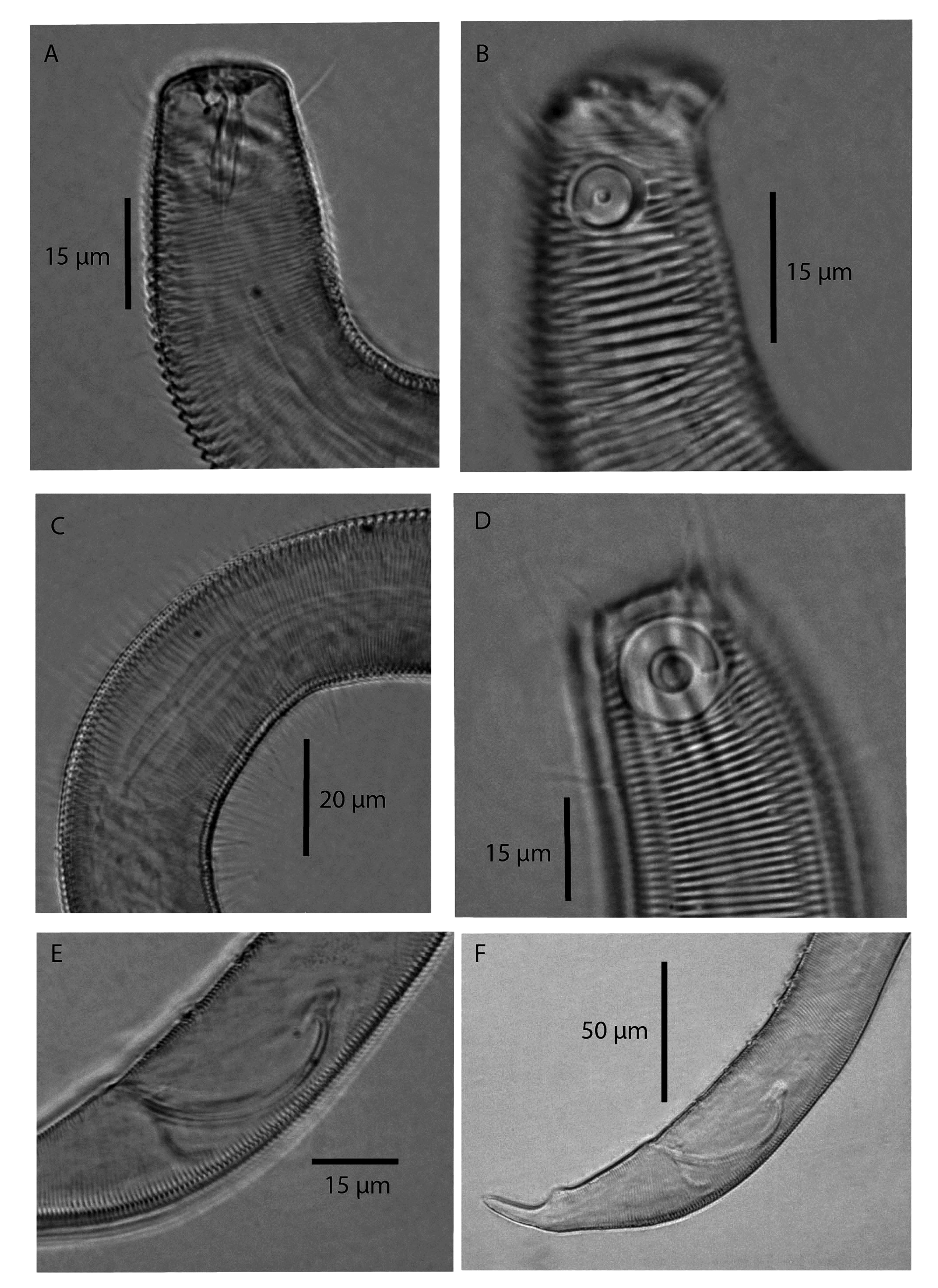
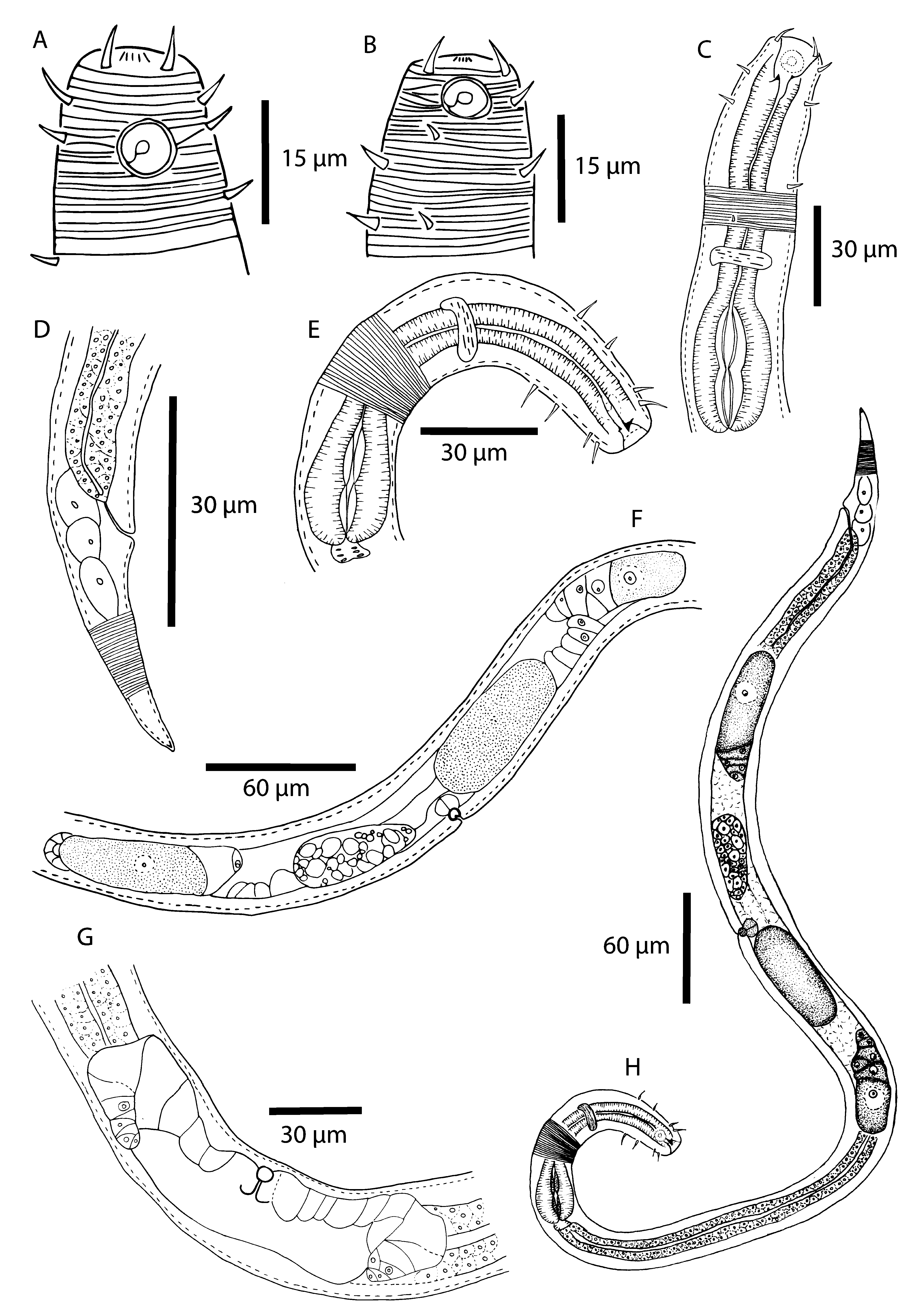
(measurements in Table 3, Figs 5–7 View FIGURE 5 View FIGURE 6 View FIGURE 7 ).
Type material. Holotype male, adult MNRJ 384. Station ME1E3 (05°00’51”S, 036°24’30”W). Paratype female, adult, MNRJ 385. Station CME7 (05°01’20”S, 036°11’45”W). Other paratypes, nine females (199 and 203 LMZOO-UFPE), nine males (204–208 LMZOO-UFPE), nine J1 and one J4 (209 LMZOO-UFPE), same data as for holotype.
Etymology. The specific epithet “ paramphidialis ” is given because this species has many similarities to Stygodesmodora amphidialis .
Description. Holotype ( Figs 5–6 View FIGURE 5 View FIGURE 6 ). Body cylindrical, yellowish brown, tapering slightly toward each end. Cuticle annulated except in final portion of tail, no lateral differentiation. Ventral ala absent. Based on pores and some setae along the body, the somatic setae are arranged in eight longitudinal rows: two dorsal, four lateral and two ventral. Head region with annulations completely surrounding fovea amphidialis ( Fig. 5 View FIGURE 5 B). In some specimens, labial region folded inward. Anterior sensilla arrangement: six outer labial papillae (difficult to see), four cephalic setae and 12 cervical setae. Cervical setae are arranged in two circles: four at the anterior edge of the fovea amphidialis and six just below the fovea amphidialis ( Fig. 5 View FIGURE 5 B). Fovea amphidialis cryptospiral, occupying 62% of diameter of head, located on cuticularized amphideal plate. Oral cavity with one dorsal tooth and one ventral tooth. Pharynx cylindrical, with muscular and slightly elongated endbulb. Lumen bipartite ( Figs. 5 View FIGURE 5 E and 6C). Nerve ring located 77 µm from anterior region. Ventral gland and excretory-secretory pore not observed. Cardia well developed, inserted in intestine. Reproductive system monorchic, with single anterior testis situated to left of intestine, outstretched. Spicules arched, proximal region rounded. Velum present, gubernaculum well developed and slightly arched. Seven tubular precloacal supplements ( Fig. 5 View FIGURE 5 A). Supplements irregularly distributed in precloacal region. Caudal papillae present. Tail conical, with three small caudal glands and spinneret.
Paratype female ( Figs 6–7 View FIGURE 6 View FIGURE 7 ). Similar to male in body size, with minor differences: fovea amphidialis smaller than in male and with different cephalic arrangement (i.e. two sexual dimorphisms). Female with eight cervical setae, in two circles: four between cephalic setae and fovea amphidialis and four at anterior edge of fovea amphidialis . Reproductive system with paired ovaries, opposite and reflexed, situated to right of intestine. Vulva as transverse slit. Vagina vera short, vagina uterina with sphincter muscle. Egg found in uterus ( Fig. 7 View FIGURE 7 F). Globular spermatic cells visible in uterus within a spermatheca. Vulva comprising 62% of total body length (545 µm from anterior end). Tail conical.
Sex Male Female Juvenile Juveniles. Very similar to adults, except lacking some characteristics such as supplements, immature reproductive system, and size of fovea amphidialis (smaller than males). Juveniles in stage 1 lack cervical setae and have very few somatic setae along the body. Juveniles in stage 4 are very similar to adults. Ten juveniles were found, nine in stage 1 and one in stage 4.
Diagnosis. Stygodesmodora paramphidialis sp. n. is characterized by a cephalic arrangement with four cephalic setae and 12 cervical setae (for males). Sexually dimorphic in size of fovea amphidialis (larger in male than in female) and the number of cervical setae (12 in males and eight in females). Slightly elongated endbulb with bipartite lumen. With 6–8 tubular supplements, approximately equally spaced, without clustering.
Differential diagnosis. Stygodesmodora paramphidialis sp. n. is similar to Stygodesmodora amphidialis sp. n. Both species have four cephalic setae, one dorsal and one ventral tooth, arched spicules, developed capitulum, velum, tubular supplements and caudal papillae. The species can be distinguished by the size of the fovea amphidialis (larger in S. amphidialis sp. n., occupying 83% of the head diameter, than in S. paramphidialis sp. n., occupying 62% of the head diameter in males). Stygodesmodora paramphidialis sp. n. has a slightly elongated endbulb with a bipartite lumen; this feature is also observed in the female and juvenile, and is the main difference between the females in the two taxa, since S. amphidialis sp. n. has a pyriform endbulb and a simple lumen. Another difference is the cephalic arrangement: S. amphidialis sp. n. has four cephalic setae and 16 cervical setae; whereas S. paramphidialis sp. n. has four cephalic and 12 cervical setae. The last major difference is in the position of the supplements. Stygodesmodora amphidialis sp. n. has a group of anteriorly aggregated supplements and the remaining supplements are more separated from each other, whereas in S. paramphidialis sp. n. all the supplements are irregularly distributed.
Stygodesmodora paramphidialis sp. n. can be distinguished from other species by means of the above comparisons with S. amphidialis sp. n.

No known copyright restrictions apply. See Agosti, D., Egloff, W., 2009. Taxonomic information exchange and copyright: the Plazi approach. BMC Research Notes 2009, 2:53 for further explanation.