
Neotachidius, 2005
|
publication ID |
https://doi.org/ 10.1111/j.1096-3642.2004.00148.x |
|
DOI |
https://doi.org/10.5281/zenodo.5700763 |
|
persistent identifier |
https://treatment.plazi.org/id/038387C0-FF8A-FFF1-7B86-FD8BFF1D7252 |
|
treatment provided by |
Carolina |
|
scientific name |
Neotachidius |
| status |
sp. nov. |
NEOTACHIDIUS COREANUS SP. NOV.
Type material: Holotype ♀ ( NMNH reg. no. 251948) dissected and mounted on slides . Paratypes are 1 ♀ ( NMNH reg. no. 251949) and 4 ♂♂ ( NMNH reg. nos. 251950–53) dissected and mounted on slides ; 20 ♀♀ and 20 ♂♂ in alcohol ( NMNH reg. no. 251954) ; 11 ♀♀ and 5 ♂♂ in alcohol ( NHM reg. nos. 2003.755–770) .
Type locality: Station 5 in a small river discharging into Kwangyang Bay, South Korea (34∞57.1¢N, 127∞36.4¢E), salinity 11.10‰ (see Ohtsuka et al. 1992) .
Body length: ♀: 690 ± 40 mm (N = 53); ♂: 600 ± 40 mm (N = 51).
Description
Based on NMNH paratypes (reg. nos. 251949–53) and NHM paratypes (reg. nos. 2003.755–770) .
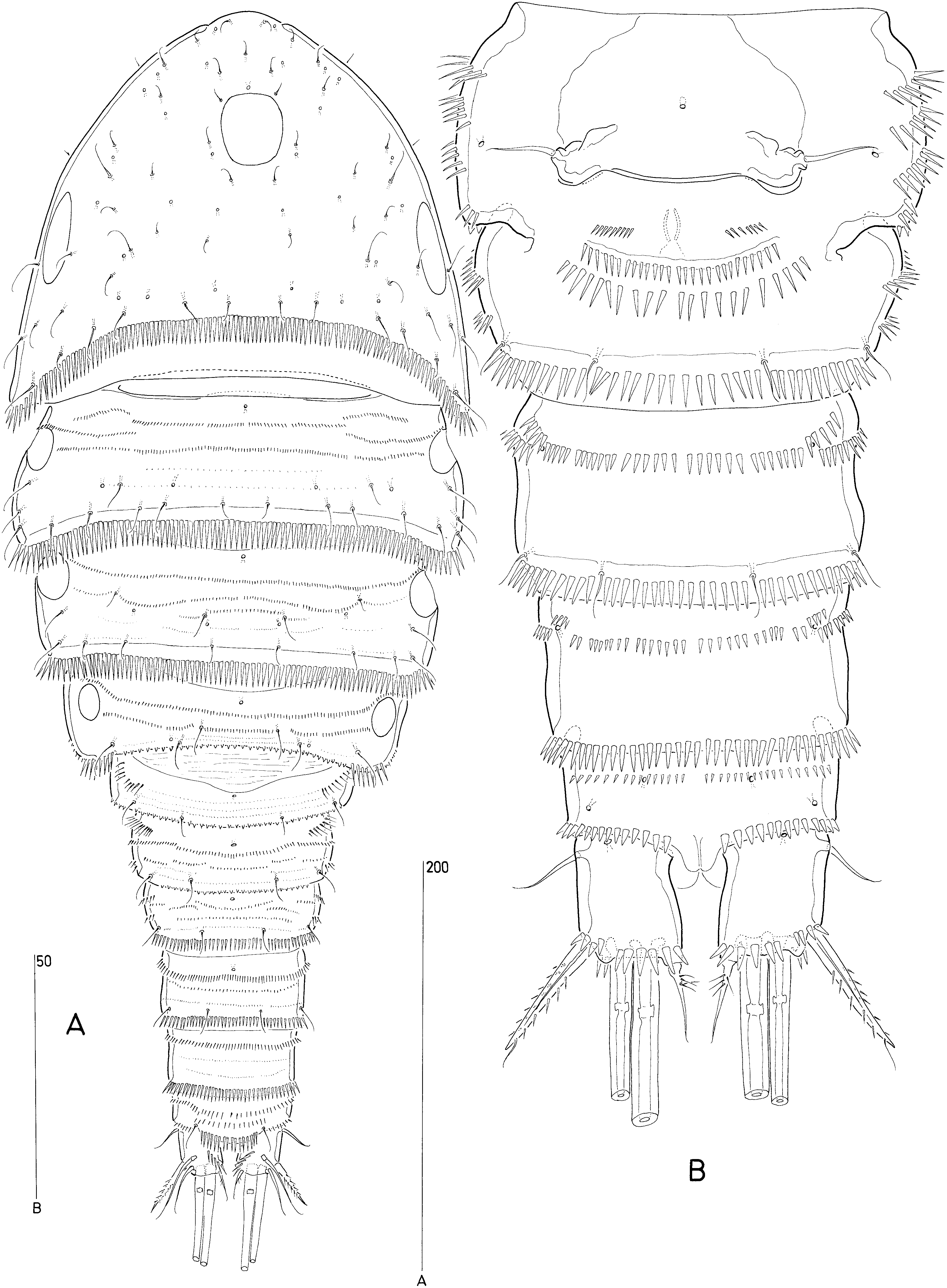
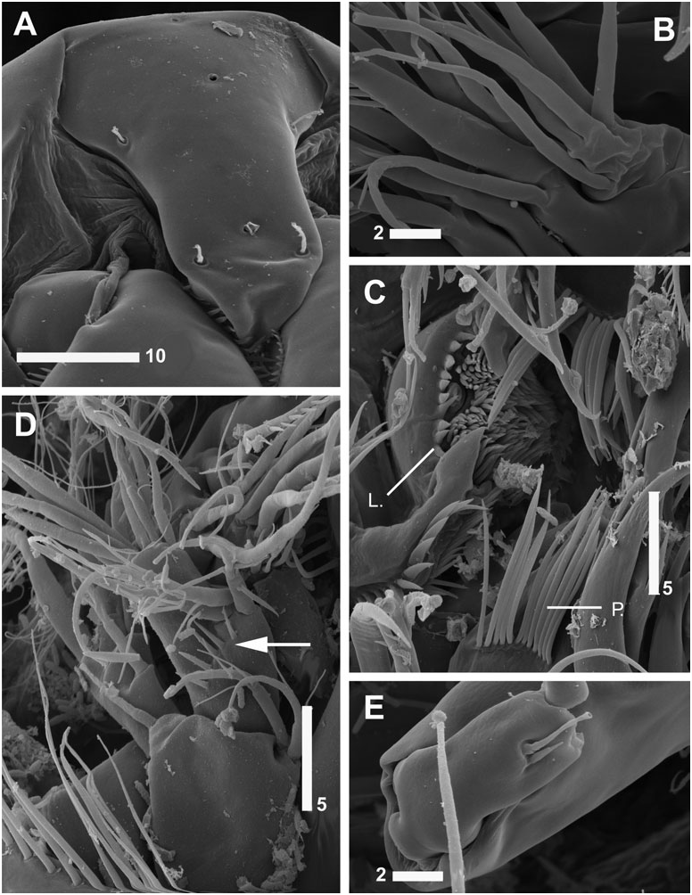
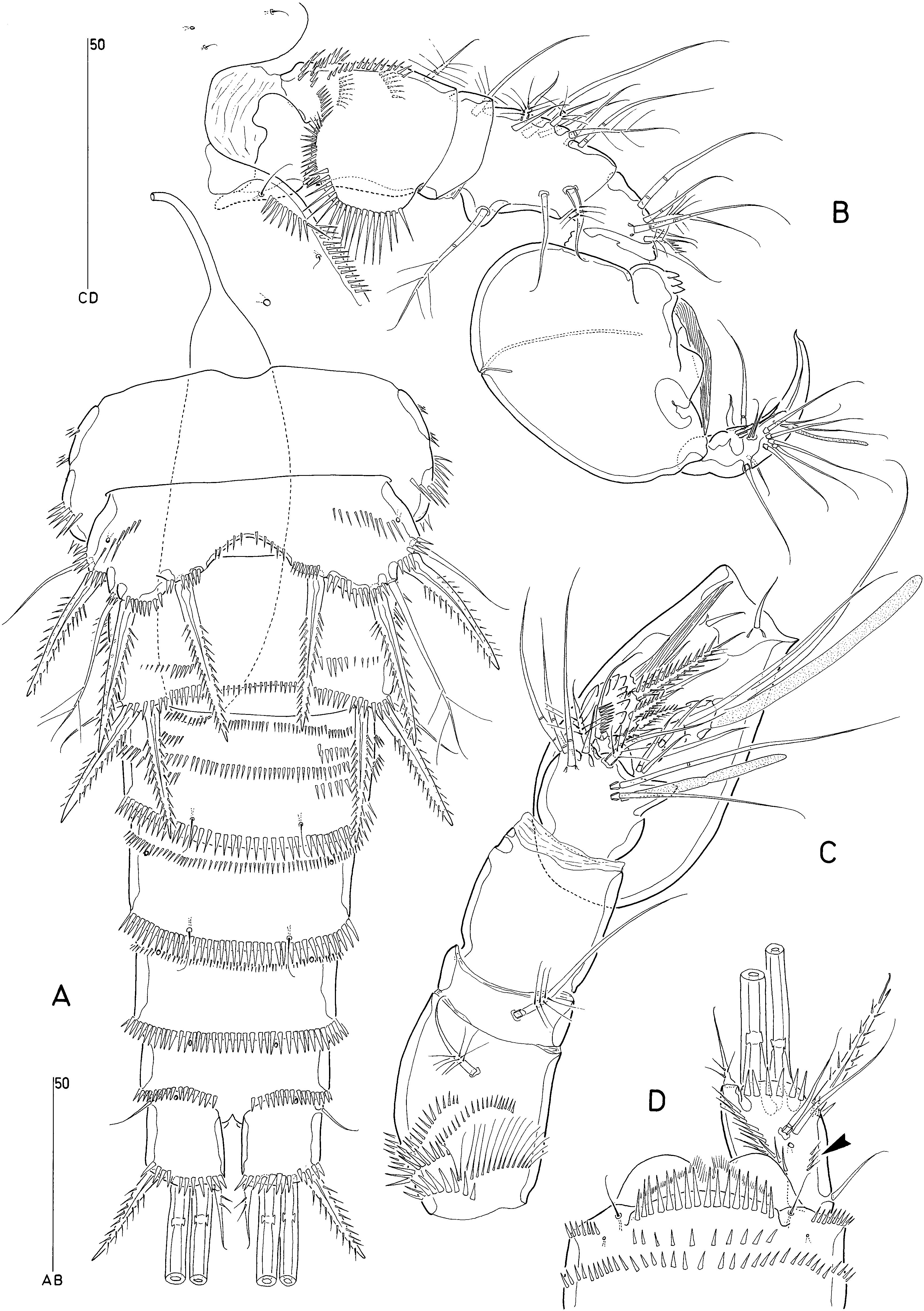
Female: Body robust ( Fig. 1A View Figure 1 ), cyclopiform, with distinct separation between prosome and urosome.
Rostrum ( Figs 4B View Figure 4 , 5A View Figure 5 ) weakly defined at base, ventrally directed, not discernible in dorsal aspect ( Fig. 1A View Figure 1 ); elongate-ovoid with slightly constricted tip; dorsal surface with two pairs of sensillae and three median pores.
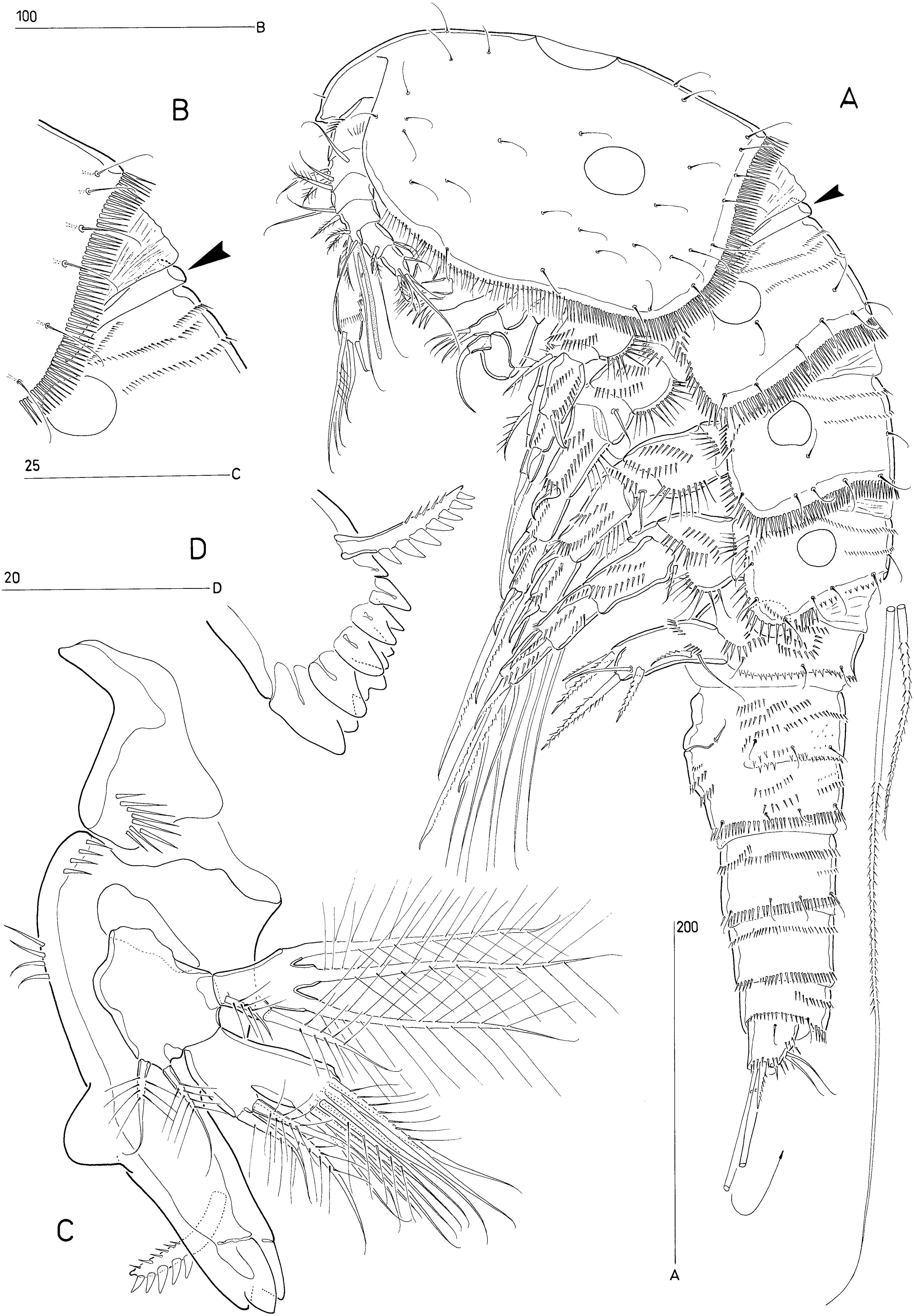
Cephalosome ( Figs 1A View Figure 1 , 2A View Figure 2 , 3A View Figure 3 ) with spinules along posterior margin and setules around lateroventral margins; with median dorsal and paired lateral integumental windows; sensillar and pore patterns as figured in Figure 1A View Figure 1 . Tergite of first pedigerous somite rudimentary, represented by a transverse sclerite ( Figs 1A View Figure 1 , 2B View Figure 2 ; arrowed in Figs 2A View Figure 2 , 3A, B View Figure 3 ) which is partly fused along its lateral sides to that of the second pedigerous somite.
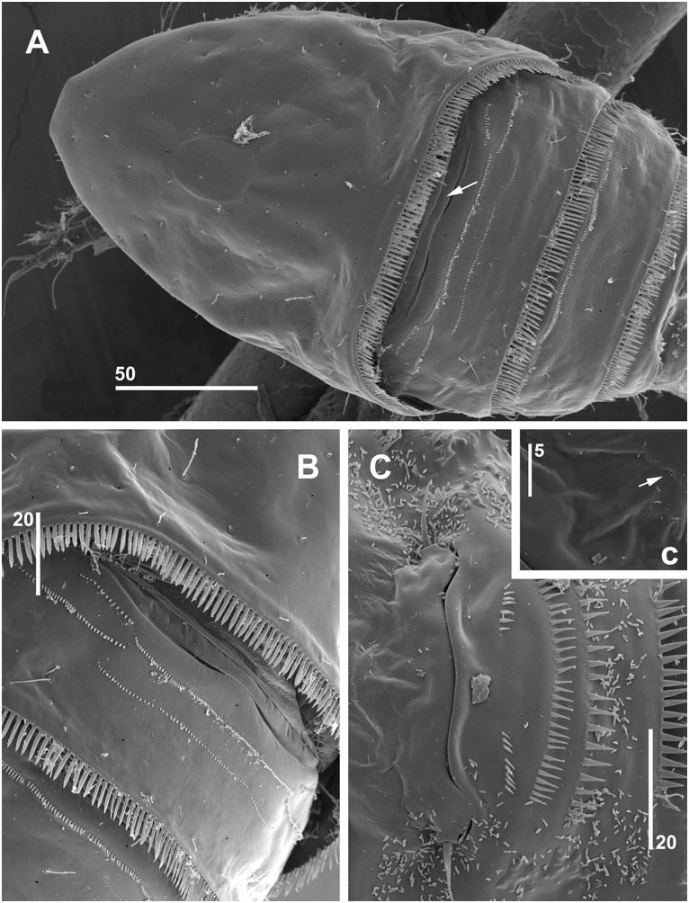
Somites bearing P2–P5 ( Fig. 3A View Figure 3 ) with paired lateral integumental windows; free margins of tergites with long spinules all around except for denticulate dorsal margin of P4-bearing somite ( Fig. 1A View Figure 1 ); dorsal surfaces with minute spinule rows, pores and sensillae as figured. P5-bearing somite with serrate posterior margin, with serrations being larger laterally than dorsally ( Fig. 9C View Figure 9 ); lateral integumental windows largely concealed beneath tergite of preceding somite ( Fig. 3A View Figure 3 ) and surrounded by spinule rows ( Fig. 9C View Figure 9 ).
Original segmentation of genital double-somite marked by bilateral constriction and dorsal serrate surface ridge. Genital field positioned ventrally on raised anterior half of double-somite (see lateral aspect; Fig. 9C View Figure 9 ). Genital apertures fused medially forming common genital slit ( Figs 1B View Figure 1 , 2C View Figure 2 ), closed off on either side by unisetose operculum derived from P6 ( Figs 1B View Figure 1 , 9C View Figure 9 ). Isolated copulatory pore not discernible, probably located medially within genital slit ( Figs 1B View Figure 1 , 2C View Figure 2 ). Single median seminal receptacle present ( Fig. 9C View Figure 9 ). Raised ventral surface posterior to genital field with three spinule rows (anteriormost paired) ( Figs 1B View Figure 1 , 2C View Figure 2 ). Median integumental pore present anterior to genital slit (arrowed in Fig. 2C View Figure 2 ). Remaining urosomites with spinules around posterior margin and surface ornamentation as figured ( Fig. 1A, B View Figure 1 ). Anal somite with two spinule rows dorsally and spinulose operculum ( Fig. 10D View Figure 10 ).
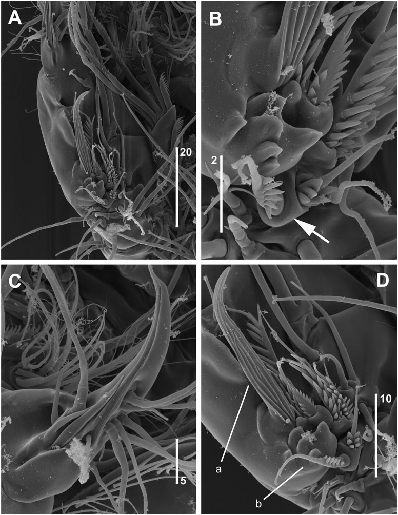
Caudal rami ( Figs 1B View Figure 1 , 10D View Figure 10 ) slightly convergent and slightly longer than wide, with oblique spinule row on dorsal surface and short row of fine spinules dorsolaterally (arrowed in Fig. 10D View Figure 10 ). With seven setae: I well developed, bare; II bare, displaced to near seta VII; III bipinnate and spiniform, with subapical flagellate extension; IV and V bipinnate and with fracture planes; VI swollen at base and typically with few spinules at inner proximal margin; VII bi-articulate at base and naked.
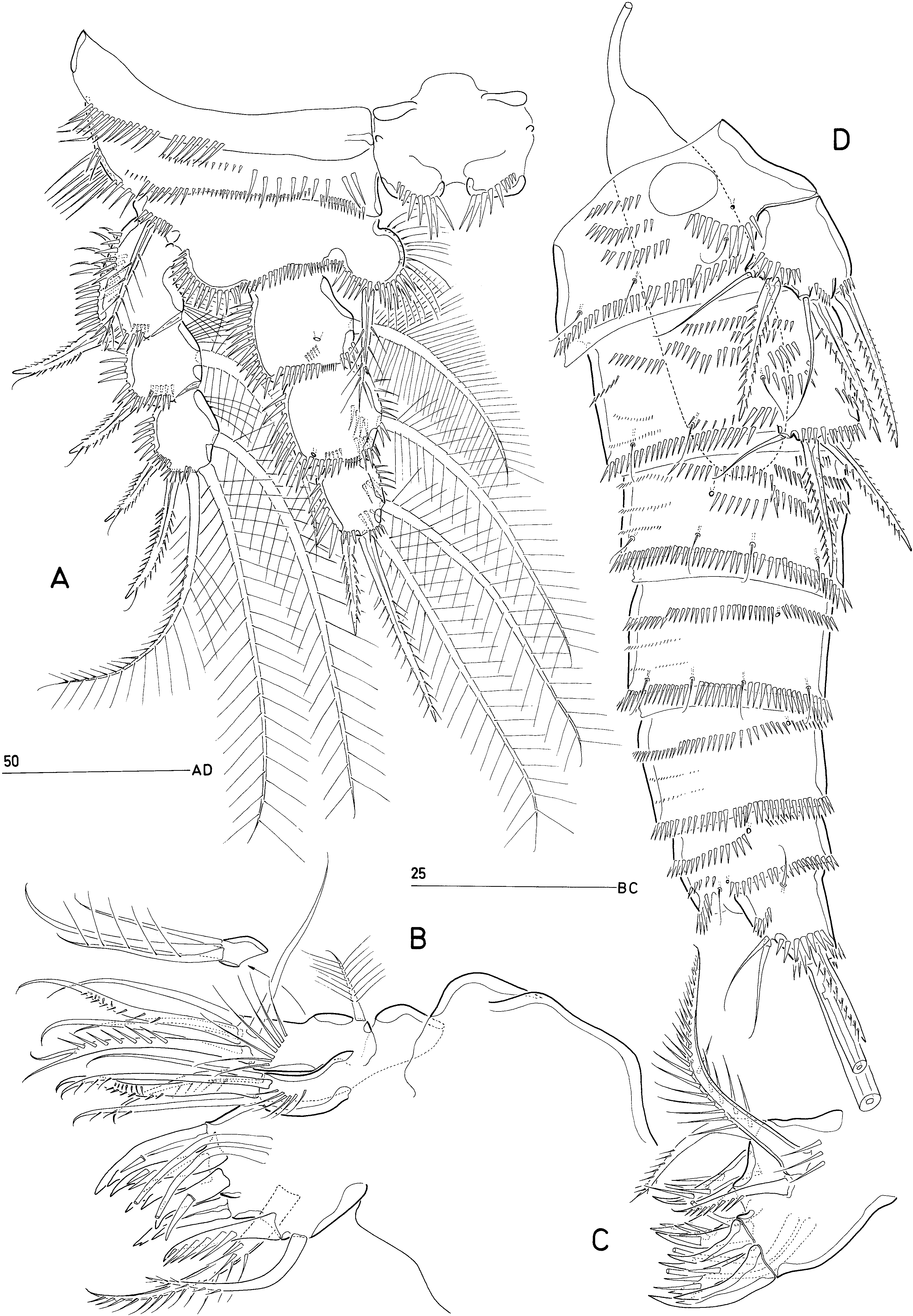
Antennule ( Fig. 4A View Figure 4 ) short, 7-segmented. Segment 1 with spinule rows around posterior and anterior margins. Armature formula: 1-[1 pinnate], 2-[1 naked +7 pinnate], 3-[5 naked +3 pinnate], 4-[3 pinnate + (1 naked + ae)], 5-[2 pinnate], 6-[2 naked +6 pinnate], 7- [5 naked +1 pinnate + acrothek]. Apical acrothek consisting of aesthetasc and plumose seta.
Antenna ( Fig. 4C, D View Figure 4 ) with spinule rows on abexopodal margin of basis and proximal endopod segment. Exopod incompletely 2-segmented; exp-1 shortest, with one pinnate spine; exp-2 with short pinnate spine fused to lateral margin and two unequal pinnate spines apically; few coarse spinules present around outer distal corner of exp-2. Distal endopod segment laterally with one naked spine in proximal third and one plumose seta plus one unipinnate spine in middle third ( Fig. 4D View Figure 4 ); both lateral spines with subapical tubular extension. Apical armature of enp-2 consisting of one unipinnate spine and four geniculate setae; longest geniculate seta with long setules and fused at base to short pinnate seta; segment with various spinule rows as figured.
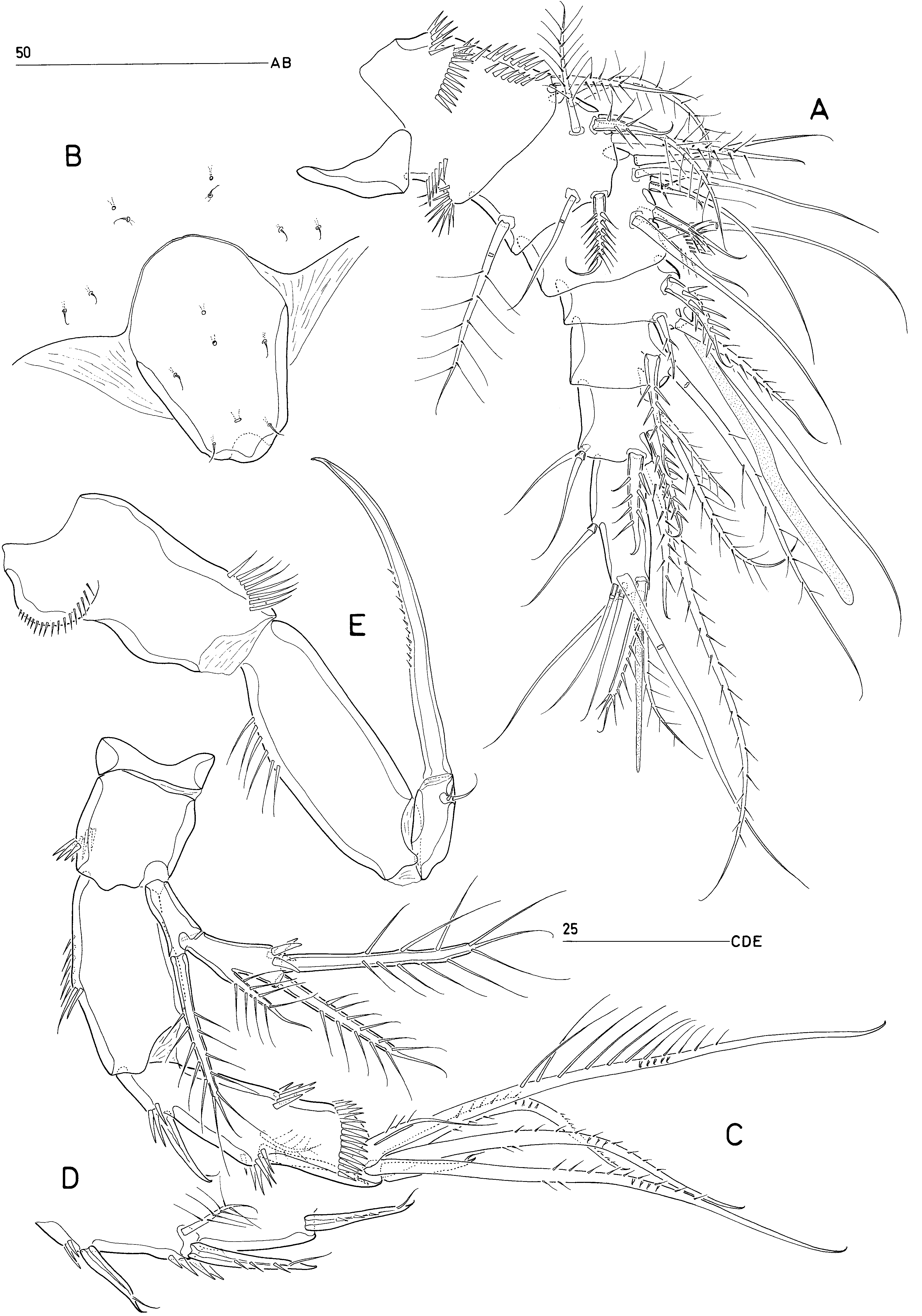
Labrum ( Fig. 5C View Figure 5 ) strongly developed, weakly trilobate; median lobe with short strong spinules along free margin and densely packed spinules plus a large median pore on posterior surface; lateral lobes each with double row of long spinules.
Mandible ( Fig. 3C, D View Figure 3 ). Gnathobase with series of blunt, multicusped teeth; dorsal corner with strong spine bearing minute spinules along dorsal margin and very coarse spinules along ventral margin. Basis relatively small, with two short plumose setae. Endopod longer than exopod, without surface spinules; with three plumose setae laterally (two fused at base) and nine setae apically (seven naked, two pinnate), several of which fused to segment. Exopod 1- segmented, with one short seta near proximal margin and four plumose setae along lateral margin and apex; distalmost three setae fused to segment.
Paragnaths ( Fig. 5C View Figure 5 ) strongly developed lobes with medially directed long spinules.
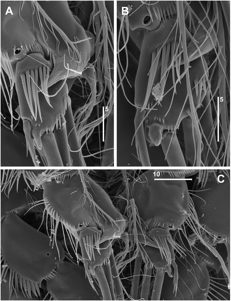
Maxillule ( Fig. 6B, C View Figure 6 ). Praecoxal arthrite with six spines, one pinnate seta and one minute tube-seta around distal margin; anterior surface with two juxtaposed setae; posterior surface with two pinnate setae, innermost very large and typically curved ( Fig. 6C View Figure 6 ). Coxal endite with long spinules on anterior surface; with two smooth and three pinnate (of which one geniculate) setae. Basis with long setules on anterior surface; armature consisting of four naked and two pinnate (of which one geniculate) setae. Endopod a small segment with one plumose and two naked setae. Exopod represented by a single plumose seta.
Maxilla ( Fig. 9A View Figure 9 ). Syncoxa with three endites and long spinules along outer margin; proximal endite expanded distally, with one large and three shorter pinnate setae; middle and distal endites cylindrical, each with one naked and two pinnate setae. Allobasis with long setules along outer margin; drawn out into pinnate claw; accessory armature consisting of one naked and two pinnate setae. Endopod ( Fig. 5B View Figure 5 ) indistinctly 2-segmented with one geniculate spine, two pinnate and three naked setae.
Maxilliped ( Figs 4E View Figure 4 , 5E View Figure 5 ). Syncoxa with spinular row near medial distal corner and smaller spinules around proximal outer margin. Basis outer margin with slender spinules in proximal half. Endopod with curved claw and two minute accessory setae, one of which is tubular ( Fig. 5E View Figure 5 ).
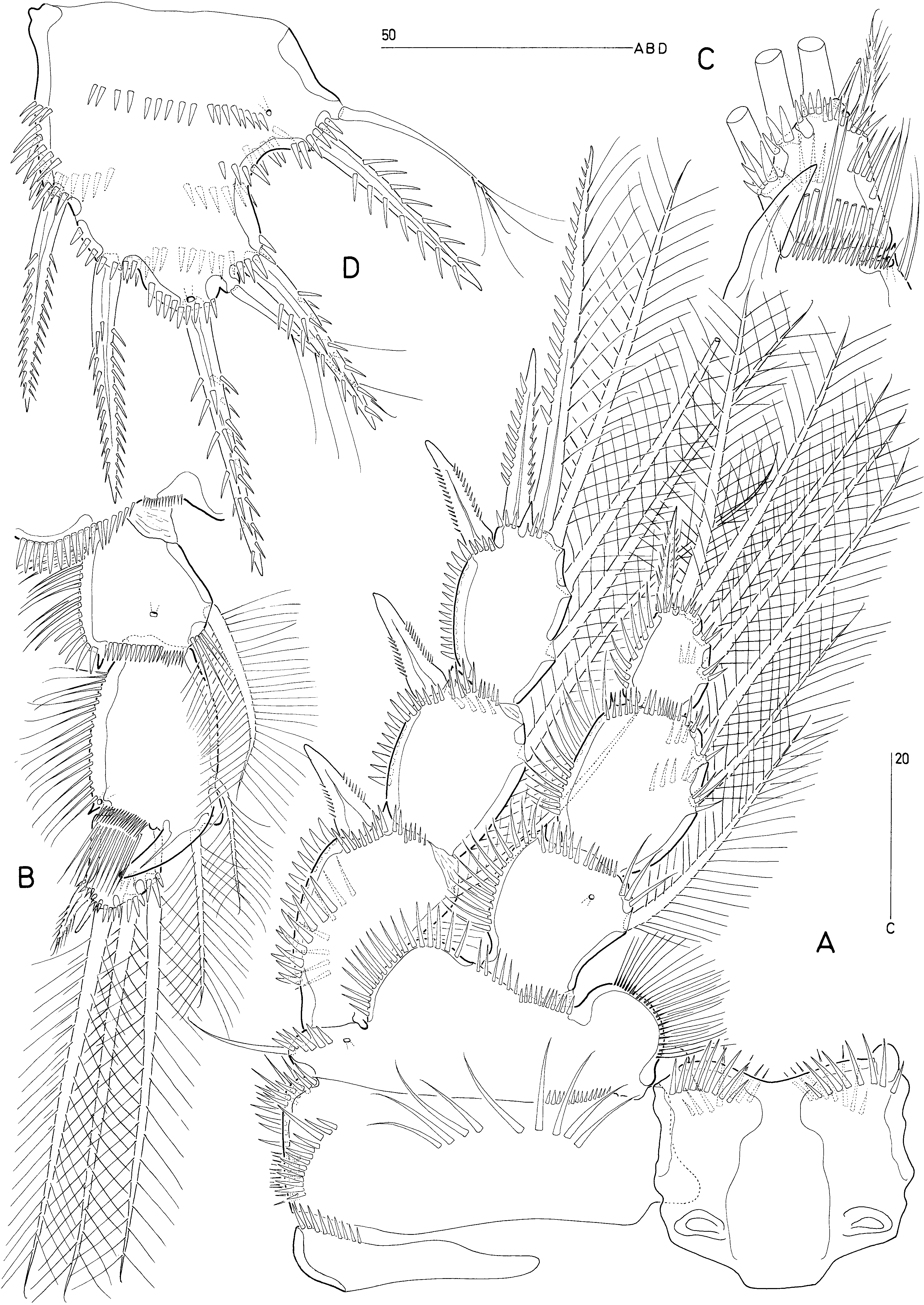
P1 ( Fig. 6A View Figure 6 ). Intercoxal sclerite bilobate, with strong spinules on anterior surface. Praecoxa well developed, with long spinules on anterior surface. Coxa with various anterior spinule rows as figured. Basis with produced lobate inner process bearing spinules anteriorly and setules posteriorly; outer and inner basal setae bipinnate. Rami 3-segmented. Exopod shorter than endopod; inner margin of segments with setules, outer margin with spinules; outer spines with subapical flagellate extension. Endopod with small enp-3; distal and outer margins of all segments with spinules; few spinules also present on proximal inner margin of enp-2 and -3; outer distal element of enp-3 spiniform and bipinnate. Posterior surface of endopod segments and exp-1 and -2 typically with spinule patches.
P2-P4 ( Figs 7A View Figure 7 , 8A View Figure 8 , 9B View Figure 9 ). Intercoxal sclerites with spinules anteriorly (P2–P4) and posteriorly (P2–P3). Praecoxa a small sclerite with anterior spinules. Coxa typically with very long spinules anteriorly and various spinule rows arranged around outer margin. Inner margin of basis forming lobate setulose expansion; with spinules around distal and outer margin as figured; outer basal seta bare (P2) or sparsely plumose (P3–P4). Endopods shorter than exopods; rami 3- segmented. Exp-1 and -2 (and - 3 in P4), and enp-2 and -3 typically with posterior spinule patches. Inner margin of exp-1 and -2 (and - 3 in P4) with few long setules; spinular ornamentation around distal and outer segment margins as figured. Exp-3 of P2–P3 forming spinous extension between bases of distal outer spine and outer apical spine. Armature formula of P1–P4 as for genus.
P5 ( Fig. 7D View Figure 7 ) about 1.05 times as long as maximum width; typically with distinct outer concavity, separating outer lobe from distal portion; with numerous spinular rows on both posterior and anterior surfaces as figured; outer lobe with basal plumose seta and pinnate spine; distal portion with plumose seta flanked by strong pinnate spines around apex, and two pinnate spines along inner margin; anterior surface with three secretory pores (one marginal).
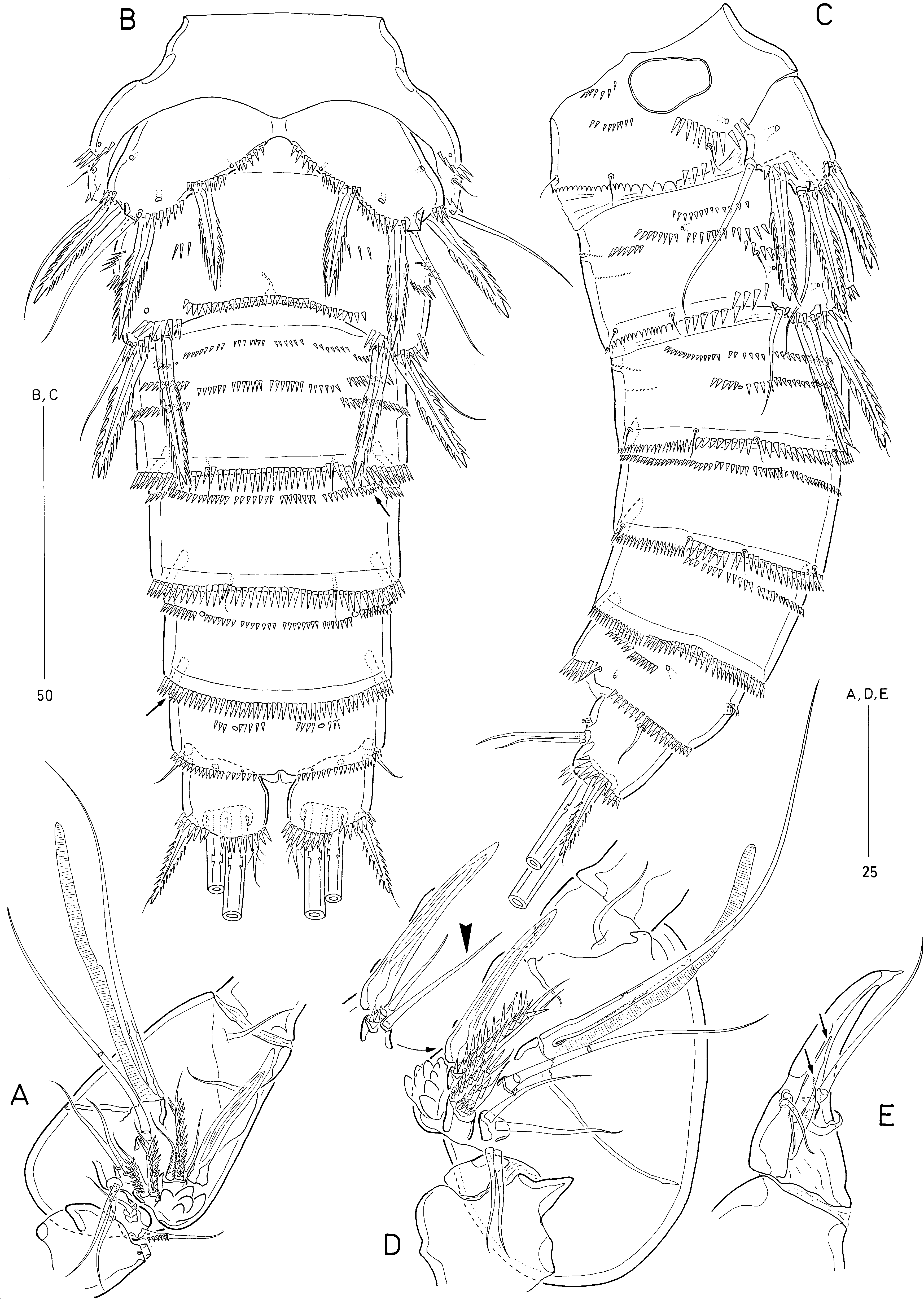
Male: Sexually dimorphic in size, urosome ornamentation and segmentation, antennule, P2 endopod, P3 exopod, P5 and P6.
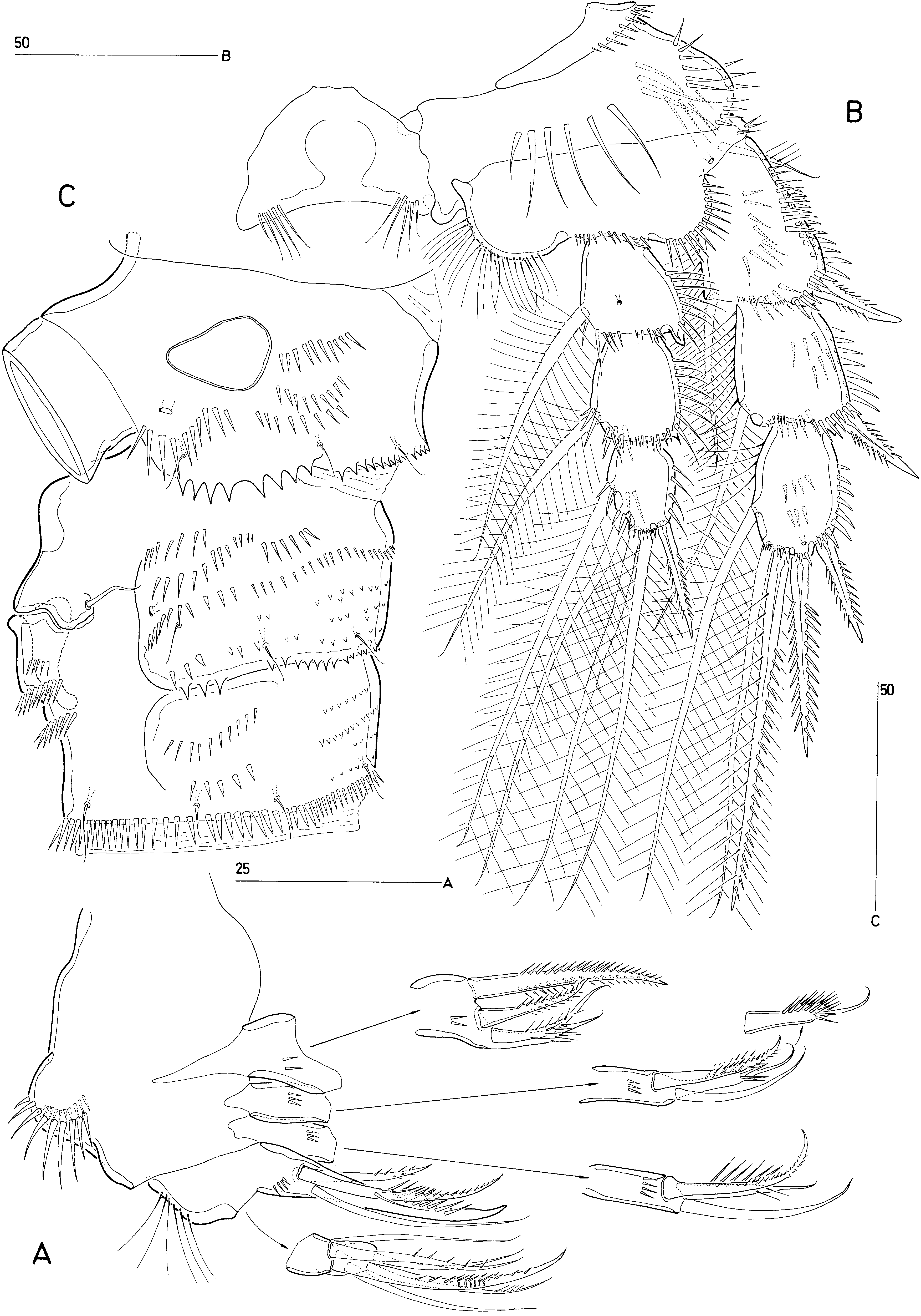
Antennule ( Fig. 10B View Figure 10 ) 7-segmented, with one segment distal to geniculation (chirocer condition). Segment 1 with sclerite around base; posterior margin forming lobate extension bearing long spinules; with numerous spinule rows around anterior margin as figured ( Fig. 10B, C View Figure 10 ). Segment 3 with bulbous process on proximal posterior margin bearing plumose seta. Segment 4 largely membranous around posterior margin; anterior margin with dorsal spinous process. Segment 5 minute, represented by a small sclerite on anterior margin ( Fig. 14D View Figure 14 ; arrowed in Fig. 15B View Figure 15 for N. parvus ). Segment 6 ( Fig. 14D View Figure 14 ) very large and swollen; with incomplete transverse surface suture ventrally and posteriorly; anterior margin forming multicuspidate structure (typically with eight cusps) in proximal half; with modified longitudinally striated element often closely adpressed to anterior surface ( Fig. 14D View Figure 14 ; cf. Fig. 15D View Figure 15 for N. parvus ). Segment 7 ( Figs 10B View Figure 10 , 14E View Figure 14 ) hook-shaped, without surface sutures marking original segmentation; apex weakly chitinized, recurved; anterior surface forming longitudinal furrow containing two basally fused elements (arrowed in Fig. 14C View Figure 14 ) as in N. parvus ( Fig. 15C View Figure 15 ). Armature formula: 1-[1 pinnate], 2-[1 pinnate], 3-[6 bare +5 pinnate], 4-[6 bare +2 pinnate + ae], 5-[2 pinnate], 6-[9 bare +3 pinnate +1 striated element + (1 + ae)], 7-[12 +2 modified + acrothek]. Apical acrothek consisting of short aesthetasc and small naked seta. Many setae on segments 3–4 with proximal flexure zone.
P2 endopod ( Figs 7B View Figure 7 , 16A, C View Figure 16 ). Middle segment enlarged on inner margin, forming outwardly directed spinous apophysis partly overlying enp-3 anteriorly; inner setae markedly shorter than in ♀. Enp-3 small ( Figs 7C View Figure 7 , 16A View Figure 16 ), with row of long setules on anterior surface, typically overlying apex of spinous apophysis; outer distal seta strongly reduced, represented by minute, basally fused spine with recurved tip; outer spine reduced.
P3 exopod ( Fig. 8B View Figure 8 ) distinctly longer and more slender than in ♀. Inner setae of exp-1 and -2 smaller than in ♀. Exp-2 elongate; posterior spinules absent. Exp-3 longer than in ♀ inner distal seta vestigial.
P5 ( Figs 6D View Figure 6 , 10A View Figure 10 ) medially fused, each with three pinnate spines and two setae; outermost seta naked, representing outer basal seta, other seta sparsely plumose. Anterior surface with fine spinule rows and pore.
P6 ( Figs 6D View Figure 6 , 10A View Figure 10 ) symmetrical; each member with two pinnate spines and naked outer basal seta; posterior margin with coarse spinules bi-laterally and fine spinules medially. Spermatophore large, about 100 mm in length.
Ornamentation of urosome essentially as in ♀ except for more elaborate spinular patterns on first abdominal somite ( Figs 6D View Figure 6 , 10A View Figure 10 ).
Etymology: The specific name refers to Korea, the country where the type locality of the new species is situated.

| NMNH |
Smithsonian Institution, National Museum of Natural History |
| NHM |
University of Nottingham |
| V |
Royal British Columbia Museum - Herbarium |
| VI |
Mykotektet, National Veterinary Institute |
No known copyright restrictions apply. See Agosti, D., Egloff, W., 2009. Taxonomic information exchange and copyright: the Plazi approach. BMC Research Notes 2009, 2:53 for further explanation.