
Brachysomus (Formanek, 1905)
|
publication ID |
https://doi.org/ 10.11646/zootaxa.5193.1.1 |
|
publication LSID |
lsid:zoobank.org:pub:78BDA3C9-8B2E-444F-AB50-1A64FB3F8786 |
|
DOI |
https://doi.org/10.5281/zenodo.7144612 |
|
persistent identifier |
https://treatment.plazi.org/id/0383A324-4620-FFC7-FF6C-A085FA9A6CF4 |
|
treatment provided by |
Plazi |
|
scientific name |
Brachysomus |
| status |
|
General morphology of Brachysomus View in CoL
Head
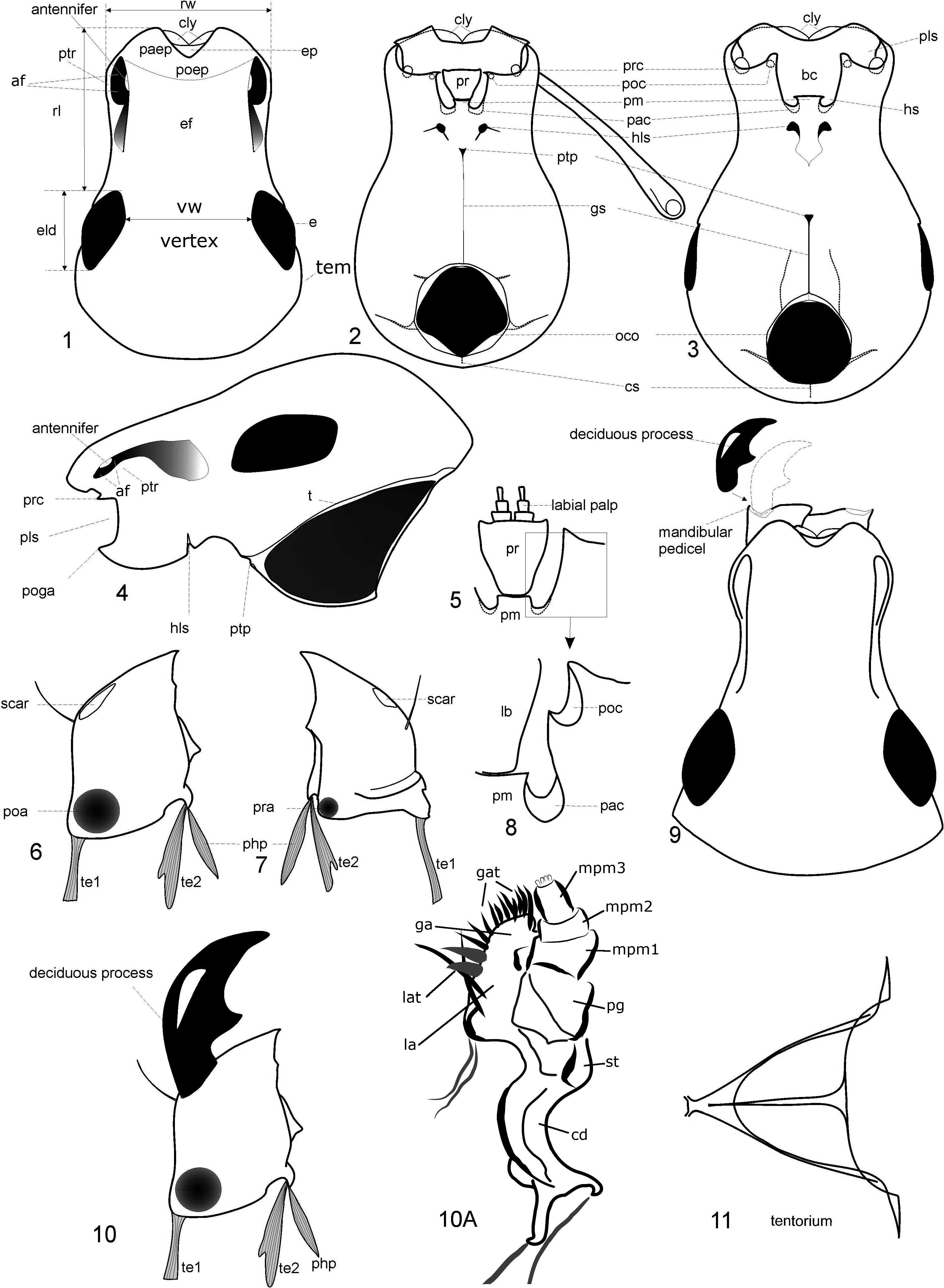
( Figs. 1–4, 10, 11 View FIGURES 1–11 , 18 View FIGURES 18–21 )
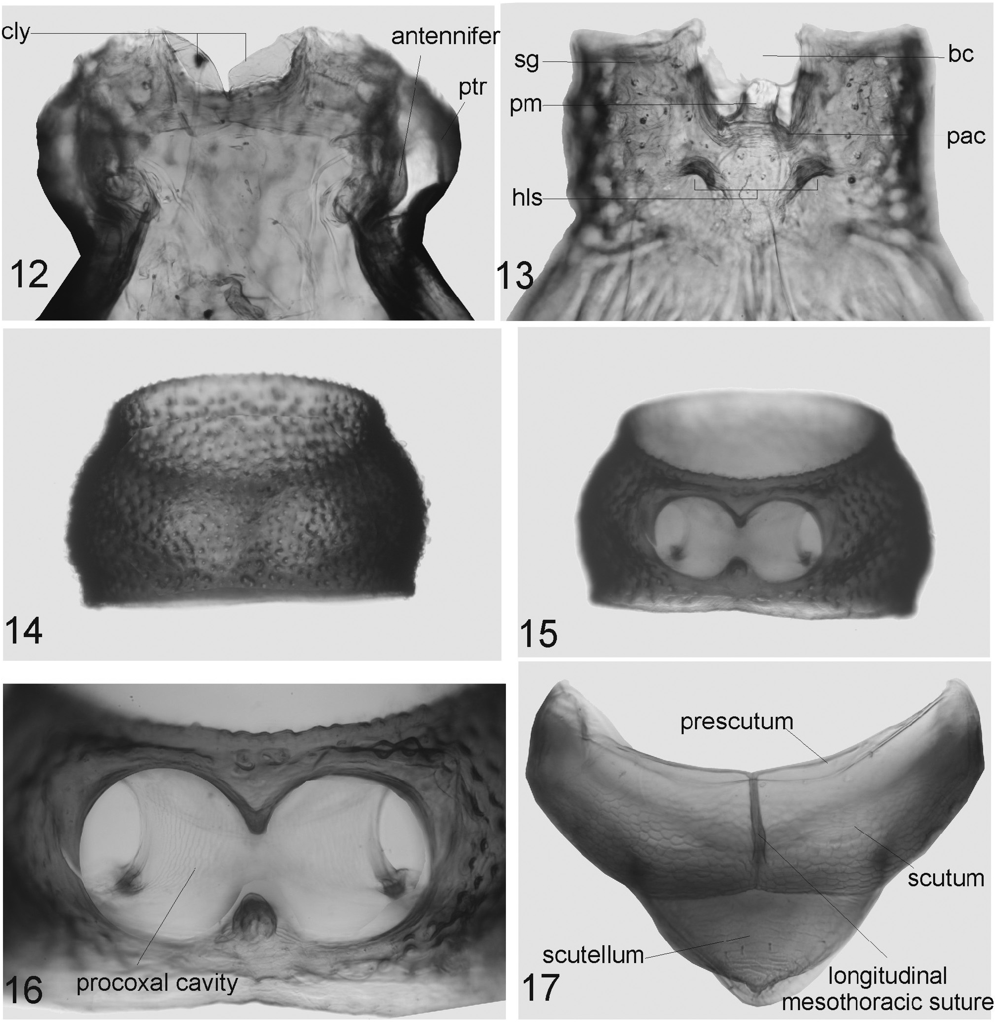
Head capsule and tentorium ( Figs. 1–4, 11–13 View FIGURES 1–11 View FIGURES 12–17 ). Rostrum as long as wide or transverse, separated from vertex by moderately deep transverse depression. Temples usually as long as ELD. Vertex flat or slightly convex.Anterior part of the epifrons ( Fig. 1 View FIGURES 1–11 , ef) consists of the flat epistome ( Fig. 1 View FIGURES 1–11 , ep) and its neighbouring areas: lateral—parepistome (paep) and posterior—postepistome (poep) ( Morimoto et al. 2006); the latter part is flat ( Brachysomus s. str.) or depressed (subgen. Hippomias ) and can be scaled, pilose or bare. Sometimes epistome (in males of Brachysomus mihoki group) bears process in lateroapical angle. These areas of the head capsule are not demarcated from each other by sulci. Eyes large, round or elliptic, moderately convex. Pterygia ( Figs. 1, 4 View FIGURES 1–11 , ptr) slightly projecting from outline of rostrum, surrounding the antennal fossa ( Figs. 1, 4 View FIGURES 1–11 , af) and protecting the antennal articulation from below.Only a small part of the antennifer (an articulating process from the head capsule that contacts the antennal scape) is visible from above, or it is completely hidden by lateral margins of epifrons.
The ventral surface of the head contains the entire set of structures that can be present in Entiminae. Ventral surface of rostrum contains following parts: subgenae (sg) and postmentum (pm) surrounding buccal cavity (bc) covered by prementum (pr). The head capsule contains occipital opening (oco), gular suture (gs), cervical suture (cs), and posterior tentorial pit (ptp). Gular suture distinct ( Figs. 2–3 View FIGURES 1–11 , gs), short or long, with single median triangular posterior tentorial pit ( Figs. 2–3 View FIGURES 1–11 , ptp) (in Entiminae and other advanced Curculionidae the commonly known paired posterior tentorial pits generally merged into a single pit (Lyal 1995)). The pair of pits located further anteriorly on the ventral side of the rostrum are part of the hypostomal-labial suture ( Figs. 2–3 View FIGURES 1–11 , hls, after Lyal 1995) or midventral suture (as accepted by Doyen 1966 for Tenebrio molitor ) but not anterior pits ( Girón & Franz 2010). The anterior tentorial pits, i. e. the external indications of the origin of the anterior tentorial arms, are paired pits sometimes visible in most Coleoptera on the apical parts of the head, where they partially delimit the clypeus postero-laterally (being part of the epistomal sulcus, as quite usual in insects). Anterior tentorial arms and pits have been lost in advanced Curculionidae including Entiminae in the course of strong transformations of external and internal exoskeletal structures of the head capsule ( Fig. 4, t, 11 View FIGURES 1–11 ) (Lyal 1995). The hypostomal-labial sutures ( Figs. 2–3 View FIGURES 1–11 , hls) are situated antero-laterad of the posterior tentorial pit and just posteriad of the maxillary articulation, and are mainly visible as the abovementioned pits that are part of the hls suture.
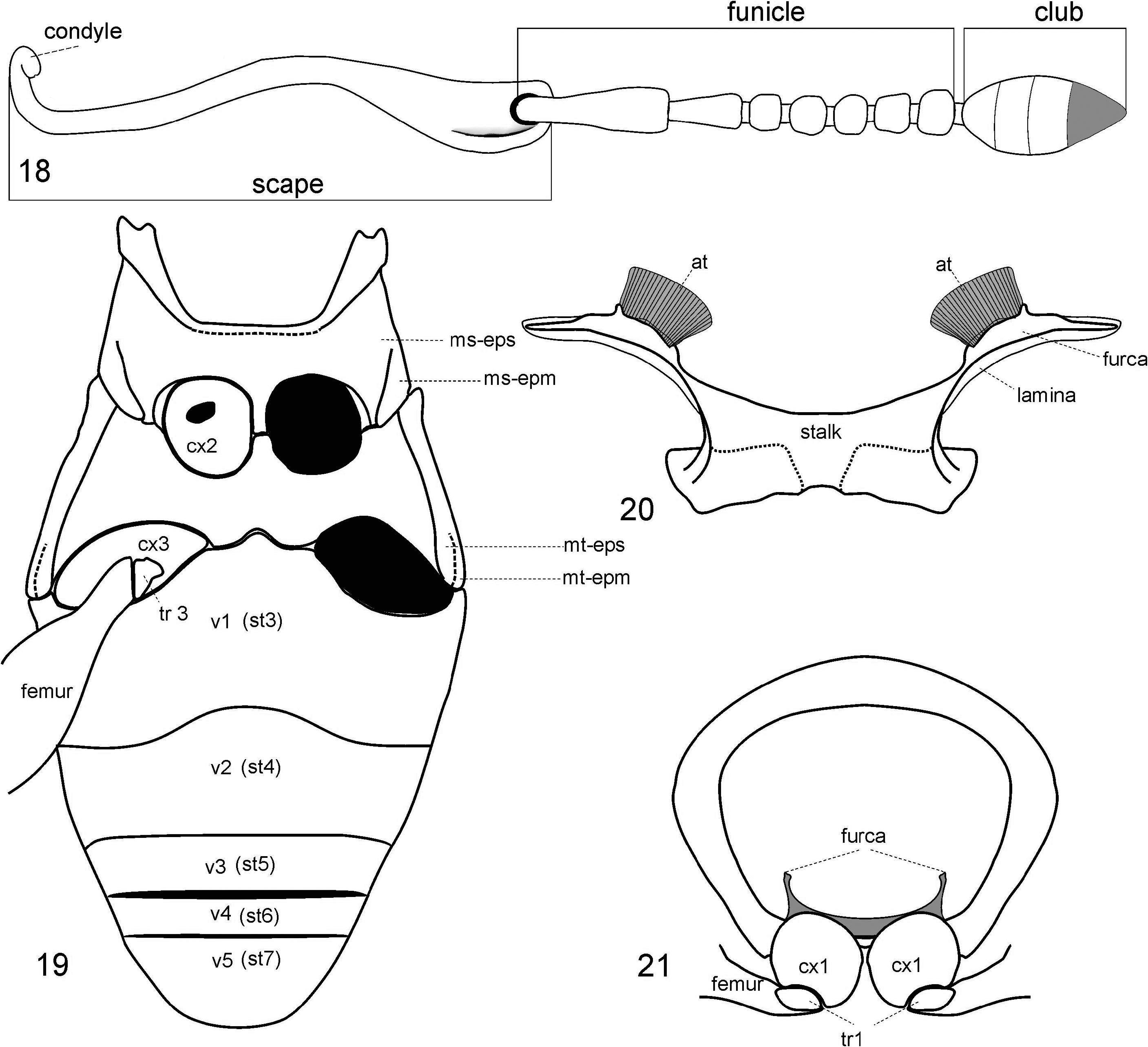
Antennae ( Fig. 18 View FIGURES 18–21 ). Scape moderately curved and widened in distal 1/3, sometimes straight and evenly widened distally, with unciform condyle at base. The underside at the apex of the scape bears a large excision for the condyle of the 1st funicular antennomere (pedicel).A short scrobe (for harbouring the base of the funicle) extends proximally from the apical excision of the scape ( Fig. 18 View FIGURES 18–21 ). The funicle consists of 7 antennomeres; the 1st and 2nd usually rather elongate, longer than other ones; 1st at least 1.5x as long as 2nd; 3rd–6th as long as wide or transverse; 7th transverse. Club egg-shaped, oblong-egg-shaped or broadly spindle-shaped, slightly set off from funicle; 1st clubantennomere trapezium-shaped, evenly widened distally.
Mandibles ( Figs. 6, 7, 10 View FIGURES 1–11 ). Subtriangular, asymmetric regarding dentition and size (left mandible somewhat larger than right one), divided into base (including articulations) and cutting (mesal), dorsal (= morphologically anterior) and ventral (= morphologically posterior) surfaces. Anterior surface convex, with a lateral scar shortly beyond midlength (left by the deciduous mandibular process) and a pair of mandibular chaetae. The origins of the pharyngeal process and of the tendons for the adducting (te2) and abducting (te1) muscles of the mandibles are situated along the base. Prearthris (anterior articulation of the mandible; Fig. 6 View FIGURES 1–11 , pra) large, circular depression, situated in laterobasal angle on anterior surface of mandible and attaches the mandible to the precoila ( Fig. 2-3 View FIGURES 1–11 , poc). Postarthris (posterior articulation of the mandible; Fig. 6 View FIGURES 1–11 , poa) small, shaped as a concave disk, situated in laterobasal angle on posterior surface of mandible, attaches the mandible to the postcoila (poc). In the laterobasal angle, the mandibular sclerotisation forms a convex extension to which the tendon of musculus abductor mandibulae (te 1) is attached. In the mediobasal angle of the ventral surface a small extension is situated from which the tendon of musculus adductor mandibulae (te 2) and the pharyngeal process (php) originate. The tendon of the adductor is larger than that of the abductor. The deciduous mandibular process (= mandibular appendage or pupal process of some authors; Fig. 9, 10 View FIGURES 1–11 ) is sickle-shaped with a short mesal dens at midlength. It is usually broken in older beetles, but often present in freshly emerged imagines.
Maxillae. Galea and lacinia fused. Cardo strongly elongate, at base with two extensions, to which tendons are attached. Maxillary palps composed of three palpomeres ( Fig. 10A View FIGURES 1–11 ).
Labium ( Figs. 5, 8 View FIGURES 1–11 ). Prementum triangular or trapezoidal, bears 4 setae. Base of labial palp on dorsal surface of the prementum near its apex; 3 palpomeres. Ligula reduced, carinate, represented by a transverse fold on distal surface of prementum. Cavities on each side of ligula serve for insertion of maxillary lobes (fused galea and lacinia).
Thorax
( Figs. 14–17 View FIGURES 12–17 , 19, 21 View FIGURES 18–21 )
Hindwings very small or entirely absent. This has resulted in reductive trends in many structures of the pterothorax that in winged entimines are involved in flight (aptery). Similar trends also occur in other wingless Entiminae. One of these trends is that in wingless taxa the pterothorax is relatively shorter than in groups possessing well-developed functional hindwings.
Prothorax. Transverse, evenly convex across disc and along sides, more or less strongly constricted at apex and base. Disc densely finely punctate, with gradual transition to tuberculate sides ( Fig. 14 View FIGURES 12–17 ). Anterior foramen of prothorax smaller than posterior one. Procoxal cavities open ( Figs. 15, 16 View FIGURES 12–17 ). Precoxal part of prosternum as long as postcoxal part. Notosternal suture very short, indistinct, obliterated anteriorly. Prothorax trapeziform in transverse section ( Fig. 21 View FIGURES 18–21 ).
Mesothorax. Mesonotum ( Fig. 17 View FIGURES 12–17 ) moderately sclerotized. Scutum with a well developed longitudinal mesothoracic suture. Scutellum small, not forming externally visible scutellar shield ( Fig. 17 View FIGURES 12–17 ). Suture between mesepisternum and mesoventrite obsolete. Suture between mesepisternum and mesepimeron visible. Mesoventrite strongly sclerotized. Mesocoxal cavities medially directed ( Fig. 19 View FIGURES 18–21 ).
Metathorax. Metanotum membranous. Metepisternum distinctly separated from metaventrite by a suture. Metepimeron separated from metepisternum only in posteroventral part, by a short pleural suture ( Fig. 19 View FIGURES 18–21 ).
Metendosternite ( Fig. 20 View FIGURES 18–21 ). Stalk very short.Anterior tendon displaced laterally, rectangular, slightly sclerotized. Furcal arms marginate, nearly perpendicular to longitudinal axis of body, not bifurcate at apex, without carina and processes. Lamina of furcal arms vestigial.
Elytra. Ovate or broadly-ovate, without humeral and apical prominences, slightly convex along sides and across disc, wider in the middle part (EL/EW = 1.16–1.23). Base of elytra weakly sinuate. Number of complete striae 10. Striae narrow, punctures small, shallow, weakly separated; interstriae slightly convex, 1.5–2x wider than striae.
Legs. Femora, swollen in middle part, unarmed. Tibiae with straight outer (= dorsal) margin, or slightly widened at apex; inner (= ventral) margin S-shaped, sometimes C-shaped on fore tibiae. Hind tibiae of male in some species with internal margin acute (knife-shaped), their mucro developed to a varied extent. Second tarsomere strongly transverse, 3rd one with two distinct lobes. Ultimate tarsomere of fore tarsus (not including claws) extending beyond apical lobes of 3rd by 0.6–1.14x of their length. Claws connate in basal half.
Abdomen
( Figs. 19 View FIGURES 18–21 , 22–24 View FIGURES 22–24 )
Ventrites. 1st–3rd ventrites ( Fig. 19 View FIGURES 18–21 ) fused but with distinct immovable sutures, covered with hairs and piliform scales. Posterior margin of 1st ventrite sinuate. Surface of male 5th ventrite moderately convex, with or without depression near the apex, apical margin straight or sinuate.
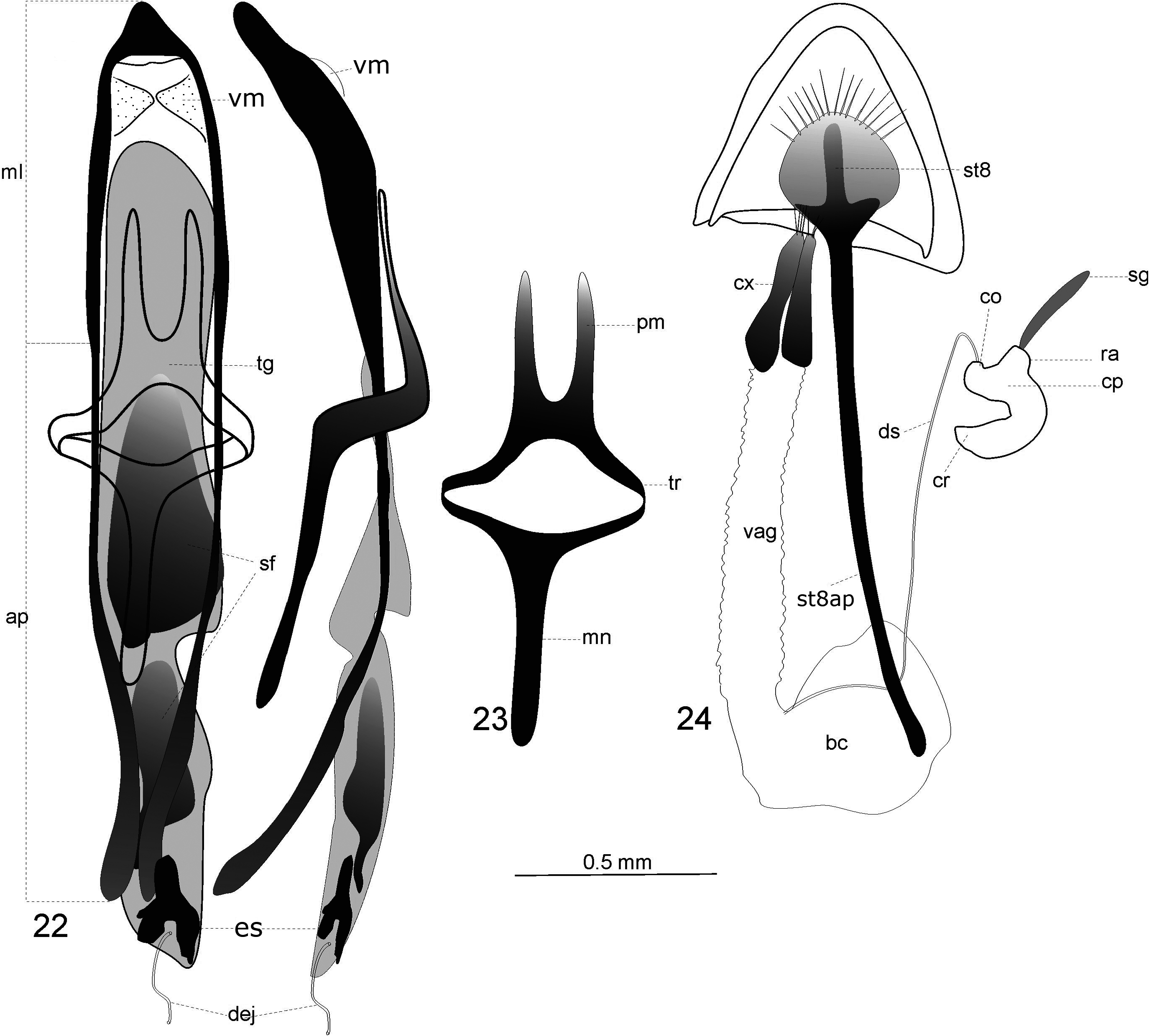
Male genitalia ( Figs. 22, 23 View FIGURES 22–24 ). Aedeagus strongly or weakly sclerotized, apodemes 1–3x as long as median lobe. Apex of median lobe acutely or widely rounded. Apical region covered with paired ventral membranes ( Girón & Chamorro 2020) which are also called ligulae ( Yunakov 2012), that are membranous or moderately sclerotized, oblong or as long as wide ( Fig. 22 View FIGURES 22–24 ). Tegmen with developed or vestigial paramera; apodeme straigth, 10x as long as wide. Endophallus covered with microscopic spiculae (in areas sf in Fig. 22 View FIGURES 22–24 ), its distal area with strongly sclerotized, endophallic sclerite (es). This sclerite is occasionally transformed into a strong baculiform process of various size and shape. Proximal area of endophallus occasionally with one or two dorsal sclerites.
Female genitalia ( Fig. 24 View FIGURES 22–24 ). Coxites of segment IX weakly sclerotized, undivided, evenly covered with pores and microscopic sensilla, in apical part with long sensilla, styli absent. Spermatheca moderately sclerotized, with well developed or reduced ramus (ra) and developed collum (co; = nodulus sensu Borovec 2006) as long as ramus or shorter. Corpus (cp) vestigial or swollen. Sternite VIII ( Fig. 24 View FIGURES 22–24 , st8) weakly sclerotized; lamina membranous, subtriangular, rhomboid or subrotundate, weakly sclerotized, posterior margin with 1–2 rows of thin cirri; apodeme of sternite VIII ( Fig. 24 View FIGURES 22–24 , st8ap) thin, straight or weakly curved.
Vestiture
Overall varying from very sparse to very dense depending on ecological preferences of species. The vestiture includes one or several types of scales. There can be piliform, lanceolate, ovate or round scales covering the dorsal and lateral surfaces of the body. Apical margin of scales straight, bifurcate, excised, or multidentate. Apically excised and multidentate scales present in species from xerothermic forests. Coloration of scales usually uniform, in Hippomias species grey-brownish, often conforming with the color of the litter. In Brachysomus s. str. species inhabiting xerothermic habitats scales with metallic shine. Many species show spotty coloration of the vestiture, forming staggered dark-drown spots against light integument.
The setae of the elytral striae are narrow or distinctly widened apically, suberect or erect, situated in dense or sparse, simple or double rows. Spaces between the setae are ≤ 1x the length of a seta, and 0.2–1.0x (usually 0.3–0.5x) the width of an interstria.
The ventral surface of both legs and body (compared to the dorsal surface) are nearly always scaled more sparsely. Dorsal surface of femora and tibiae and (in Hippomias ) antennae, densely covered with scales. Compared with the vestiture of the body, scales are usually narrower on the legs. The funicular antennomeres and the tarsi are covered with thin hairs or setae only, scales are absent. The antennal club is densely covered with sensilla.
No known copyright restrictions apply. See Agosti, D., Egloff, W., 2009. Taxonomic information exchange and copyright: the Plazi approach. BMC Research Notes 2009, 2:53 for further explanation.
|
Kingdom |
|
|
Phylum |
|
|
Class |
|
|
Order |
|
|
Family |