
Microsarimodes flavomaculata Wang & Bourgoin, 2020
|
publication ID |
https://doi.org/10.11646/zootaxa.4858.4.7 |
|
publication LSID |
lsid:zoobank.org:pub:A1D9D589-0CF3-47CE-9D3B-E66E0E81A73F |
|
DOI |
https://doi.org/10.5281/zenodo.4538751 |
|
persistent identifier |
https://treatment.plazi.org/id/0383DD52-FFCC-FFE2-FF40-89B4FABDF84E |
|
treatment provided by |
Plazi |
|
scientific name |
Microsarimodes flavomaculata Wang & Bourgoin |
| status |
sp. nov. |
Microsarimodes flavomaculata Wang & Bourgoin View in CoL , sp. nov.
ZooBank registration: LSID urn:lsid:zoobank.org:act:
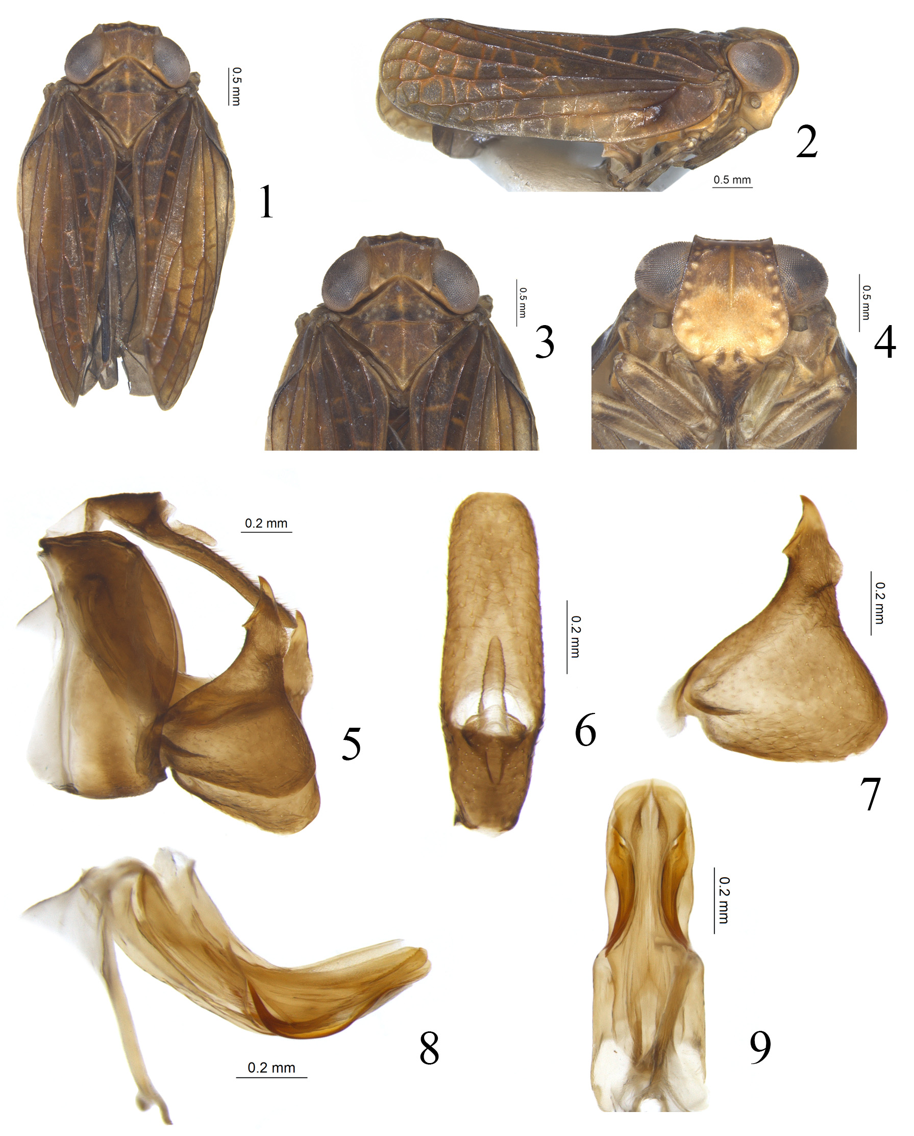
( Figs 1–9 View FIGURES 1–9 )
Diagnosis. This new species is similar to Microsarimodes tumida Chang & Chen, 2019 , but differs by: 1) Pronotum with several tubercles on the disc ( Figs 1, 3 View FIGURES 1–9 ), but without tubercles in M. tumida ( Chang et al., 2019, fig. 18); 2) The yellow patches on frons intersperse in basal half of frons ( Fig. 4 View FIGURES 1–9 ), while only basal 1/3 on frons in M. tumida ( Chang et al., 2019, fig. 20); 3) Male anal tube in dorsal view with lateral margins paralleled in apical 2/3, anal opening located at basal 1/3 of anal tube ( Fig. 6 View FIGURES 1–9 ), while lateral margins gradually narrower from middle part to the apex, anal opening located at middle of anal tube in M. tumida ( Chang et al., 2019, fig. 24); 4) Capitulum of gonostylus much longer, the tumefied protuberance located in the postero-lateral area with its apex exceeding to the apical margin ( Fig. 7 View FIGURES 1–9 ), while capitulum of gonostylus short, the tumefied protuberance located in the anterior-lateral area with its apex reaching to the middle in M. tumida ( Chang et al., 2019, fig. 25: b).
Etymology. Combination of Latin words “ flavus ” meaning yellow and “macula” meaning patch, referring to the yellow marking on the basal half of the frons.
Description. Length: male (including forewings) (N=1): 5.2 mm.
Coloration. Vertex brown, margins brown, disc with weak yellow median carina ( Figs 1, 3 View FIGURES 1–9 ). Compound eyes grayish brown, supported by tawny callus ( Figs 1, 3 View FIGURES 1–9 ). Frons light yellow in basal half and brown in apical half, the boundary between the light yellow and brown areas M-shaped ( Fig. 4 View FIGURES 1–9 ); apical margin black, lateral margins mostly black with basal slightly yellow ( Fig. 4 View FIGURES 1–9 ); median carina yellow, only elevated and visible at apical half ( Fig. 4 View FIGURES 1–9 ). Frons with around 11 to 14 large irregular yellowish tubercles on apical and lateral areas ( Fig. 4 View FIGURES 1–9 ). Antennae brown ( Fig. 4 View FIGURES 1–9 ). Postclypeus yellow, with two broad interrupted brownish longitudinal bands ( Fig. 4 View FIGURES 1–9 ). Genae tawny ( Fig. 2 View FIGURES 1–9 ). Pronotum brown, anterior and posterior margins brown; median carina yellow, only visible at middle, lateral areas with several grayish inconspicuous tubercles ( Figs 1, 3 View FIGURES 1–9 ). Mesonotum brown, median carina yellow, sublateral carinae brown ( Figs 1, 3 View FIGURES 1–9 ). Forewings tawny, longitudinal veins mostly brown and transverse veins tawny ( Figs 1, 2 View FIGURES 1–9 ). Hindwings brown ( Fig. 1 View FIGURES 1–9 ). Legs light yellow ( Fig. 4 View FIGURES 1–9 ).
Head and thorax. Vertex 1.6 times wider in width at middle than length at middle, anterior margin slightly angularly convex, posterior margin roundly concave with the level deeper than anterior margin, lateral margins slightly concave inward at middle, then convex near base, postero-lateral angles rounded ( Figs 1, 3 View FIGURES 1–9 ). Frons 1.0 times wider at widest part than long in midline, 1.5 times wider at widest part than apical margin ( Fig. 4 View FIGURES 1–9 ); apical margin straight ( Fig. 4 View FIGURES 1–9 ). Pronotum 2.5 times wider in posterior margin than long in midline, 1.2 times longer in midline than vertex ( Figs 1, 3 View FIGURES 1–9 ); anterior margin sharply angularly convex, posterior margin straight ( Figs 1, 3 View FIGURES 1–9 ). Mesonotum with anterior margin 1.7 times wider than long in midline, 1.6 times longer in midline than pronotum, median carina from anterior margin to the base but not elevated, sublateral carinae elevated from anterior margin but not reaching to the posterior margin ( Figs 1, 3 View FIGURES 1–9 ). Forewings with MP 1+2 bifurcate at apical 1/6, MP 3+4 single ( Fig. 2 View FIGURES 1–9 ). Metatibiotarsal spinulation formula unknown (hind legs missing).
Male genitalia. Anal tube in lateral view with ventral margin straight ( Fig. 5 View FIGURES 1–9 ); in dorsal view long cylindrical, 3.3 times longer in midline than widest part, lateral margins paralleled at apical 2/3, then gradually narrower to base, apical margin convex with middle part almost straight ( Fig. 6 View FIGURES 1–9 ); anal opening located at basal 1/3 of anal tube ( Fig. 6 View FIGURES 1–9 ). Pygofer in lateral view rectangular, 2.4 times longer in midline than width at middle; dorsal margin straight, posterior margin slightly convex caudad ( Fig. 5 View FIGURES 1–9 ). Gonostylus triangular in lateral view, highest near middle, dorsal margin of gonostylus almost straight, oblique toward to capitulum, ventral margin with postero-ventral angle strongly convex ( Figs 5, 7 View FIGURES 1–9 ). Capitulum of gonostylus long and broad, triangular, gradually narrowing from base to apex, tip pointed, antero-lateral margin with a spinous process, postero-lateral margin with a roundly tumefied protuberance at base with its apex exceeding to the apical margin ( Figs 5, 7 View FIGURES 1–9 ). Periandrium dorsal lobe rounded in the apex in lateral view ( Fig. 8 View FIGURES 1–9 ); periandrium lateral lobe bearing density of minute teeth on dorsal margin ( Fig. 8 View FIGURES 1–9 ); periandrium ventral lobe slightly shorter than lateral lobes ( Fig. 8 View FIGURES 1–9 ), rounded in the apical margin in ventral view ( Fig. 9 View FIGURES 1–9 ). Aedeagus with a pair of large hook-like processes originated from apical 1/5 along the ventral margin, then curved to dorso-anterior, reaching to the middle of periandrium ( Fig. 8 View FIGURES 1–9 ), in ventral view, this pair of processes curved to outward ( Fig. 9 View FIGURES 1–9 ).
Type materials. Holotype: ♂, China, Yunnan Province, Mangshi , 30 iv 2012, coll. Menglin Wang.
No known copyright restrictions apply. See Agosti, D., Egloff, W., 2009. Taxonomic information exchange and copyright: the Plazi approach. BMC Research Notes 2009, 2:53 for further explanation.
|
Kingdom |
|
|
Phylum |
|
|
Class |
|
|
Order |
|
|
Family |
|
|
Tribe |
Sarimini |
|
Genus |