
Eigenmannia besouro, Peixoto, Luiz Antônio W. & Wosiacki, Wolmar B., 2016
|
publication ID |
https://doi.org/10.11646/zootaxa.4126.2.6 |
|
publication LSID |
lsid:zoobank.org:pub:CF9067FD-F385-4885-8095-CD8D06C767F7 |
|
DOI |
https://doi.org/10.5281/zenodo.6063782 |
|
persistent identifier |
https://treatment.plazi.org/id/038587B2-FF85-FFB7-FF21-F8B84F76F9C4 |
|
treatment provided by |
Plazi (2016-06-17 08:02:11, last updated 2024-11-29 06:24:25) |
|
scientific name |
Eigenmannia besouro |
| status |
sp. nov. |
Eigenmannia besouro View in CoL , new species
( Figure 1 View FIGURE 1 )
Holotype. MZUSP 57890, 91.9 mm LEA, Brazil, Bahia, São Desidério, Sítio Grande, rio São Francisco basin, rio Grande, 12°25'40.09"S 45°05'10"W, O. Oyakawa, A. Akama & V. Garutti, 8 Jul 1998.
Paratypes. All from rio São Francisco basin, Brazil. Bahia: MZUSP 57889, 3, 94.6–105.4 mm LEA, São Desidério, rio São Desidério, O. Oyakawa, A. Akama & V. Garutti, 7 Jul 1998; MZUSP 57895, 1, 117.93 mm LEA, Formosa do Rio Preto, rio Preto, 11°3'15"S 45°12'6"W, O. Oyakawa, A. Akama & V. Garutti, 3 Jul 1998. MZUSP 83792, 6+1 CS, 55.8–68.8 mm LEA, Formosa do Rio Preto, rio Preto, 11°8'28"S 46°4'1"W, C. Moreira & J. Nolasco, 15 Nov 2002; MZUSP 84036, 7, 44.3–61.3 mm LEA, Formosa do Rio Preto, rio Preto, 11°8'28"S 46°4'1"W, C. Moreira, J. Nolasco & M. Avila, 3 Aug 2002; MZUSP 84070, 6, 13.9–100.1 mm LEA, Formosa do Rio Preto, rio Sapão, tributary to rio Preto, 10°55'39"S 45°41'54"W, C. Moreira & J. Nolasco, 6 Aug 2002; MZUSP 84113, 1, 75.9 mm LEA, Formosa do Rio Preto, rio Preto, 11°2'59"S 45°11'31"W, C. Moreira & J. Nolasco, 20 Nov 2002; MZUSP 98748, 2, 65.6–81.0 mm LEA, Barra, rio Grande, 11°6'8"S 43°9'26"W, O. Oyakawa, A. Akama, V. Garutti & J. Nolasco, 10 Apr 2001; MZUSP 114281, 1, 80.6 mm LEA, Luís Eduardo Magalhães, rio Veredinha, rio Grande, 11°10'1.5"S 44°17'9.3"W, J. Birindelli, F. Dagosta, M. Loeb & C. Santos, 4 Dez 2012; MZUSP 114520, 2, 10.9–26.8 mm LEA, São Desidério, rio Galheirão, rio Grande, 12°29'29.3"S 45°12'7.2"W, O. Oyakawa, A. Zanata, P. Camelier & M. Melo, 4 Dec 2012; MZUSP 115398, 1, 50.3 mm LEA, Barreiras, rio de Ondas, 12°7'37.00"S 45°8'18"W, O. Oyakawa, A. Zanata, P. Camelier & M. Melo, 3 Dec 2012; MZUSP 119104, 5 + 1 CS, 69.6–106.1 mm LEA; MPEG 33920, 1, 87.9 mm LEA, same data as the holotype. MPEG 33919, 1 CS, 86.0 mm LEA, Formosa do Rio Preto, rio Sapão, tributary to rio Preto, 10°55'39"S 45°41'54"W, C. Moreira & J. Nolasco, 6 Aug 2002. MPEG 33921, 1, 93.2 mm LEA, São Desidério, rio São Desidério, O. Oyakawa, A. Akama & V. Garutti, 7 Jul 1998.
Diagnosis. Eigenmannia besouro can be distinguished from its congeners, except species of the Eigenmannia trilineata species-group, by the presence of the superior midlateral stripe (vs. absence). Eigenmannia besouro differs from species of the Eigenmannia trilineata species-group, except E. vicentespelaea and E. waiwai , by the subterminal mouth (vs. terminal). Eigenmannia besouro can be further differentiated from the striped species of Eigenmannia , except E. vicentespelaea , by the more anterior origin of the superior midlateral stripe, at vertical between base of 5th to 15th anal-fin ray in specimens larger than 65 mm LEA (vs. origin at vertical between 18th to 35th anal-fin ray). Eigenmannia besouro can be differentiated from E. vicentespelaea by: the number of pectoral-fin rays, ii,13–14 (vs. ii,15–17); the dentition pattern of dentary, 19–30 teeth distributed in two or three rows [outermost row with 7–14 teeth; median row with nine; innermost row with 7–13 teeth] (vs. 38–45 distributed in three or four rows [outermost row with 12–21 teeth; the second row with 14–16 teeth; the third row with ten teeth; the innermost row with 2–10 teeth]); and the depth of posterodorsal expansion on infraorbitals 1+2 less than 40% of the length of infraorbitals 1+2 ( Fig. 2 View FIGURE 2 ) (vs. corresponds approximately to total length of infraorbitals 1+2). Eigenmannia besouro is distinguished from E. waiwai by: the anterodorsal process of the maxilla equal to the width of the posterior nostril ( Fig. 3 View FIGURE 3 a) (vs. equal to 1.5 times of the width of the posterior nostril; Fig. 3 View FIGURE 3 b); the anterodorsal process of the premaxilla abruptly directed anteroventrally ( Fig. 3 View FIGURE 3 a) (vs. by the anterodorsal process of the premaxilla gradually directed anteroventrally; Fig. 3 View FIGURE 3 b); the dentition pattern of the premaxilla, 18–29 teeth distributed in three or four rows [outermost row with 4–10 teeth; second row with 5–11 teeth; third row with 6–8 row; innermost row with six teeth] ( Fig. 4 View FIGURE 4 ) (vs. 35–40 distributed in five rows [outermost row with 4–10 teeth; second row with seven or eight teeth; third row with eight or nine teeth; fourth row with 7–9 teeth; innermost row with six teeth]); and the dentition pattern of the dentary, 19–30 teeth distributed in two or three rows [outermost row with 7–14 teeth; median row with nine; innermost row with 7–13 teeth] (vs. 37–38 distributed in four rows [outermost row with seven teeth; second row with 11–15 teeth; third row with 8–15; innermost row with 4–8 teeth]).
Description. Species of medium size for Eigenmannia , maximum length 147.6 mm TL. Morphometric data in Table 1. Body elongate and laterally compressed. Dorsal profile of body nearly straight from rear of head to vertical through middle of anal fin, and then posteroventrally aligned to distal portion of caudal filament. Ventral profile of body slightly concave along anterior half of abdominal cavity; then posterodorsally aligned to last analfin ray. Ventral profile of caudal filament straight. Greatest body depth at vertical through pectoral-fin distal margin.
Head laterally compressed, greatest width at opercular region and greatest depth at posteior margin of supraoccipital. Dorsal profile of head convex from upper lip to vertical through branchial opening. Ventral profile of head slightly concave from anterior margin of lower lip to branchial opening. Snout rounded in lateral profile. Mouth subterminal. Upper lip slightly overlapping lower lip. Premaxilla teeth, 18(1) 21(1) or 29(1) in three or four rows [outermost row with 4(1), 6(1) or 10(1) teeth; second row with 5(2) or 11(1); third row with 6(1), 7(1) or 8(1) teeth; innermost row with 6(1) teeth]. Maxilla with sickle-shaped anterodorsal process equal to width of posterior nostril. Dentary teeth, 19(1), 23(1) or 30(1) in two or three rows [outermost row with 7(1), 10(1) or 14(1) teeth; median row teeth, 9(1); innermost row teeth, 7(1), 12(1) or 13(1)]. Dentary teeth similar in size. Coronomeckelian bone equal to 30% of Meckel’s cartilage length. Endopterygoid teeth, 10(1) or 11(2) in two rows. Mouth rictus at vertical through anterior nostril or in region between nares. Anterior naris tube-like, posterior margin at vertical through posterior margin or in median portion of rictus. Posterior nares elliptical, without tube, closer to anterior margin of eye than snout tip. Eye approximately circular, covered by skin, positioned laterally on anterior half of head. Antorbital and infraorbitals 1–4 as enlarged, partial cylinders with slender bony arches. Fifth and sixth infraorbitals slender and tubular. Depth of posterodorsal expansion on infraorbitals 1+2 equals 40% of length of infraorbitals 1+2. Branchial opening moderately elongate. Branchial membrane joined to isthmus. Anus and urogenital papilla shifting anteriorly ontogenetically. Anus and urogenital papilla at vertical through posterior margin of orbit in mature specimens.
Holotype Min Max Mean SD N Total length (mm) 127.0 70.5 147.6 – – 20 Length to end of anal fin (mm) 91.9 49.4 106.1 – – 20 Head length (mm) 12.9 7.7 13.7 – – 20
Percentage of LEA
Head length 14.0 12.5 15.6 14.0 0.8 20 Preanal distance 18.8 16.3 19.4 18.0 0.8 20 Prepectoral distance 14.7 14.1 16.4 15.1 0.6 20 Snout to anus 8.6 6.1 13.1 9.2 2.1 20 Body depth at pectoral fin 17.9 13.6 17.9 15.1 0.9 20 Body depth at anal fin 15.8 13.0 16.1 14.4 0.8 20 Body width 6.2 5.2 7.5 6.2 0.6 20 Anal-fin length 82.1 79.3 87.0 83.6 2.1 20 Pectoral-fin length 10.3 8.1 11.0 10.0 0.7 20 Caudal filament length 36.3 26.7 53.8 41.9 6.7 20 Caudal filament depth 1.6 0.6 2.1 1.3 0.4 20 Caudal filament width 0.8 0.4 1.0 0.6 0.1 20
Pecentage of HL
Snout length 26.7 23.7 31.9 28.0 2.2 20 Internasal distance 8.2 6.0 10.5 8.4 1.0 20 Snout to posterior naris distance 16.9 16.9 23.4 20.3 1.7 20 Posterior naris to orbit distance 7.4 4.4 9.2 6.6 1.1 20 Internarial width 15.5 10.7 22.6 14.6 2.3 20 Orbital diameter 24.6 16.9 25.1 20.5 2.3 20 Postorbital distance 54.5 46.9 55.9 50.8 2.5 20 Opercular opening 28.8 20.1 29.1 25.2 2.6 20 Suborbital depth 23.2 18.3 24.8 22.3 1.7 20 Interorbital distance 29.8 25.6 38.6 31.5 3.6 20 Head width at opercle 53.4 50.3 67.0 56.3 3.9 20 Head width at orbits 45.1 30.6 50.1 40.6 4.9 20 Head depth at supraoccipital 74.7 67.0 80.9 74.2 3.4 20 Head depth at orbits 56.6 49.8 59.5 55.0 3.1 20 Maxilla length 18.4 14.3 23.8 19.0 2.8 20 Oral width 16.1 9.5 19.1 13.4 2.6 20 Cycloid scales from immediately posterior to head to distal portion of caudal filament. Lateral line complete, 96(1), 98(3), 99(1), 100(1), 101(1), 102*(2), 104(1), 105(1), 107(1), 110(1), 111(1), 113(2), 114(1), 116(2), 118(1) or 120(1) perforated scales to vertical through end of anal fin. Longitudinal series of scales above lateral line at midbody, 7(4), 8*(12), 9(3) or 10(1). Scales over anal-fin pterygiophores approximately half size of others.
Pectoral-fin rays, ii,13(6) or ii,14*(15); distal margin slightly rounded; tip reaching vertical through 20th to 25th anal-fin rays. Anal-fin origin posterior to vertical through pectoral-fin base; total anal-fin rays, 1 50–181 [168*] (N= 21); distal margin of anal fin slightly convex. Caudal filament cylindrical, tapering gradually distally, moderate size, approximately 45% of LEA in mature specimens.
Precaudal vertebrae, 14(3). Anterior vertebrae, 11(2) or 12(1), transitional vertebrae, 2(1) or 3(2). Displaced hemal spines, 3(3).
Coloration in alcohol. Background color dark brown. Dorsal region of head dark brown; gradually becoming lighter ventrally. Lips and suborbital region light brown. Dorsal region of body dark brown, gradually lighter in region overlying anal-fin pterygiophores. Four longitudinal dark stripes along body. Lateral-line stripe thin, one or two scales deep, extending from first perforated lateral-line scale to distal portion of caudal filament. Superior midlateral stripe thick, two or three scales deep, tapering from vertical between base of 5th to 15th anal-fin ray to posterior two-thirds of body in specimens over 65 mm LEA; or tapering from vertical between base of 25th to 36th anal-fin ray to posterior two-thirds of body in specimens up to 60 mm LEA. Inferior midlateral stripe moderately thick, two or three scales deep, extending from vertical between base of 15th to 28th anal-fin ray to posterior onethird of body. Anal-fin base stripe thick, two scales deep, extending from vertical between base of first and 26th anal-fin ray to last anal-fin ray. Pectoral and anal fins hyaline, with scattered tiny chromatophores on interradial membranes.
Remarks. Recently, Peixoto et al. (2015) redescribed E. microstoma and recorded the rio São Francisco basin as its range, the same hydrographic basin of E. besouro . Aiming to clarify the differences between the two species, some diagnostic characters are presented. Eigenmannia besouro differs from E. miscrostoma by: mouth subterminal (vs. terminal); suborbital depth, 18.3–24.8% HL (vs. 29.9–40.8%); number of scale series above lateral line, 7–10 (vs. 11-15); depth of the posterodorsal expansion on infraorbitals 1+2 less than 40% of the length of infraorbitals 1+2 (vs. approximately equal to the total length of infraorbitals 1+2); and length of the coronomeckelian bone equivalent to 30% of the length of Meckel’s cartilage (vs. equals 45% of the length of Meckel’s cartilage).
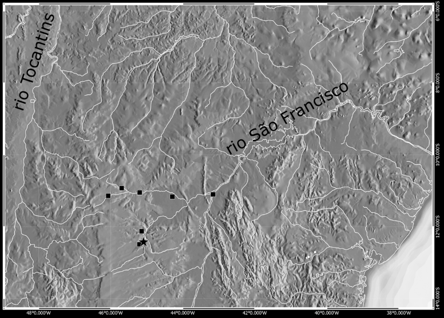
Distribution. Eigenmannia besouro is known from tributaries of the left margin of rio São Francisco, Bahia, northeastern Brazil ( Fig. 5 View FIGURE 5 ).
Etymology. The species name, besouro (‘beetle’ in Portuguese) is in honor of Manoel Henrique Pereira, known as Besouro Mangangá (‘The Mangangá Beetle’), a native of the Recôncavo region of Bahia, and a legendary figure in the Afro-Brazilian martial art capoeira; a noun in opposition.
Discussion. Given the absence of proposed synapomorphies for Eigenmannia ( Mago-Leccia, 1994; Albert & Campos-da-Paz, 1998; Albert, 2001), we consider it more appropriate to assign E. besouro provisionally as a member of this genus because it does not have most of the synapomorphies present in the remaining genera of Eigenmaniinae.
Vari et al. (2012) proposed six synapomorphies for Japigny : (1) color pattern composed by alternating dark bands; (2) presence of single row teeth at the base of the upper oral valve; (3) lateral process of the parapophysis of the second vertebrae ventrally curved; (4) distinct separation of the lateral process of the second vertebrae from the parapophysis of the fourth vertebrae; (5) lateral process of the parapophysis of the fourth vertebrae ventrally curved; and (6) retroarticular not included in the socket of the lower jaw with the quadrate. In Eigenmannia besouro , the color pattern is composed of four longitudinal dark stripes without alternating dark bands on flanks. Additionally, the single row of teeth at the base of the upper oral valve is absent, the contact of the lateral process of the second vertebrae with the parapophysis of the fourth vertebrae is present, and the retroarticular is included in the socket of the lower jaw with the quadrate, distinct from the states described by Vari et al. (2012) for Japigny . These features argue against the inclusion of the new species in Japigny . However, the ventral curvature of the lateral process of the parapophysis of the second and fourth vertebrae is present in E. besouro , as well as in most genera of the Eigenmanniinae, and may represent a plesimorphic condition in the subfamily.
In the same study, Vari et al. (2012) proposed four synapomorphies for Archolaemus : (1) a free orbital rim; (2) anterobasal margins of the teeth of the first tooth row attached to the dentigerous surface of the premaxilla; (3) pronounced gap equal to approximately one-half the width of the eye between the anterior margin of the premaxilla and the posterior margin of the upper lip; and (4) ventral surface of the upper lip porous. In E. besouro the orbit is covered by skin and the anterobasal margins of the teeth of the first row are immobile and firmly attached on the ventral surface of the premaxilla, thus distinct from the state described for species of Archolaemus . However, the latter two characters are observed in E. besouro and may represent homoplasies, as discussed by these authors when considering the same condition present in some species of the E. trilineata species-group.
Recently, Dutra et al. (2014) diagnosed Distocyclus by four autapomorphies: (1) a conical snout; (2) a small nasal capsule, with the internarial distance equal to the diameter of the posterior nostril; (3) the absence of endopterygoid teeth; and (4) a single tooth row limited to the anterior portion of dentary. Eigenmannia besouro shows a rounded snout in profile, the nasal capsule is moderate in size, with the internarial distance equal to two and a half times the diameter of the posterior nostril, the endopterygoid has ten or 11 teeth distributed in two rows, and the dentary has 19–30 teeth distributed in two or three complete rows. Therefore, E. besouro does not fit into the definition of Distocyclus provided by Dutra et al. (2014).
Finally, Correa et al. (2006) defined Rhabdolichops by: (1) absence of scales above the lateral line on the anterior portion of the body; (2) premaxilla trapeizodal and elongate; (3) presence of two prootic foramina; (4) extrascapular fused with the neurocranium; (5) gill rakers long and bony; and (6) gill rakers ossified. In E. besouro , the region above the lateral line on the anterior portion of the body is covered by scales and the premaxilla is approximately rectangular. Additionally, the new species shows only one prootic foramen, the extrascapular is independent from the neurocranium, and the gill rakers are short and not ossified. It is, therefore, distinct from species of Rhabdolichops .
The E. trilineata species-group is defined by the presence of the superior midlateral stripe, which is an exclusive synapomorphy for this clade ( Peixoto et al., 2015). In view of the presence of this character, E. besouro is hypothesized as a member of this clade. Within this group, E. besouro shares with E. viscentespelaea and E. waiwai the subterminal position of the mouth, so it is possible that these three species comprise a monophyletic group, since the remaining species present a terminal mouth. Moreover, E. besouro shares with E. waiwai the depth of the posterodorsal expansion on infraorbitals 1+2 less than 40% of the length of infraorbitals 1+2, and this character may represent a synapomorphy for these two species.
Peixoto, L. A. W, Dutra, G. M. & Wosiacki, W. B. (2015) The Electric Glass Knifefishes from the Eigenmannia trilineata speciesgroup (Gymnotiformes: Sternopygidae): monophyly and description of seven new species. Zoological Journal of Linnean Society, 175, 384 - 414. http: // dx. doi. org / 10.1111 / zoj. 12274
Dutra, G. M, de Santana, C. D, Vari R. P. & Wosiacki, W. B. (2014) The South American Electric Glass Knifefish genus Distocyclus (Gymnotiformes: Sternopygidae): Redefinition and Revision. Copeia, 2014 (2), 345 - 354. http: // dx. doi. org / 10.1643 / CI- 13 - 066
Correa, S. B., Crampton, W. G. R. & Albert, J. S. (2006) Three new species of the neotropical electric fish Rhabdolichops (Gymnotiformes: Sternopygidae) from the Central Amazon, with a new diagnosis of the genus. Copeia, 2006 (1), 27 - 42. http: // dx. doi. org / 10.1643 / 0045 - 8511 (2006) 006 [0027: tnsotn] 2.0. co; 2
Albert, J. S. & Campos-da-Paz, R. (1998) Phylogenetic systematics of American knifefishes: a review of the available data. In: Malabarba, L. R., Reis, R. E, Vari, R. P., Lucena, Z. M. & Lucena, C. A. S (Eds.), Phylogeny and Classification of Neotropical Fishes. Edipucrs, Porto Alegre, pp. 419 - 446.
Albert, J. S. (2001) Species diversity and phylogenetic systematics of American knifefishes (Gymnotiformes, Teleostei). Miscellaneous Publication, Museum of Zoology, University of Michigan, 190, 1 - 127.
Mago-Leccia, F. (1994) Electric Fishes of the Continental Waters of America. Classification and Catalogue of the Electric Fishes of the Order Gymnotiformes (Teleostei: Ostariophysi) with Descriptions of New Genera and Species. Biblioteca de la academia de ciencias fisicas, matematics y naturales, 29, 1 - 225.

FIGURE 1. Eigenmannia besouro, MZUSP 57890, holotype, 91.9 mm LEA, Brazil, Bahia, São Desidério, rio São Francisco, rio Grande. (A) Left lateral view of body; (B) Head.
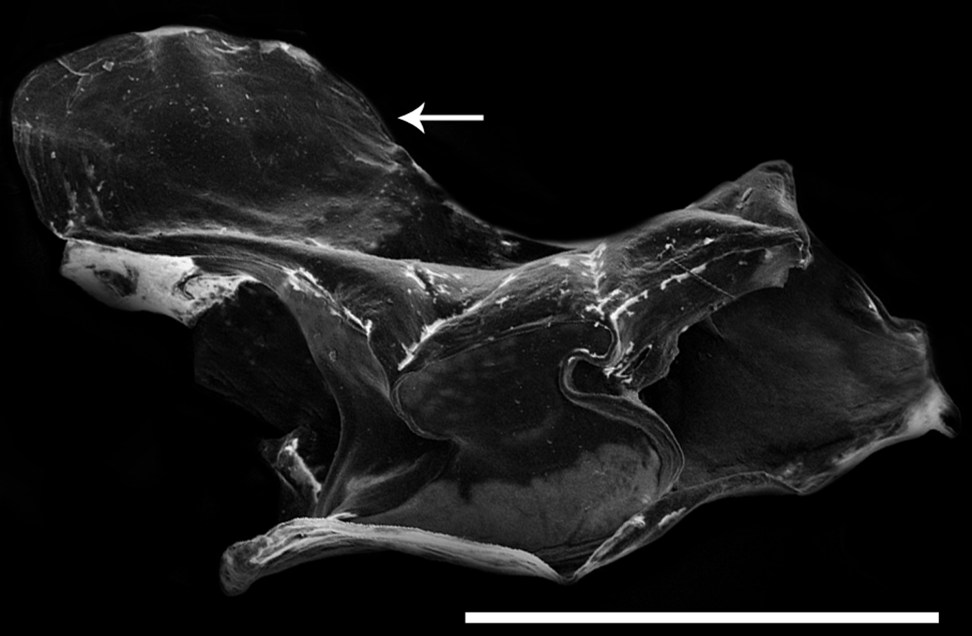
FIGURE 2. Scanning electron micrographs, lateral view, anterior to right, of right infraorbital 1 + 2 of E. besouro (MZUSP 119104, 92.8 mm LEA). Arrow indicates the posterodorsal expansion. Scale bar: 1 mm.

FIGURE 3. Scanning electron micrographs, lateral view, anterior to left, of left maxilla of: A) Eigenmannia besouro (MZUSP 119104, 92.8 mm LEA) and B) E. waiwai (INPA 37597, 154.8 mm LEA). Arrow indicates anterodorsal process. CT = connective tissue on leading edge of maxilla. Black line delimits the bony portion of maxilla. Scale bar: 1 mm.

FIGURE 4. Scanning electron micrographs, ventral view of left premaxilla, showing the dentition pattern of Eigenmannia besouro (MZUSP 119104, 92.8 mm LEA). Scale bar: 0.5 mm.
No known copyright restrictions apply. See Agosti, D., Egloff, W., 2009. Taxonomic information exchange and copyright: the Plazi approach. BMC Research Notes 2009, 2:53 for further explanation.
|
Kingdom |
|
|
Phylum |
|
|
Class |
|
|
Order |
|
|
Family |
|
|
Genus |
1 (by plazi, 2016-06-17 08:02:11)
2 (by ImsDioSync, 2017-01-30 22:59:12)
3 (by ImsDioSync, 2017-01-30 22:59:44)
4 (by ExternalLinkService, 2019-09-26 09:10:47)
5 (by ExternalLinkService, 2022-01-29 21:57:15)
6 (by ExternalLinkService, 2022-02-13 17:35:00)
7 (by plazi, 2023-10-30 03:20:03)