
Saprinillus paromaloides Kryzhanovskij, 1974
|
publication ID |
https://doi.org/ 10.5281/zenodo.4272127 |
|
DOI |
https://doi.org/10.5281/zenodo.4342025 |
|
persistent identifier |
https://treatment.plazi.org/id/0385915E-FF52-09EE-6001-FF3DCEFFFA0D |
|
treatment provided by |
Felipe |
|
scientific name |
Saprinillus paromaloides Kryzhanovskij, 1974 |
| status |
|
Saprinillus paromaloides Kryzhanovskij, 1974 View in CoL
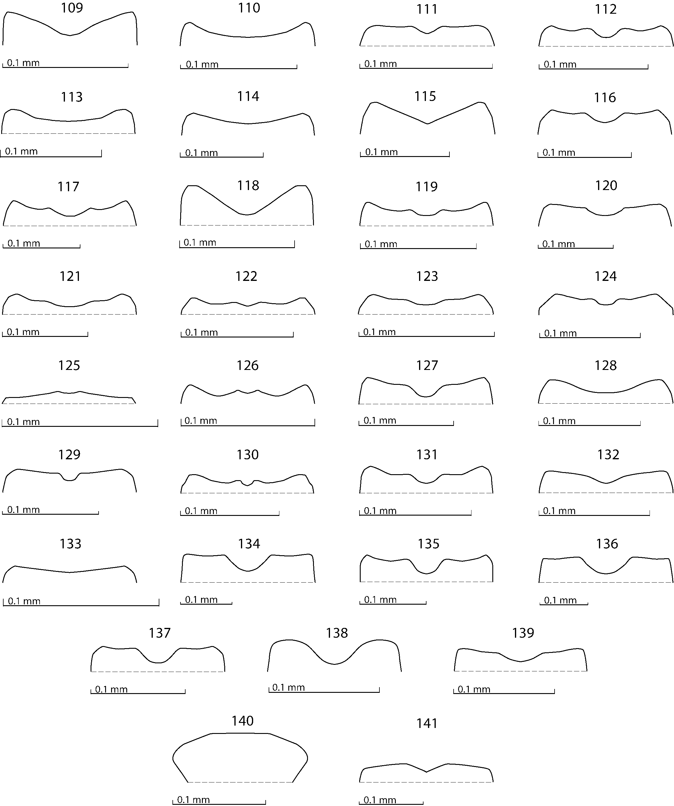
( Figs. 133 View Figs , 611–626 View Figs View Figs )
Saprinillus paromaloides Kryzhanovskij, 1974: 107 View in CoL , Figs. 7–10 View Figs View Fig View Figs .
Saprinillus paromaloides: KRYZHANOVSKIJ & REICHARDT (1976) View in CoL : 216 (partim); MAZUR (1984): 83; MAZUR (1997): 249; MAZUR (2004): 96; LACKNER (2009a): 113, 117, Figs. 8–13, 14 View Fig View Figs :A–G.
Type locality. Mongolia, Khushu Sayr, South Gobi Aimak.
Type material examined. PARATYPES: 1 ♁ 1 ♀, ‘MNR [= Mongolian People’s Republic], Yu. [Yuzhno = Southern] Gob. [= Gobijskij] Khushu / Sayr, 25 km South of Khailastyn-Gobi / 21.vi.1971 / Kerzhner [written] //, Solonchak [= salt-marsh], v opade [in debris] / pod [= under] Kalidium [written] // Paratypus 1971 / Saprinillus / paromaloides sp.n. / Kryzhanovskyi det. [red label, printed-written] // St. Petersburg / Zool. Inst [yellow label, printed] // D07–070 [written pink label, added by the author] ( ZIN)’.
Redescription. Body length: PEL: 1.50 mm; APW: 0.50 mm; PPW: 1.00 mm; EL: 1.00 mm; EW: 1.10 mm.
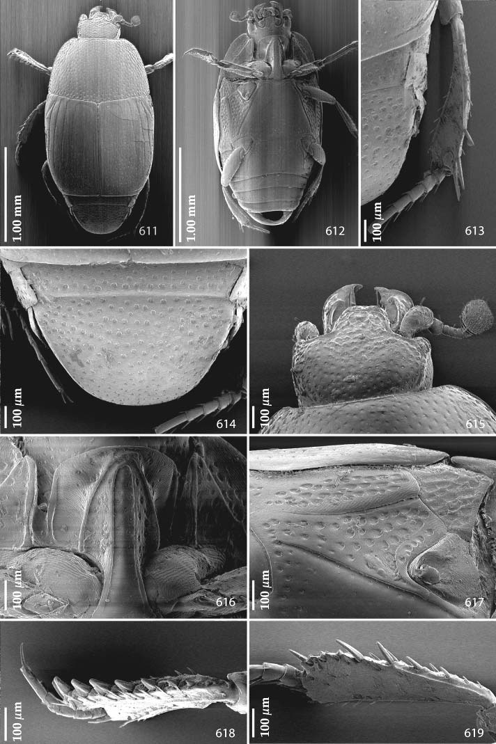
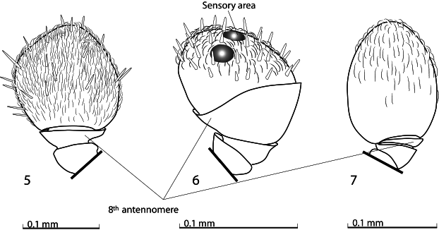
Body ( Figs. 611–612 View Figs ) cylindrical, convex, without metallic luster; cuticle light brown, legs, antennae and mouthparts light brown. Antennal scape ( Fig. 615 View Figs ) with two short setae; club without visible articulation, entire surface covered with short sensilla intermingled with much sparser erect sensilla; sensory structures of antennal club not examined.
Mouthparts. Mandibles with almost straight outer margin, outer margin carinate, strongly curved inwardly, mandibular apex acutely pointed. Disc of labrum not examined, two well impressed labral pits present, with two well-sclerotized setae arising from each; terminal labial palpomere elongated, its width about one-third its length; mentum square-shaped, without emargination in middle of anterior margin ( Fig. 133 View Figs ); antero-lateral corners with several short setae, lateral margins with a single row of short ramose setae; disc of mentum laterally covered with few short setae; medially imbricate, glabrous. Cardo of maxilla on lateral margin with few short setae; stipes triangular, with three much longer setae; terminal maxillary palpomere elongated, its width about one-third its length, approximately twice as long as penultimate; remaining mouthparts not examined.
Clypeus ( Fig. 615 View Figs ) flat, rugulose-lacunose; frontal stria widely interrupted anteriorly, forming acute angles above eyes; supraorbital stria vaguely impressed; frontal disc ( Fig. 615 View Figs ) anteriorly with a shallow depression, shallowly rugulose-lacunose, posteriorly with regular round moderate punctures; eyes flattened, visible from above.
Pronotum ( Fig. 611 View Figs ) weakly convergent anteriorly; apical angles blunt; marginal pronotal stria complete, slightly carinate laterally; disc laterally with coarse and dense deep punctures becoming sparser and finer medially; antescutellar area with vague depression; pronotal hypomeron glabrous.
Elytral epipleuron with scattered fine punctures; marginal epipleural stria thin, complete; marginal elytral stria complete, slightly carinate, for short distance continuous along elytral apex; apical elytral stria absent.
Elytra with four well impressed dorsal elytral striae 1–4, in punctures, first three about the same length, reaching about two-thirds of elytral length apically, fourth dorsal elytral stria slightly shortened, reaching about elytral half apically, basally united with sutural elytral stria; sutural elytral stria well impressed, on basal half in sparse punctures, reaching elytral apex; between it and elytral suture a row of fine punctures present; humeral elytral stria finely impressed on basal third; inner subhumeral stria present as short median fragment; elytral disc on apical half with deep scattered punctation, punctures separated by about 2–4 times their own diameter, becoming somewhat denser apically; basal half of elytral disc with much finer and sparser punctation.
Propygidium ( Fig. 614 View Figs ) transverse, almost completely exposed, with several rows of punctures; pygidium ( Fig. 614 View Figs ) with round regular punctures, separated by about their own to twice their diameter, becoming finer apically; interspaces in both cases imbricate.
Anterior margin of median portion of prosternum ( Fig. 616 View Figs ) evenly rounded; marginal prosternal stria present laterally and as short vague anterior fragment; pre-apical foveae well-impressed, deep; prosternal process flattened, surface between carinal prosternal striae shallowly imbricate-punctate; laterally coarsely imbricate-punctate; carinal prosternal striae carinate, slightly divergent on prosternal apophysis, subparallel, united anteriorly; lateral prosternal striae strongly carinate, convergent anteriorly, vaguely united in front of united carinal prosternal striae.
Anterior margin of mesoventrite deeply emarginate medially, discal marginal mesoventral stria well impressed, carinate; disc of mesoventrite with sparse fine punctures; meso-metaventral sutural stria inconspicuous, meso-metaventral suture well observable, thin.
Intercoxal disc of metaventrite almost smooth, with fine sparse punctures; area around hind coxae with few coarser punctures; lateral metaventral stria ( Fig. 617 View Figs ) well impressed, straight, carinate, almost reaching hind coxa; lateral disc of metaventrite ( Fig. 617 View Figs ) with shallow large punctures, interspaces imbricate; metepisternum ( Fig. 617 View Figs ) with similar, even coarser and denser punctures, fused metepimeron smooth; surface around lateral margins of metepisternum imbricate.
Intercoxal disc of the first abdominal sternite completely striate laterally, lateral stria distinctly carinate, disc smooth, only in anterolateral corners with sparse round punctures of various sizes.
Protibia ( Fig. 618 View Figs ) on outer margin with eight short denticles diminishing in size in proximal direction, teeth absent; setae of outer row sparse, regular and short; setae of median row not inspected; protarsal groove moderately deep; anterior protibial stria shortened apically, two tarsal denticles present apically; protibial spur not inspected. Apical margin of protibia not inspected; posterior surface of protibia not inspected.
Mesotibia ( Fig. 619 View Figs ) slender, outer margin with two sparse rows of thin denticles growing in size apically; setae of outer row worn off; setae of median row inconspicuous; posterior mesotibial stria inconspicuous; anterior surface of mesotibia imbricate with scattered minuscule punctures with microscopic setae; anterior mesotibial stria complete, terminating in two tiny inner anterior denticles; mesotibial spur stout, moderately long; apical margin with several tiny denticles; claws of apical tarsomere shorter than half its length; metatibia ( Fig. 613 View Figs ) basically similar to mesotibia, but slenderer and denticles of outer margin much sparser than those of mesotibia.
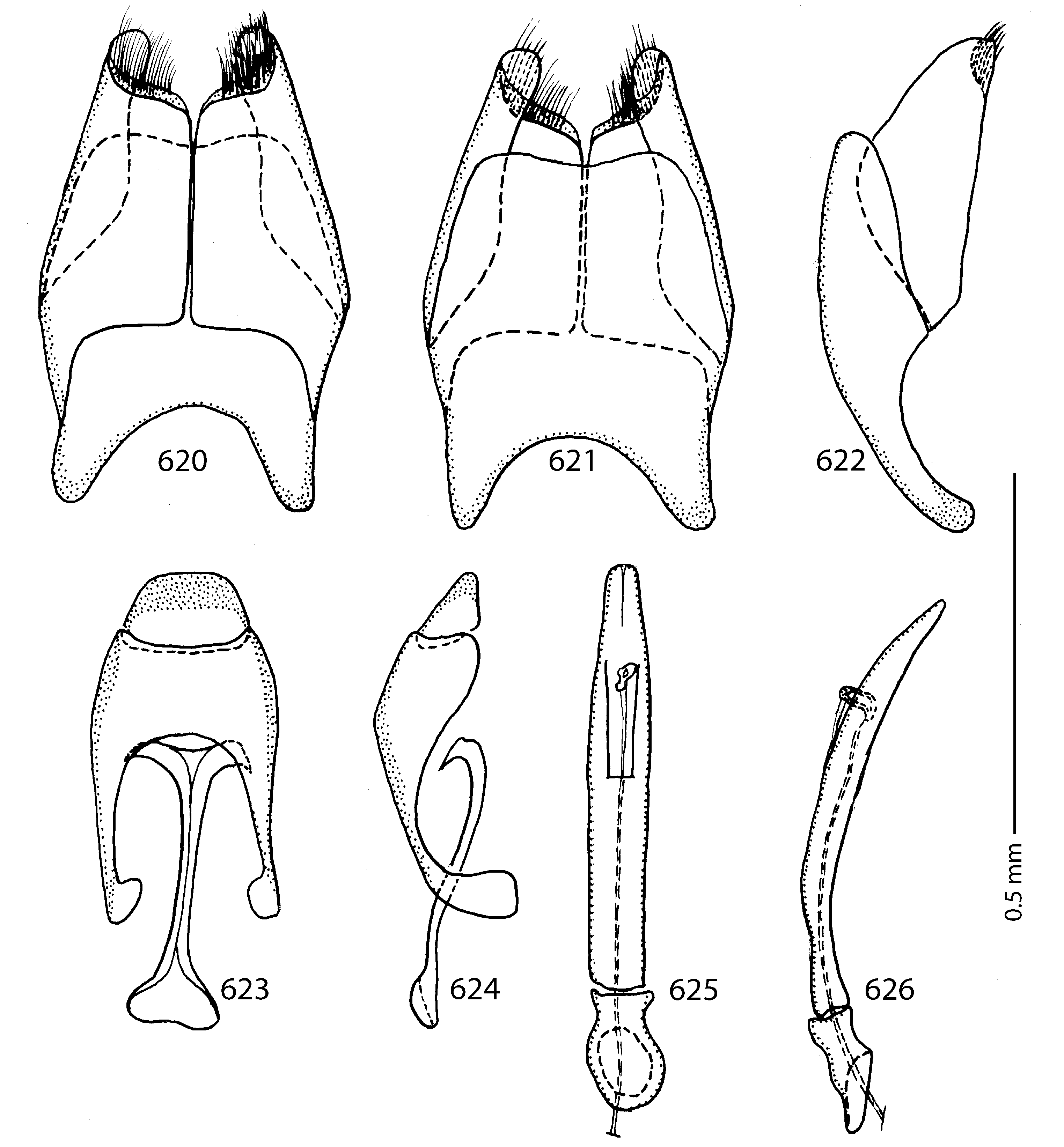
Male genitalia. Eighth sternite ( Figs. 620–621 View Figs ) longitudinally separated medially, apically with tiny inflatable membrane (velum) with dense brush of moderately long setae; eighth tergite and eighth sternite not fused laterally ( Fig. 622 View Figs ). Morphology of 9 th tergite ( Figs. 623–624 View Figs ) typical for the subfamily; spiculum gastrale ( Fig. 623 View Figs ) expanded on both ends. Basal piece of aedeagus ( Figs. 625–626 View Figs ) rather short, ratio of its length: length of parameres 1: 4; parameres fused along their basal half; aedeagus slightly curved ventrad ( Fig. 626 View Figs ).
Remarks. Redescription of this taxon has been recently published by the author ( LACKNER 2009a). For the sake of consistency and clarity of the terminology used it is repeated in the present paper. This re-description is likewise focused more in detail on several morphological structures omitted in the afore-mentioned publication, namely legs, mouthparts and male genitalia.


| ZIN |
Russian Academy of Sciences, Zoological Institute, Zoological Museum |
No known copyright restrictions apply. See Agosti, D., Egloff, W., 2009. Taxonomic information exchange and copyright: the Plazi approach. BMC Research Notes 2009, 2:53 for further explanation.
|
Kingdom |
|
|
Phylum |
|
|
Class |
|
|
Order |
|
|
Family |
|
|
Genus |
Saprinillus paromaloides Kryzhanovskij, 1974
| Lackner, Tomáš 2010 |
Saprinillus paromaloides:
| LACKNER T. 2009: 113 |
| MAZUR S. 2004: 96 |
| MAZUR S. 1997: 249 |
| MAZUR S. 1984: 83 |
| KRYZHANOVSKIJ O. L. & REICHARDT A. N. 1976: 216 |
Saprinillus paromaloides
| KRYZHANOVSKIJ O. L. 1974: 107 |