
Xenonychus tridens ( Jacquelin-Duval, 1852 ), 1899
|
publication ID |
https://doi.org/ 10.5281/zenodo.4272127 |
|
DOI |
https://doi.org/10.5281/zenodo.4341991 |
|
persistent identifier |
https://treatment.plazi.org/id/0385915E-FF72-09C0-6036-FF3DCECFFCAD |
|
treatment provided by |
Felipe |
|
scientific name |
Xenonychus tridens ( Jacquelin-Duval, 1852 ) |
| status |
|
Xenonychus tridens ( Jacquelin-Duval, 1852) View in CoL
( Figs. 28 View Figs , 72 View Figs , 106 View Figs , 139 View Figs , 711–729 View Figs View Figs )
Saprinus tridens Jacquelin-Duval, 1852: 703 View in CoL .
Saprinus tridens: MARSEUL (1855) View in CoL : 501, t. XIX, Fig. 118 View Figs ; SCHMIDT (1885a): 309.
Saprinus ciliaris Mulsant & Rey, 1853: 99 View in CoL . Synonymized by KRAATZ (1858): 131.
Saprinus serripes Marseul, 1855: 677 View in CoL . Synonymized by MARSEUL (1862): 482.
Xenonychus fossor Wollaston, 1864: 181 View in CoL . Synonymized by SCHMIDT (1887): 354.
Saprinus (Hypocaccus) tridens: SCHMIDT (1885a) View in CoL : 309.
Styphrus tridens: BICKHARDT (1910) : 107; G. MÜLLER (1931): 102.
Xenonychus tridens: GANGLBAUER (1899) View in CoL : 394; REITTER (1910): 13; REICHARDT (1941): 334; THÉROND (1963): 69; KRYZHANOVSKIJ & REICHARDT (1976): 112, 242, Fig. 471 View Figs ; VIENNA (1980): 115, 198, Fig. 70 View Figs ; MAZUR (1984): 108; MAZUR (1997): 267; YÉLAMOS (2002): 245, 340, Figs. 12A View Figs , 161H View Figs , 170B, 171 View Figs ; MAZUR (2004): 101.
Note. Sensory structures of antennal club and spermatheca were studied by DE MARZO & VIENNA (1982a,b).
Type locality. France, Le Grau de Roi.
Type material. Xenonychus tridens . NEOTYPE (here designated): ♁, ‘ FRANCE / Le Grau de Roi / (13) 8.viii.1962 [written] // tamissage au / pied des / Graminées [written] // Y. Gomy [printed] // Collection Y. Gomy [printed] // NEO- TYPUS / Xenonychus / tridens / ( Jacquelin-Duval, 1852) / Des. T. Lackner 2009 [red label, written]’ ( MNHN).
Comment. The type specimen of this species was not found in the collection of Jacquelin-Duval housed in MNHN. According to Mrs. Azadeh Taghavian, the type specimen of this species is probably lost, since there is an empty mounting card with the label ‘ Saprinus tridens ’. Although the author has inspected the fragments of various histerids on the bottom of the box, none of them contained pieces of Xenonychus tridens . The neotype is designated here to fix the identity of the type species of Xenonychus .
Additional material examined. ALGERIA: Ghardaia, 1.v.1987, 1♁, A.Olexa lgt .; Sahara, Béni Abbès , 20.x.1980, 1 ♀, A. Olexa lgt .; 1 ♁, ditto, but 27.iv.1987 ; Ain Sefra , 26.iv.1987, 1 ♁, A. Olexa lgt . MOROCCO: Mogador [= Essaouira], 2 ♀♀, Quedenfeldt lgt. ( TLAN) .
Redescription. Body length: PEL: 2.175–2.80 mm; APW: 0.825 –1.075 mm; PPW: 1.775 – 2.125 mm; EL: 1.425–1.70 mm; EW: 1.975 –2.475 mm.
Body ( Figs. 711–712 View Figs ) ovoid, moderately convex from above, underside strongly convex, cuticle light to dark brown with feeble bronze metallic tinge; legs, mouthparts and antennal scape rufopiceous, antennal flagellum yellow.
Antennal scape ( Fig. 715 View Figs ) with long numerous setae; club ( Fig. 714 View Figs ) rather small, without visible articulation, surface (apart from two sensory areas on apical part of club furnished with thick short sensilla, intermingled with thicker sparse erect sensilla) glabrous; sensory structures of antennal club ( Fig. 28 View Figs ) in form of stipe-shaped vesicle situated under circular sensory area on internal distal margin of the ventral side of antennal club.
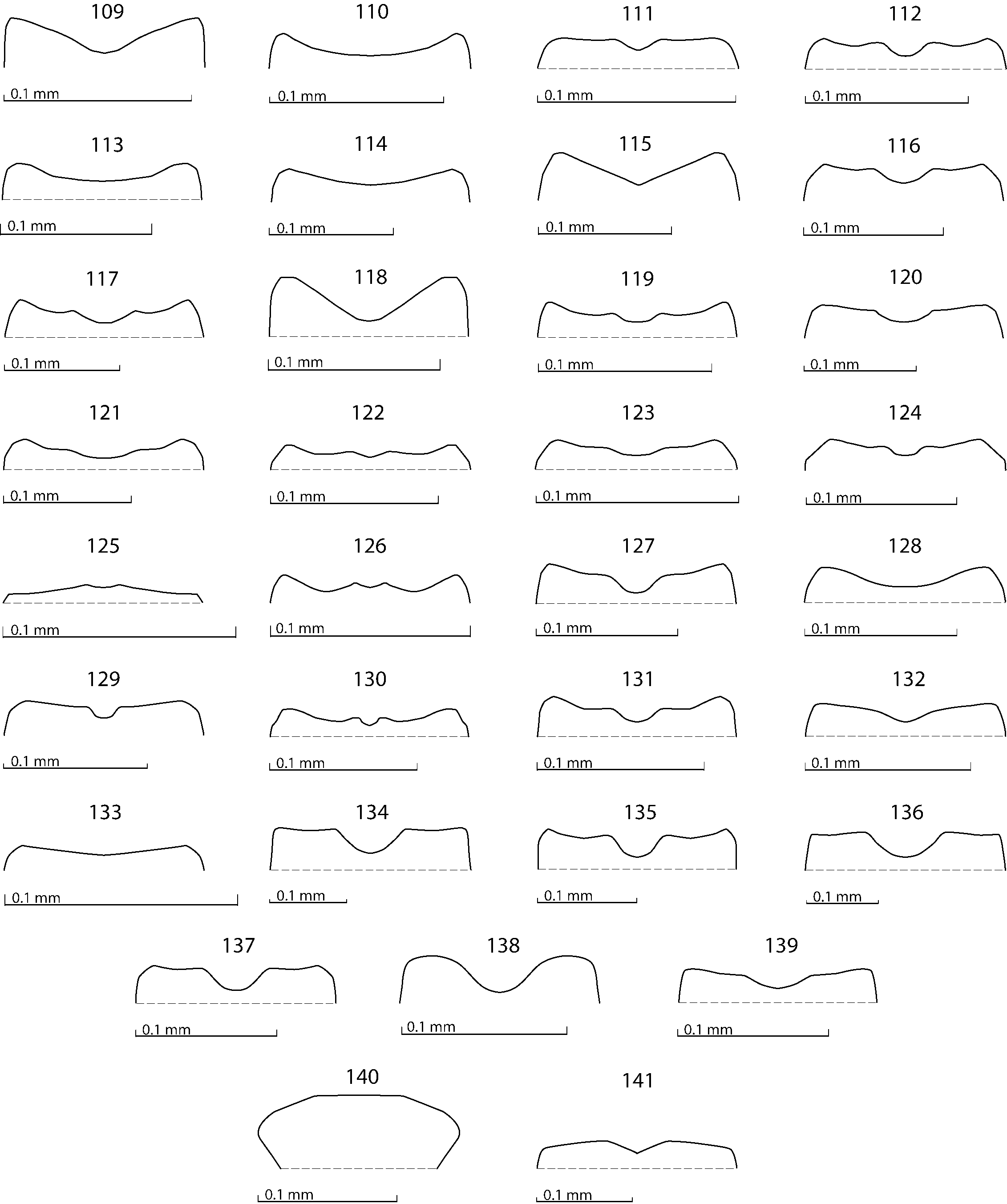
Mouthparts. Mandibles ( Fig. 106 View Figs ) with straight carinate outer margin strongly curved inwardly; mandibular apex acutely pointed; sub-apical tooth on left mandible prominent, acute; labrum ( Fig. 72 View Figs ) almost smooth, slightly convex; labral pits with single weakly sclerotized seta; epipharynx almost completely hidden under labral fold; terminal labial palpomere elongated, its width about one-third its length; mentum sub-trapezoid, anterior margin ( Fig. 139 View Figs ) with shallow median notch, surrounded with several moderately long ramose setae, disc of mentum with few similar setae, lateral margins with single row of shorter ramose setae; cardo of maxilla on lateral margin with several long setae; stipes triangular, with numerous much longer setae; terminal maxillary palpomere elongated, its width about one-third its length, approximately twice as long as penultimate.
Clypeus ( Fig. 715 View Figs ) rectangular, broader than long, slightly depressed laterally, with scattered microscopic punctation, almost smooth; frontal stria well impressed, thin, almost straight (occasionally weakened medially or interrupted), continued as well impressed carinate supraorbital stria; frontal disc ( Fig. 715 View Figs ) flat, almost smooth, broader than long, laterally exceptionally with vague interrupted rugae; eyes very flattened, invisible from above.
Pronotal sides ( Fig. 711 View Figs ) moderately convergent forwardly; apical angles prominent; anterior incision for head shallow; marginal pronotal stria well impressed, complete; disc entirely covered with shallow round regular punctation, becoming denser laterally; pronotal base with a row of oval dense punctures; pronotal hypomeron with long yellow setae; scutellum small, conspicuous.
Elytral epipleuron with round, rather deep punctures fringed with long setae; marginal epipleural stria complete; marginal elytral stria deeply impressed, continued as weakly impressed (at times intermittent) apical elytral stria. Humeral elytral stria distinctly impressed on basal third; inner subhumeral stria present medially, deep, rather long, rarely joining marginal elytral stria; elytra with four dorsal elytral striae 1–4, almost reaching elytral apex (seldom fourth dorsal elytral stria slightly shortened apically), fourth dorsal elytral stria basally connected with sutural elytral stria; sutural elytral stria well impressed, apically usually connected with apical elytral stria. Elytral punctation confined to interspace between fourth dorsal and sutural elytral striae, basally along elytral suture almost reaching elytral base, punctation irregular and shallow, becoming confluent apically.
Propygidium ( Fig. 718 View Figs ) completely exposed, with shallow confluent punctures of various sizes; pygidium ( Fig. 718 View Figs ) long, with similar punctation, punctures becoming finer and sparser apically.
Anterior margin of median portion of prosternum ( Fig. 719 View Figs ) almost straight; marginal prosternal stria present laterally and as short anterior fragment; pre-apical foveae deep; prosternal process slightly concave, compressed, surface between lateral prosternal striae substrigulate-punctate, punctures shallow, surface laterad to lateral prosternal striae substrigulate, impunctate; carinal prosternal striae divergent on prosternal apophysis, thence strongly convergent, intermittent, vaguely attaining united lateral prosternal striae; lateral prosternal striae well impressed, carinate, convergent anteriorly, united in front.
Anterior margin of mesoventrite ( Fig. 713 View Figs ) shallowly emarginate medially; discal marginal mesoventral stria well impressed, carinate, weakened anteriorly; disc with scattered deep punctures separated by about 2–3 times their diameter; meso-metaventral sutural stria ( Fig. 713 View Figs ) well impressed, almost straight, undulate, exposing meso-metaventral suture; intercoxal disc of metaventrite almost smooth, only with microscopic punctation, along base with a band of irregular deep punctation; lateral metaventral stria well impressed, carinate, obliquely arcuate, shortened apically; lateral disc of metaventrite concave, with deep setiferous punctures of various sizes; metepisternum + fused metepimeron with very dense setae, punctation unrecognizable beneath them.
Intercoxal disc of the first abdominal sternite almost completely striate laterally; basal half of disc with punctures of varied sizes, separated by about their own to twice their own diameter, apically replaced by very fine scattered microscopic punctation; along apical margin a row of larger punctures appears; lateral disc of all visible abdominal sternites setose laterally.
Protibia ( Fig. 720 View Figs ) flattened and dilated, outer margin with three large triangular teeth topped with moderately large triangular denticle, followed by five short rounded denticles diminishing in size in proximal direction; setae of outer row long, confined to basal third of protibia; setae of median row much shorter than those of outer row, but strongly sclerotized and present almost along entire length of protibia; protarsal groove rather deep; anterior protibial stria inconspicuous; single, short tarsal denticle present apically; protibial spur tiny, bent, growing out from apical protibial margin; apical margin of protibia posteriorly with two tiny apical denticles; outer part of posterior surface of protibia areolate-rugose; distinctly separated from glabrous median part of posterior surface; posterior protibial stria complete; inner-ventral denticles absent; inner margin with row of strongly sclerotized lamelliform long setae.
Mesotibia ( Fig. 716 View Figs ) slightly thickened, outer margin with two dense rows of thin denticles growing in size apically, abutting each other; posterior mesotibial surface with dense, strongly sclerotized long setae growing out near inner mesotibial margin and covering almost entire posterior surface of mesotibia, their affiliation with either dorsal or median row dubious; posterior mesotibial stria inconspicuous; anterior surface of mesotibia glabrous; anterior mesotibial stria almost complete, shortened apically; mesotibial spur long and thin; apical margin with several short denticles; claws of apical tarsomere almost straight, almost as long as apical-most mesotarsomeres itself; each tarsomere with single long strongly sclerotized seta posteriorly and anteriorly; metatibia basically similar to mesotibia, but more thickened and dilated and denticles on outer margin much longer, three rows of denticles present (as opposed to two in mesotibia).
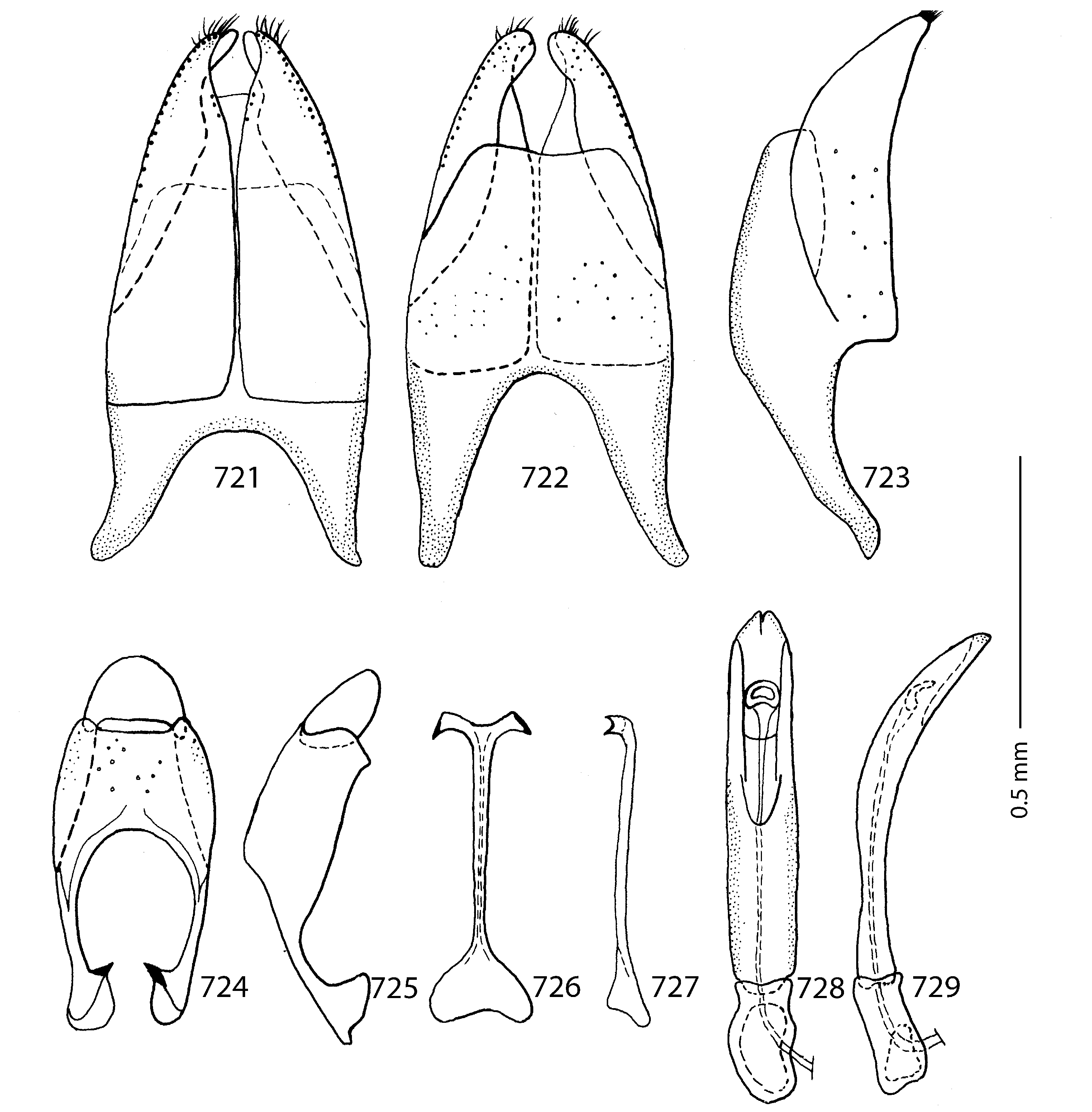
Male genitalia. Eighth sternite ( Figs. 721–722 View Figs ) segment longitudinally separated medially, apically with tiny inflatable membrane (velum); apex laterally fringed with short brush of setae; eighth tergite and eighth sternite fused laterally ( Fig. 723 View Figs ). Morphology of 9 th tergite ( Figs. 724–725 View Figs ) typical for the subfamily; spiculum gastrale ( Fig. 726 View Figs ) expanded on both ends. Basal piece of aedeagus ( Figs. 728–729 View Figs ) rather short, ratio of its length: length of parameres 1: 4; parameres fused along their basal half; aedeagus slightly curved ventrad ( Fig. 729 View Figs ).
Remarks. Xenonychus tridens is a widely distributed species and as such exhibits a large degree of variation, especially regarding punctation of dorsal surface, frontal stria and other characters.


| MNHN |
Museum National d'Histoire Naturelle |
No known copyright restrictions apply. See Agosti, D., Egloff, W., 2009. Taxonomic information exchange and copyright: the Plazi approach. BMC Research Notes 2009, 2:53 for further explanation.
|
Kingdom |
|
|
Phylum |
|
|
Class |
|
|
Order |
|
|
Family |
|
|
Genus |
Xenonychus tridens ( Jacquelin-Duval, 1852 )
| Lackner, Tomáš 2010 |
Saprinus tridens
| JACQUELIN-DUVAL M. 1852: 703 |