
Austeruseus rokuri
|
publication ID |
https://doi.org/ 10.5281/zenodo.277617 |
|
DOI |
https://doi.org/10.5281/zenodo.5627943 |
|
persistent identifier |
https://treatment.plazi.org/id/0385CF17-5C05-B607-4EEB-ABB36210CAB6 |
|
treatment provided by |
Plazi |
|
scientific name |
Austeruseus rokuri |
| status |
|
Austeruseus rokuri View in CoL nov. sp.
( Figures 16–18 View FIGURE 16 View FIGURE 17 View FIGURE 18 )
Type material. the holotype (slide FO2-009 [ZMUC-TAR-1080]) and 5 paratypes (slides FO2-010 to FO2-014 [ZMUC-TAR-1081 to ZMUC-TAR-1085]). The slides are deposited in the Zoological Museum, the Natural History Museum of Denmark, The Zoological Museum, Invertebrate Dept., University of Copenhagen, Copenhagen, Denmark.
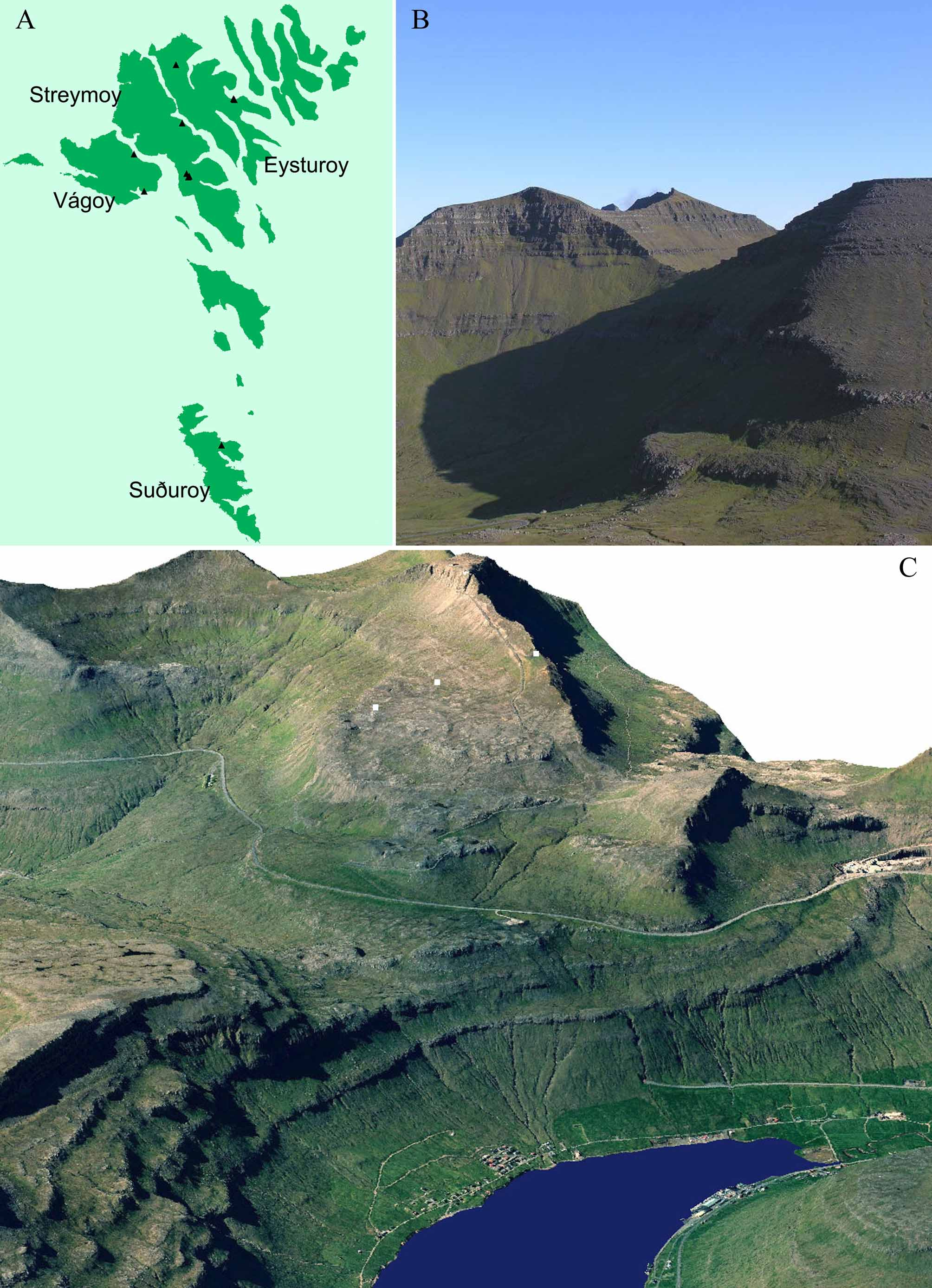
Type locality. Sornfelli mountain, Streymoy, Faroe Islands ( Fig. 1 View FIGURE 1. A C). Temporary puddle. GPS: 62°04.445’N, 6°57.252’W. Altitude 600 m a.s.l. Date: 26 October 1989. Coll.: Reinhardt M. Kristensen.
Additional material. one specimen in moult (slide FO20C-012 [ZMUC-TAR-1086]) was found in Varmakelda, Eysturoy, Faroe Islands. Homothermic spring. GPS: 62°13.097’N, 6°46.721’W. Altitude 15 m a.s.l. Date: 26 October 1989. Coll.: Reinhardt M. Kristensen.
Etymology. A. rokuri is named after BVT’s youngest son Rókur.
Specific diagnosis. Large sized animal with two macroplacoids (the first broken in the middle) and large microplacoid. Eyes present. Ventral strengthening bar, absent. Up to 6 lateral hook-shaped appendages for the insertion of stylet muscles. Three rows of teeth in the buccal cavity. Large claws of Eohypsibiidae type.
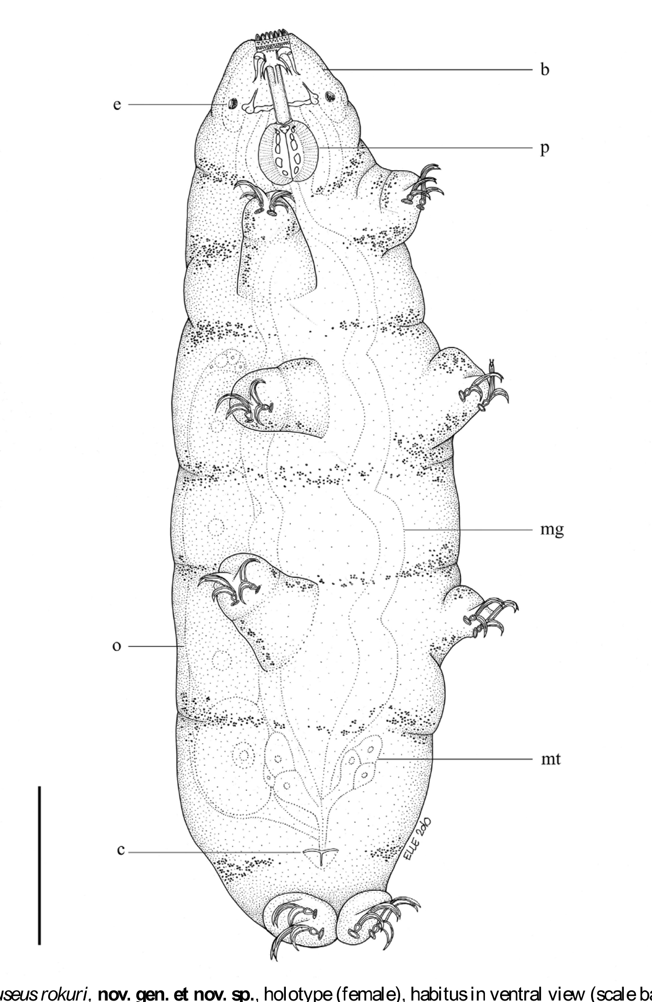
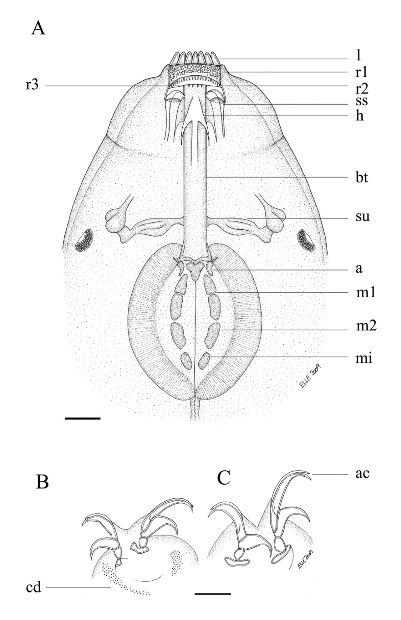
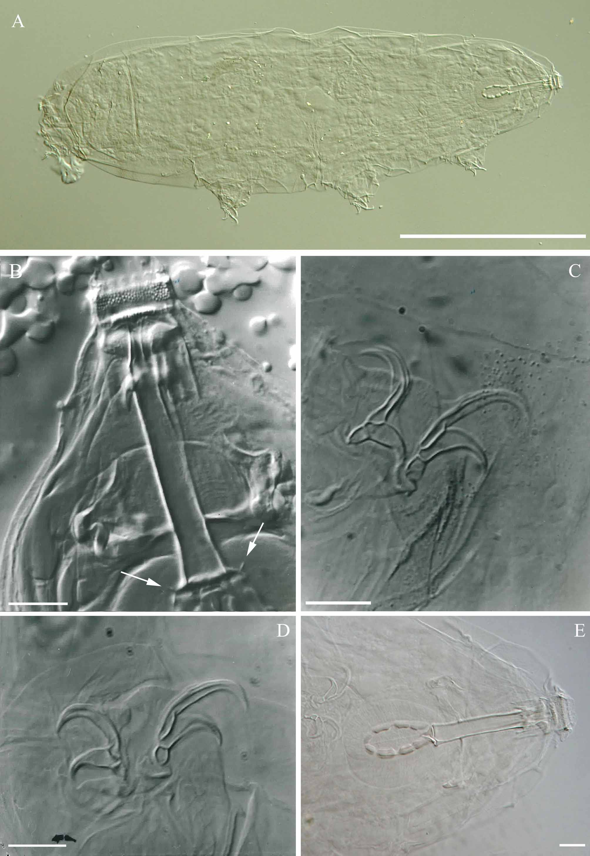
Description. Holotype (adult female) ( Fig. 16 View FIGURE 16 ): Body length, 564.1 µm (body length for paratypes, 329.4– 646.0 µm). Eyes present. Cuticle with many small dots (not “pearls”) scattered over the whole body (dorsal and ventral surfaces), but more dense at the caudal end. Mouth opening with 14 peribuccal lamellae. Buccal tube rigid, 46.4 µm long and 6.7 µm wide. Ventral strengthening bar absent. Buccal tube with three sets of wing- or hookshaped appendages (positioned dorso- and ventro-laterally) for attachment of the stylet muscles. Stylet supports inserted on the buccal tube at 35.6 µm. Furca large, not in contact with the pharyngeal bulb. Three bands of teeth present in the buccal cavity. First band of infrabuccal teeth, right behind the peribuccal lamellae and comprised of a broad band of asymmetrical arranged small teeth (mucrones) in>5 rows. Second band of infrabuccal teeth, immediately behind the first comprised of a single row of larger teeth. Third band of infrabuccal teeth, behind the second row, comprised of four teeth (infrabuccal baffles). Pharyngeal bulb oval with large triangular apophyses, and three cuticular rods close to the apophyses ( Figs. 17 View FIGURE 17 A & 18B, arrows). These rods are also present in Bertolanius and Eohypsibius , and other genera, e.g. Macrobiotus and Isohypsibius (R. Bertolani, pers. comm.). Two macroplacoids, the first, 14.4 µm long and broken in the middle, the second, 7.7 µm long, and a large microplacoid, shaped as a bent rectangle, 5.4 µm long.
Claws Eohypsibiidae type, very large. Smooth lunules present on all claws. The external claws larger than the internal, with largest on the fourth pair of legs. Two robust accessory points on primary branches ( Figs. 17 View FIGURE 17 B–C & 19C). Cuticular bars on all legs, absent. Eggs unknown.
No known copyright restrictions apply. See Agosti, D., Egloff, W., 2009. Taxonomic information exchange and copyright: the Plazi approach. BMC Research Notes 2009, 2:53 for further explanation.