
Austeruseus faeroensis
|
publication ID |
https://doi.org/ 10.5281/zenodo.277617 |
|
DOI |
https://doi.org/10.5281/zenodo.5627939 |
|
persistent identifier |
https://treatment.plazi.org/id/0385CF17-5C13-B612-4EEB-A9A46202CA95 |
|
treatment provided by |
Plazi |
|
scientific name |
Austeruseus faeroensis |
| status |
|
Austeruseus faeroensis View in CoL nov. sp.
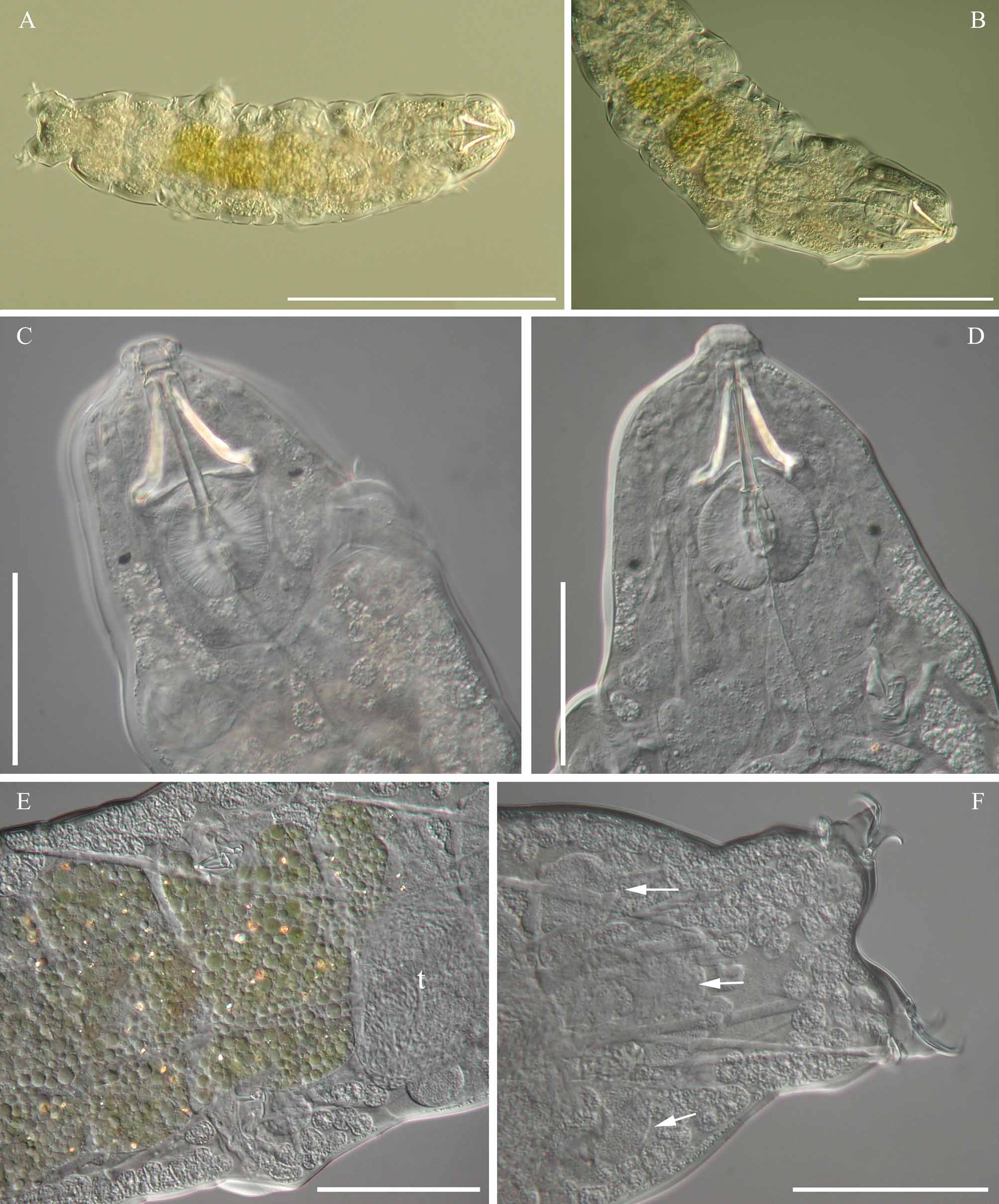
( Figures 4–8 View FIGURE 4 View FIGURE 5 View FIGURE 6 View FIGURE 7 View FIGURE 8 )
Type material. The holotype (slide FO30-005 [ZMUC-TAR 1068]), 2 paratypes (slides FO30-010c and FO30- 011-012b [ZMUC-TAR 1069 and ZMUC-TAR 1071]) and one embryonated egg (slide FO30-011b [ZMUC-TAR 1070]). The slides are deposited in the Zoological Museum, the Natural History Museum of Denmark, University of Copenhagen, Copenhagen, Denmark.
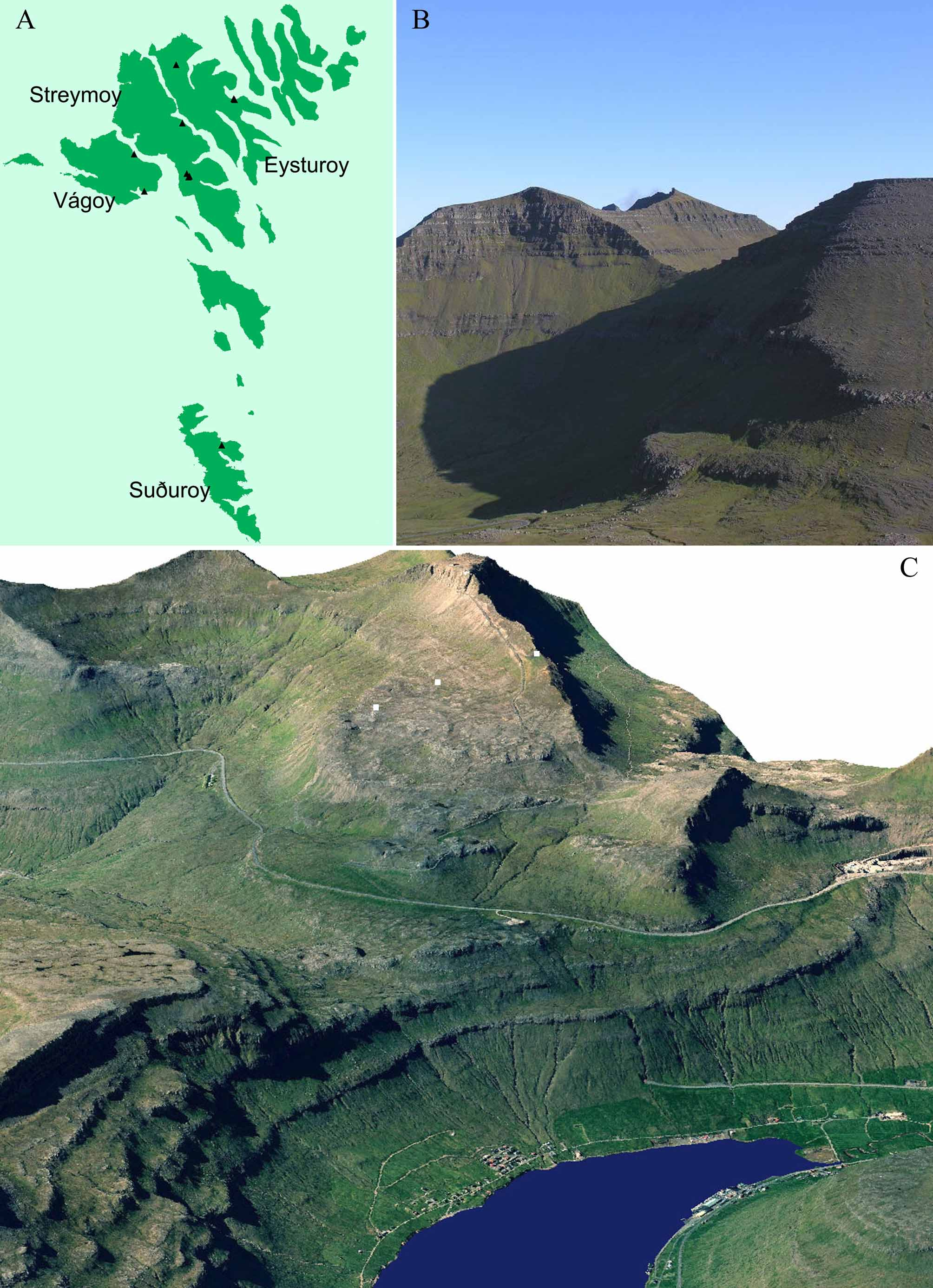
Type locality. Summit of Vaðhorn mountain, Eysturoy, Faroe Islands ( Fig. 1 View FIGURE 1. A B). The sample was a large moss cushion with underlying soil. GPS: 62°16.909’N, 7°00.349’W. Altitude 726 m a.s.l. Date: 20 November 2003. Coll.: Regin Árting.
Additional material. One specimen (F036-011 [ZMUC-TAR-1072]) was found at “Hvannhagi”, Suðuroy, Faroe Islands. The sample was a large moss cushion. GPS: 61°34.375’N, 6°49.576’W. Altitude 200 m a.s.l. Date: 24 September 2004. Coll.: Uni Árting.
Etymology. A. faeroensis is named after the type locality on Faroe Islands.
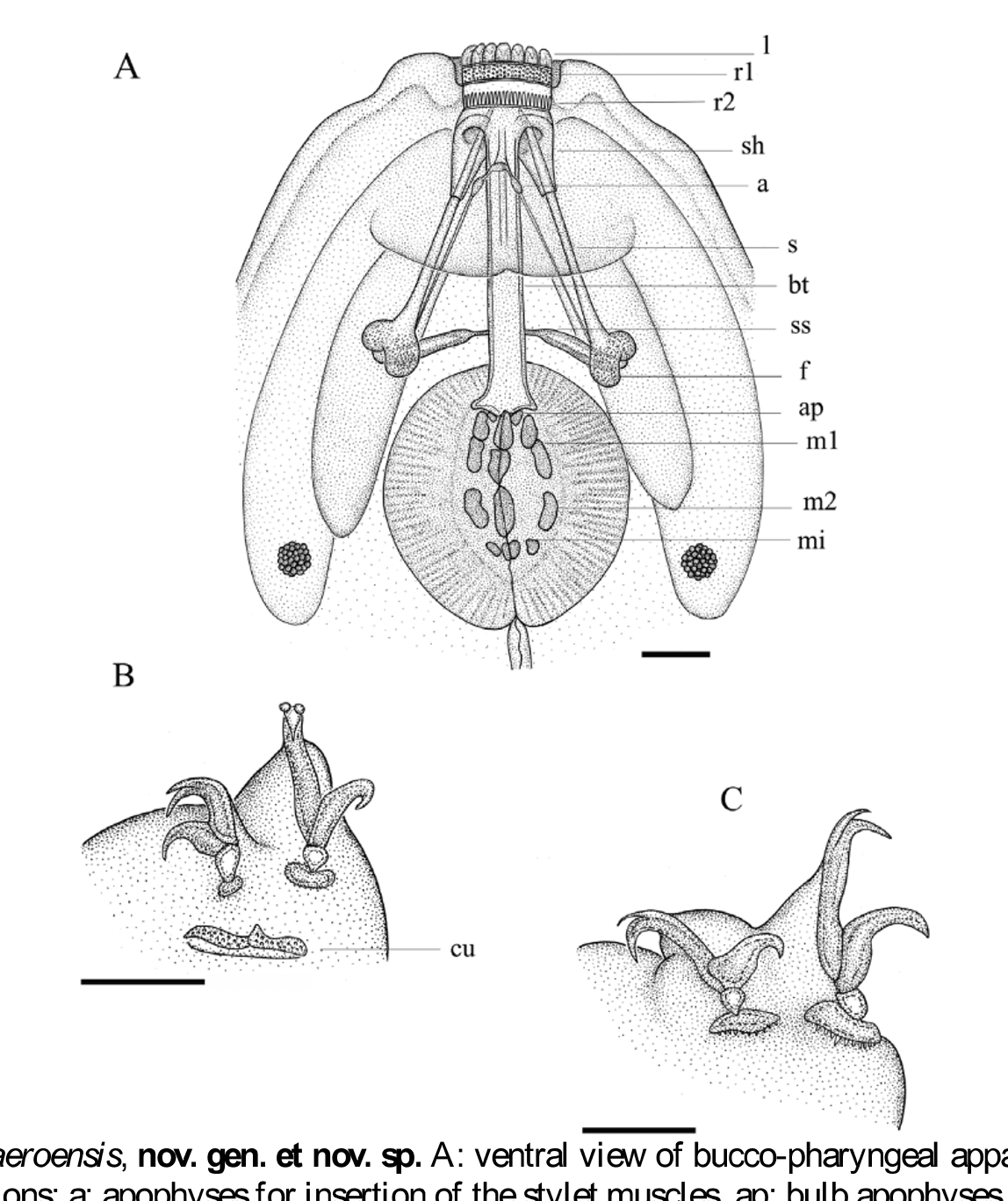
Specific diagnosis. Medium sized animals with two macroplacoids (the first broken in the middle) and microplacoid. Large black eyes present. Ventral strengthening bar absent; however, a complex of three cuticular elements and two thin filaments are attached ventral to the buccal tube. Two hook-shaped appendages for insertion of the stylet muscles attached laterally on the buccal tube. Two rows of teeth in the buccal cavity. Cuticular bars present on legs I–III (at least the first bar appears dotted). Lunules on fourth pair, dentate.
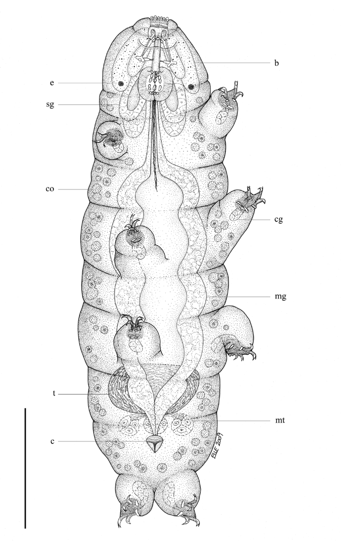
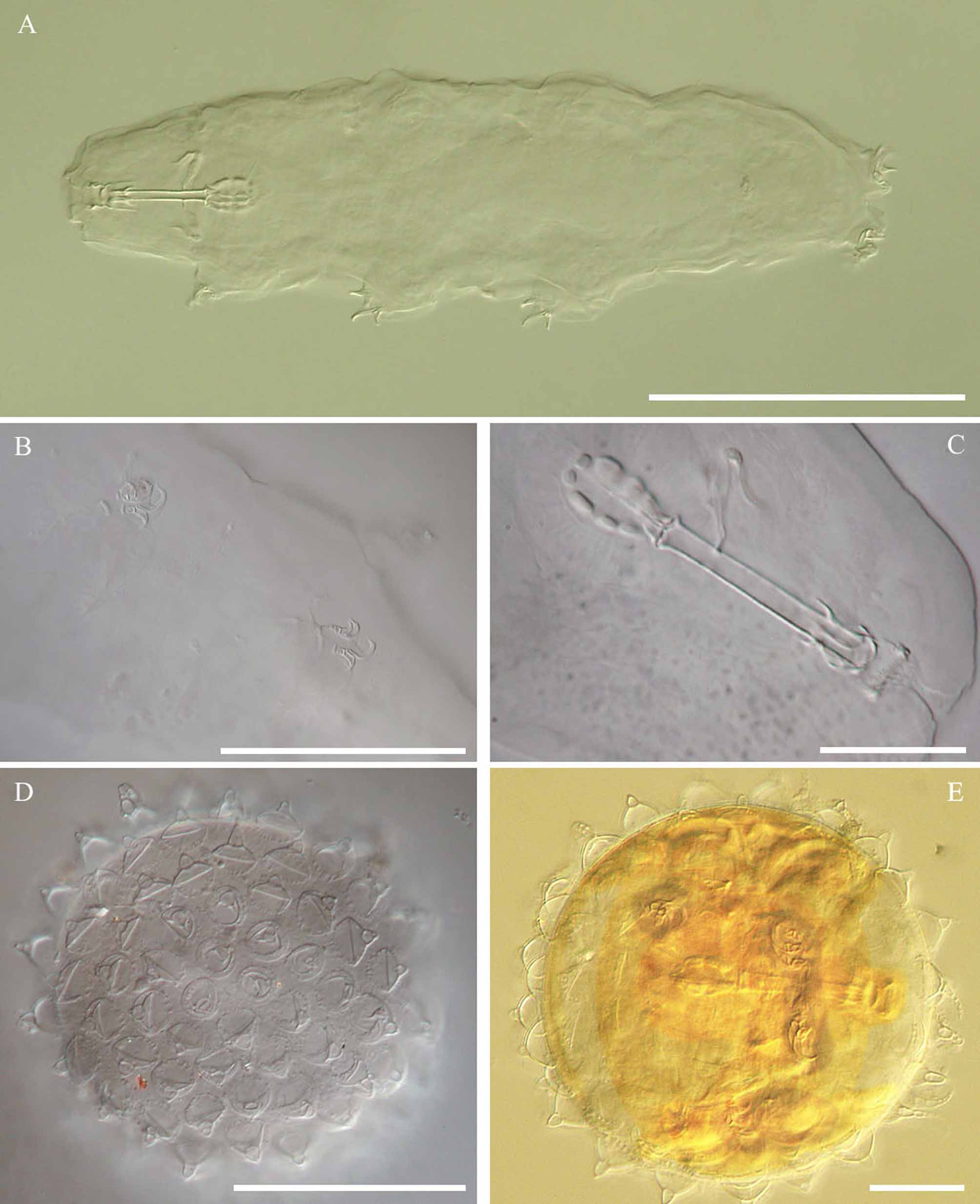
Description. Holotype (adult male) ( Fig. 4 View FIGURE 4 ): Body length, 411.7 µm (body length for paratypes range from 254.8–329.5 µm). Eyes present. The cuticule smooth. The buccal tube without ventral strengthening bar and 34.4 µm long and 4.0 µm wide. A complex of three small lens-shaped cuticular elements and two filaments replace the ventral strengthening bar ( Fig. 5 View FIGURE 5 A). The two filaments (which may be of a cuticular origin) run parallel with the stylets. The buccal tube with two lateral hook-shaped appendages. Mouth opening with 14 peribuccal lamellae, and two bands of teeth in the buccal cavity. First band of infrabuccal teeth comprised of three irregular rows of small teeth (mucrones) (one paratype with 5 irregular rows in the first row). Second band of infrabuccal teeth comprise a single row of large indented teeth, with the two largest in the middle. Third band of infrabuccal teeth absent. Stylet supports are inserted on the buccal tube at 26.7 µm of its length. Stylets are robust, straight, only bent at the caudal end (immediately before the furcae). Pharyngeal bulb oval. Triangular pharyngeal apophyses present (in one paratype the apophyses can be seen constricted near the caudal-most point, with the appearance of an uvula). Two macroplacoids and a microplacoid present. First macroplacoid, the longest (9.5 µm), was broken in the middle, second macroplacoid, 4.7 µm long. Microplacoid circular in shape, 2.1 µm long.
Claws of the Eohypsibiidae type with two accessory points on the primary branches. Claws on the fourth pair of legs were the largest. Large smooth lunules at the base of all claws on legs I–III; lunules weakly dentate on the fourth pair of legs. Cuticular bars present on legs I–III and at least on the first pair the bars appeared pitted.
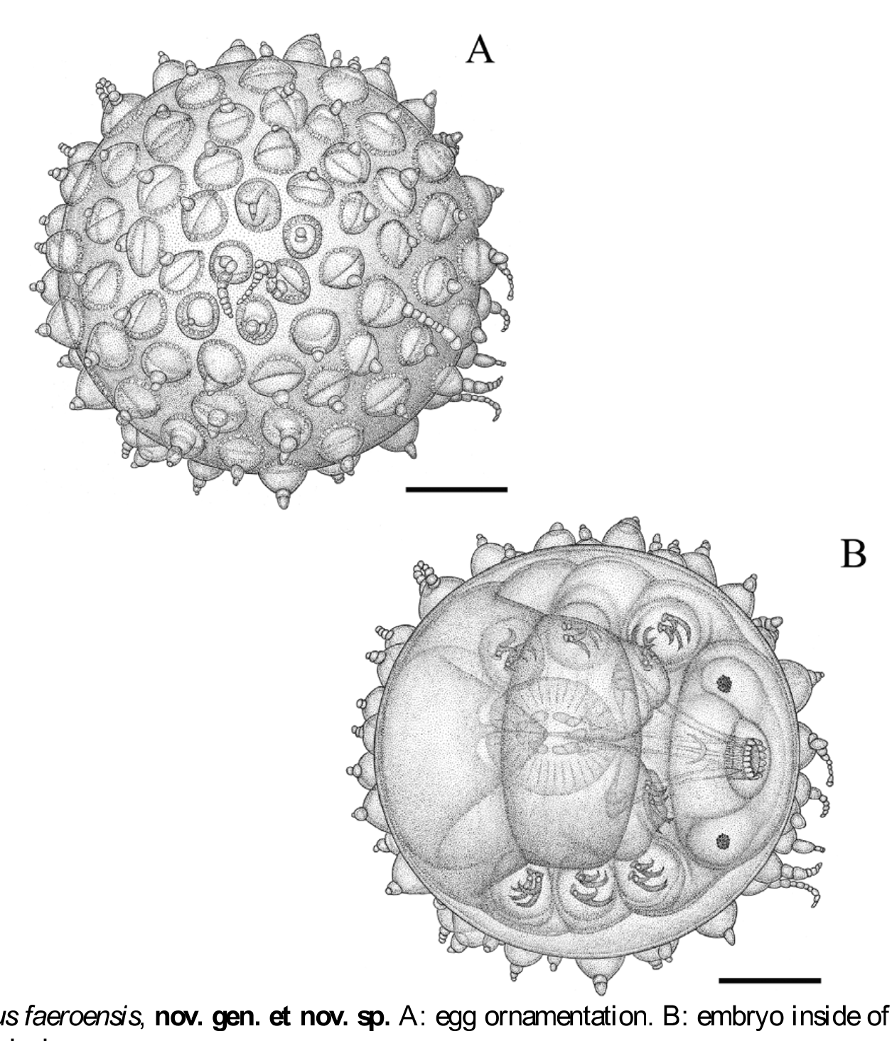
A single egg with an embryo inside, deposited free, was found and is attributed to this species ( Figs. 6 View FIGURE 6 & 7 View FIGURE 7 D– E). Egg diameter, 76.1 µm without the processes. Egg processes very variable in length, with mean length of the processes c. 8.2 µm (see illustration, Figs. 6 View FIGURE 6 A–B). Egg processes are mammiform with separate cells (from one to six) forming long tails. Base of the processes surrounded by a ring of small circles, more or less irregular in size and number. Egg surface between the processes, smooth.
No known copyright restrictions apply. See Agosti, D., Egloff, W., 2009. Taxonomic information exchange and copyright: the Plazi approach. BMC Research Notes 2009, 2:53 for further explanation.