
Austeruseus balduri
|
publication ID |
https://doi.org/ 10.5281/zenodo.277617 |
|
DOI |
https://doi.org/10.5281/zenodo.5627941 |
|
persistent identifier |
https://treatment.plazi.org/id/0385CF17-5C1F-B618-4EEB-AA52653ACADB |
|
treatment provided by |
Plazi |
|
scientific name |
Austeruseus balduri |
| status |
|
Austeruseus balduri View in CoL nov. sp.
( Figures 9–15 View FIGURE 9 View FIGURE 10 View FIGURE 11 View FIGURE 12 View FIGURE 13 View FIGURE 14 View FIGURE 15 )
Type material. The holotype (slide FO15-004 [ZMUC-TAR-1073]), 4 paratypes and 7 eggs (slides FO15-001a, - 001b, -002c, -003a, -003b, -005a, -005b and -019 [ZMUC-TAR-1074 to ZMUC-TAR-1078]). The slides are deposited in the Zoological Museum, the Natural History Museum of Denmark, University of Copenhagen, Copenhagen, Denmark.
Type locality. “Úti á Trøð”, Vágoy, Faroe Islands. The sample was a moss cushion growing on rock. GPS: 62°02.775’N, 7°07.996’W. Altitude 110 m a.s.l. Date: 10 March 2002. Coll.: Birna V. Trygvadóttir.
Additional material. Two specimens (FO1A-001a and -001b [ZMUC-TAR-1079]) found at Sornfelli mountain, Streymoy, Faroe Islands. Moss cushion. GPS: 62°04.757’N, 6°57.843’W. Altitude 640 m a.s.l. Date: 6 July 2001. Coll.: Birna V. Trygvadóttir & Reinhardt M. Kristensen.
Etymology. A. balduri is named after BVT’s eldest son Baldur.
Specific diagnosis. Small animals with two macroplacoids (the first broken in the middle) and microplacoid. Eyes present. Body broadest at the third pair of legs, tapering to rostral and caudal extremities. Ventral strengthening bar absent. Two lateral hook-shaped appendages present on the buccal tube. Small eohypsibiid type claws. Cuticular bars on legs I–III. Able to extend and retract the buccal/pharyngeal apparatus telescopically.
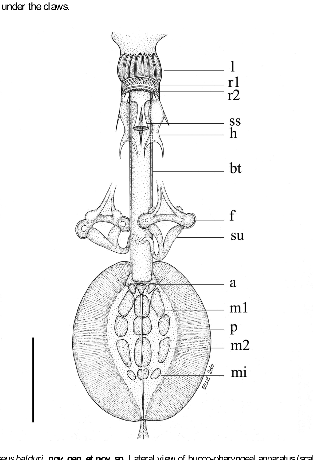
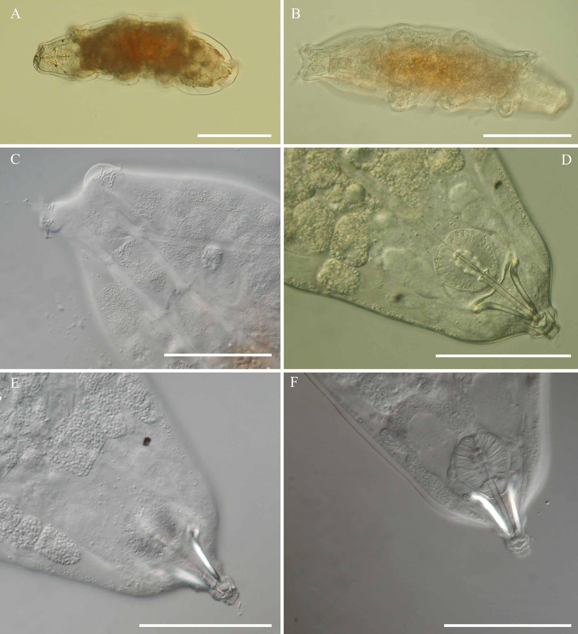
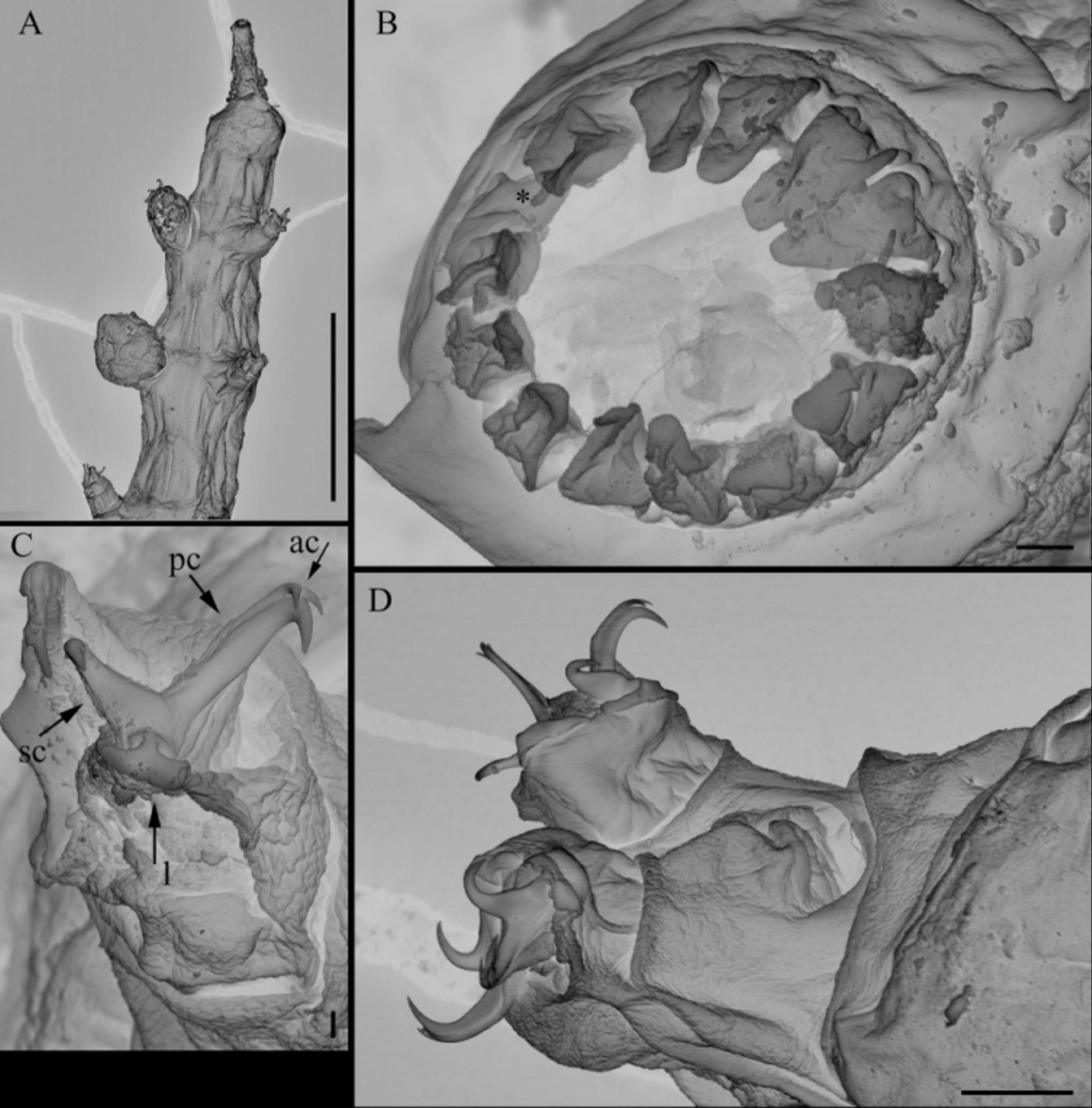
Description. Holotype (young female) ( Fig. 9 View FIGURE 9 ): Body length, 326.5 µm (length of paratypes, 236.0–376.5 µm). Eyes present. Body broadest at the level of the third pair of legs tapering to anterior and caudal extremities. Cuticle with transverse bands of minute dots. Ventral strengthening bar absent. Two lateral hook-shaped appendages for insertion of the stylet muscles. Buccal tube, 28.8 µm long and 2.4 µm wide. Mouth opening with 14 peribuccal lamellae. Two bands of teeth in the buccal cavity. First band of infrabuccal teeth, immediately behind the lamellae, comprised of five rows of small teeth (juveniles may have only three rows). Second band of infrabuccal teeth comprise a single row of teeth which forms an arch (viewed ventrally or dorsally) across the bottom of the buccal cavity ( Figs. 10 View FIGURE 10 & 12 View FIGURE 12 ). Third band of infrabuccal teeth absent. Stylet sheaths are visible even in polyvinyl lactophenol mounted specimens. Stylets are robust, straight, only bent caudally. Stylet supports are inserted on the buccal tube at 20.9 µm. Pharyngeal apophyses, two macroplacoids and a microplacoid present in the pharynx. First macroplacoid, 6.1 µm long and broken c. 60% of its length. Second macroplacoid, 3.6 µm long. Microplacoid, circular, 2.0 µm long. Pharyngeal bulb pear shaped.
This species is capable of extending and retracting the buccal apparatus, so the mouth opening protrudes forwards with a tube-like extension ( Figs. 14 View FIGURE 14 D–F & 15A), while the rest of the apparatus is drawn forward, such that the pharyngeal bulb lies in front of the eyes (the normal position of the bulb would be behind the eyes).
The claws are of the Eohypsibiidae type with smooth lunules. The external claw larger than the internal, with the largest on the fourth pair of legs ( Fig. 11 View FIGURE 11 B). Two accessory points present on primary branches. Cuticular bars present on legs I–III
Several eggs were found that do not belong to any other described species, and can probably be attributed to this species ( Figs. 11 View FIGURE 11 C, 13B). None of the eggs were embryonate, so this needs to be confirmed. Eggs deposited free. Egg diameter between 68.6–92.5 µm without processes, process length 7.8–12.5 µm. Processes mammiform, with tips comprising 4–6 small cells. Base of the processes surrounded by a ring of small circles, with smooth surface between processes. The process tips exhibit a more regular number of cells (compartments) and therefore the length of the processes were more regular than the egg belonging to A. faeroensis .


No known copyright restrictions apply. See Agosti, D., Egloff, W., 2009. Taxonomic information exchange and copyright: the Plazi approach. BMC Research Notes 2009, 2:53 for further explanation.