
Vararia yunnanensis Y.L. Deng & C.L. Zhao, 2023
|
publication ID |
https://doi.org/10.11646/phytotaxa.583.1.4 |
|
DOI |
https://doi.org/10.5281/zenodo.7622417 |
|
persistent identifier |
https://treatment.plazi.org/id/03862767-5A64-FF92-FF34-FBE88B9EF9B9 |
|
treatment provided by |
Plazi |
|
scientific name |
Vararia yunnanensis Y.L. Deng & C.L. Zhao |
| status |
sp. nov. |
Vararia yunnanensis Y.L. Deng & C.L. Zhao , sp. nov. Figs. 3 View FIGURE 3 , 4 View FIGURE 4
MycoBank no.: MB 844043 View Materials
Holotype:— CHINA. Yunnan Province, Honghe, Pingbian County, Daweishan National Nature Reserve , E 103°30′10′′, N 23°42′07′′, elev. 1500 m, on fallen branch of angiosperm, 1 August 2019, CLZhao 17725 ( SWFC!), GenBank No. (ITS ON454115 View Materials ; nLSU ON502654 View Materials ). GoogleMaps
Etymology:— yunnanensis refers to the location “ Yunnan Province ” where the type specimen was collected.
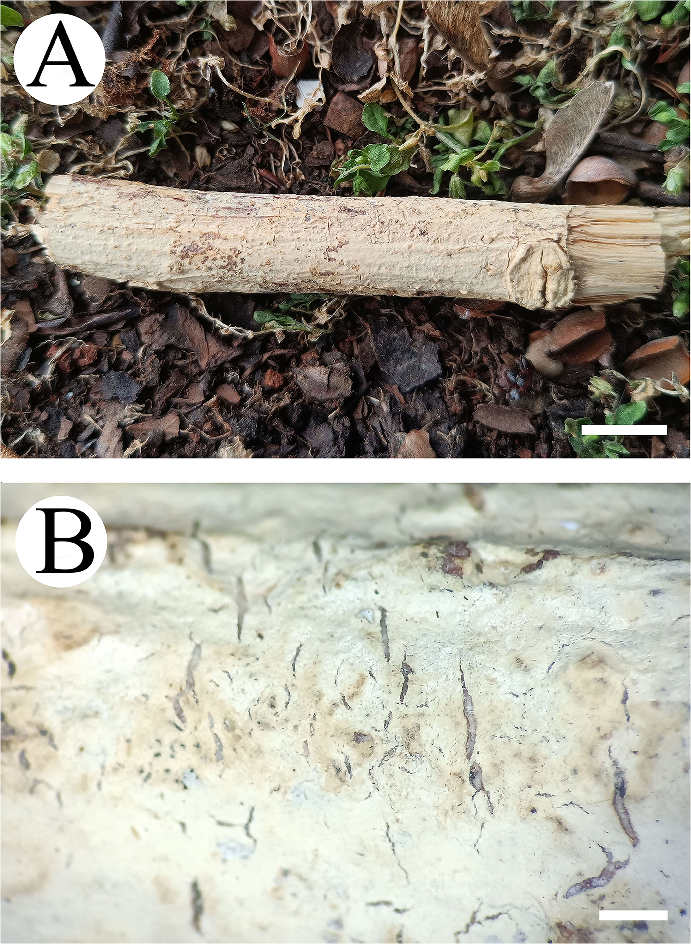
Diagnosis:— This species is characterized by the resupinate basidiomata with a cracking hymenial surface, a dimitic hyphal system with generative hyphae bearing simple-septa, and colorless, slightly thick-walled, smooth, amyloid basidiospores measuring 5–12 × 3.5–9 µm.
Basidiomata:— Basidiomata annual, adnate, resupinate, without odor or taste when fresh, up to 8 cm long, 4 cm wide, 80–100 µm thick. Hymenial surface smooth, cream (4A2/3), cracking with age. Sterile margin thin, cream (4A2/3).
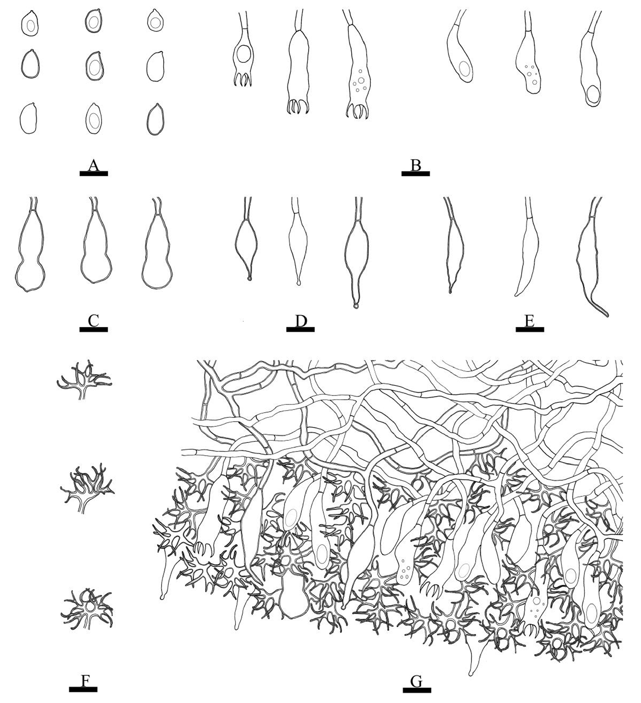
Hyphal structure: — Hyphal system dimitic, generative hyphae bearing simple-septa, dichohyphae cream (4A2/3), capillary, thick-walled, dichotomously to irregularly branched with main branches and acute tips, weakly to moderately dextrinoid in Melzer’s reagent, CB–, tissues unchanged in KOH. Subiculum composed of colorless, generative hyphae rarely branched, slightly thick-walled, 1.5–2.5 μm in diam., dichohyphae predominate, frequently branched, 1.5 µm diameter. Subhymenium composed of colorless, generative hyphae rarely branched, slightly thick-walled, 1–2 µm diameter; dichohyphae predominate, frequently branched, 0.5–1.5 µm diameter;
Hymenium: — Gloeocystidia various with three types, (i) subcylindrical cystidia, colorless, thin- to slightly thickwalled, smooth, tapered or gradually elongated apex, 16.5–58.5 × 4–10 µm; (ii) tapering cystidia, usually with a constriction at the tip, colorless, thin- to slightly thick-walled, smooth, 27.5–42 × 5.5–9 µm; (iii) fusiform cystidia, colorless, thin- to slightly thick-walled, smooth, 18.5–43.5 × 7–9 µm. Basidia cylindrical, thin-walled, with four sterigmatas and a basal simple septum, 17.5–32 × 5–9.5 µm; basidioles dominant, in shape similar to basidia, but slightly smaller.
Basidiospores:— Ellipsoid, colorless, slightly thick-walled, smooth, with oil drops, amyloid, CB–, (5.1–)5.9– 11.5(–11.8) × (4.3–)4.7–8.6(–9) µm, L = 9.14 µm, W = 6.35 µm, Q = 1.4 (n = 60/2).
Additional specimen ( paratype) examined: — CHINA. Yunnan Province, Honghe, Pingbian County, Daweishan National Nature Reserve , E 103°30′10′′, N 23°42′07′′, elev. 1500 m, on fallen angiosperm branch, 2 August 2019, CLZhao 18283 ( SWFC!), GenBank No. (ITS ON454116 View Materials , nLSU ON502653 View Materials ) GoogleMaps .
| SWFC |
Southwest Forestry College |
No known copyright restrictions apply. See Agosti, D., Egloff, W., 2009. Taxonomic information exchange and copyright: the Plazi approach. BMC Research Notes 2009, 2:53 for further explanation.
|
Kingdom |
|
|
Phylum |
|
|
Class |
|
|
Order |
|
|
Family |
|
|
Genus |