
Phasmarhabditis safricana, Ross & Pieterse & Malan & Ivanova, 2018
|
publication ID |
https://doi.org/ 10.11646/zootaxa.4420.3.5 |
|
publication LSID |
lsid:zoobank.org:pub:ADEEC06D-F245-479D-B01A-B6519B7FC826 |
|
DOI |
https://doi.org/10.5281/zenodo.5964031 |
|
persistent identifier |
https://treatment.plazi.org/id/0386982E-FF8F-FD67-FF57-FAFDDDE5FE3F |
|
treatment provided by |
Plazi |
|
scientific name |
Phasmarhabditis safricana |
| status |
|
Phasmarhabditis safricana 1 n. sp.
= Phasmarhabditis View in CoL n. sp. SA2 Ross et al., 2012
Measurements. Reference to morphometrics given in Table 1.
Description. Adults. Body 1.1–2.2 mm long; straight when relaxed, slightly tapering to anterior. Cuticle about 1 µm thick, often loose; bearing rows of transverse and longitudinal striations. Lateral fields with central band and four equal ridges running along each side of band ( Fig. 3B View FIGURE 3 ). Head truncate, lip region short, slightly offset. Six lips arranged in three groups, each lip bearing a small, short labial papilla in apical position. Each sublateral lip bearing small cephalic papilla located slightly posterior to labial one ( Fig. 3A View FIGURE 3 ). Pore-like amphids situated close to labial papillae on lateral lips. Mouth aperture triangular. Stoma tubular, relatively wide and short (about 1.5 diam. long in lip region). Cheilostom not cuticularised. Gymnostom walls nearly parallel, thickened. Stegostom with glottoid apparatus, isomorphic, isotropic, metarhabdions thickened, with three minute warts or denticles. Pharyngeal collar covering about half of stoma length. Cheilostom: gymnostom: stegostom ratio 1:1.1:1.5. Pharynx muscular, consisting of corpus expanded posteriorly, short, relatively broad isthmus and rounded or pear-shaped terminal bulb slightly wider than metacorpal expansion. Terminal bulb with valve and haustrulum. Nerve ring encircling posterior half of isthmus. Excretory pore opposite base of terminal bulb. Excretory duct weakly cuticularised. Cardia prominent; intestine with wide walls; rectal glands present. Deirids not observable.
Females. Body 1160–2120 µm long. Stoma 5.4 ± 1.2 (4–8) µm wide and 20 ± 3.2 (12–23) µm long. Cheilostom 5.5 ± 0.8 (4–7) µm long, gymnostom 6.3 ± 1.4 (4–9) µm long, stegostom 8.3 ± 1.9 (4–11) µm long. Corpus 120 ± 20 (86–150) µm long, or corresponding to 48–60 (58)% of pharynx length, 14–18 wide at anterior, metacorpal expansion 32 ± 1.6 (29–34) µm wide; isthmus 43 ± 10 (30–60) µm long and 14 ± 4 (10–20) µm wide; terminal bulb 42 ± 8 (30–54) µm long and 36 ± 5 (28–43) µm wide. Excretory pore situated at 183 ± 34 (140–237) µm from anterior body end. Ovoviviparous. Gonads amphidelphic, ovaries reflexed on dorsal side. Anterior ovary branch slightly longer than posterior one, with longer reflexion nearly reaching the vulva region in most specimens examined. Oocytes arranged in two or three rows with large, prominent nuclei. Oviduct short, composed of several large cells. Spermathecae distinct, filled with 15–20 sperm cells. Uterus spacious, containing 20– 30 eggs, 56 ± 6 (50–67) µm long and 36 ± 3 (30–40) µm wide, or juveniles. Vagina straight, muscular, less than half corresponding body diam. long or 41 ± 14 (23–65) µm. Vulva median, a wide transverse slit with flat lips. Perivulval area large, demarcated by different cuticle pattern ( Fig. 3F View FIGURE 3 ). Prerectum absent. Rectum narrow, about corresponding body diameter long. Anus an arcuate slit. Tail end dome-shaped, with spike 31 ± 6 (23–43) µm long, corresponding to 50% of total tail length ( Fig. 3G View FIGURE 3 ). Cuticle thickened at the base of the terminus, making fingerprint-like pattern ( Fig. 3H View FIGURE 3 ). Phasmids not projecting, located at spike base ( Fig. 3H View FIGURE 3 ).
Males. Slightly shorter, and much slimmer, than females. Stoma 4.2 ± 0.4 (4–5) µm wide and 20.4 ± 1.5 (18– 24) µm long. Corpus 116 ± 10 (100–130) µm long, or corresponding to 59% of pharynx length. Pharyngeal lumen often having wavy appearance throughout length of corpus. Metacorpal expansion 22 ± 2 (20–25) µm wide. Isthmus 37 ± 4 (30–45) µm long and 13 ± 2 (11–18) µm wide. Basal bulb 32 ± 2 (28–36) µm wide, and 39 ± 3 (32– 43) µm long. Excretory pore opening at 174 ± 17 (140–190) µm from anterior body end. Single testis reflexed at 314 ± 27 (280–362) µm from anterior, flexure 338 ± 32 (305–410) µm long. Sperm cells ca. 6 µm in diameter. Bursa open, wide, peloderan. Nine pairs of pedunculate genital papillae (GPs) incorporated into bursa (formula 1+1+1+2/1+3+ph) ( Fig. 3C, 3E View FIGURE 3 ). First GP pair the shortest, distanced from GP2, stronger than sequential pairs. GP8 and GP9 very close. GP4 and GP8 longest and opening dorsally. GP5–7 and GP9 not reaching the edge of bursa. Tail tip usually reaching the edge of bursa, rarely not. Phasmids short, posterior to GP9. Three pericloacal papillae present: precloacal papilla located on the anterior cloacal lip in median or submedian position, and two prominent, process-like papillae situated at lateral margins of cloacal opening ( Fig. 3D View FIGURE 3 ). Spicules nearly straight, at least twice as long as the corresponding body diameter, not distinctly cephalate, shape of manubria varying from rounded to angular. Laminae as wide as manubria. Velum extending from spicule neck to distal tips present. Distal tips rounded. Gubernaculum boat-shaped, about twice shorter than spicules; small dorsal process sometimes present.
Infective juveniles. Body short, slender, tapering gently towards both ends. Cuticle with transverse and longitudinal striations; head rounded, lip region flat, not offset from body contour; six cephalic papillae; single cephalic tooth, 1.5–2 µm high, present on dorsal sector of head; amphidial apertures inconspicuous. Stoma 2 µm long and 5 ± 2.6 (3–8) µm wide; cheilostom not cuticularised, gymnostom weakly cuticularised, stegostom slightly funnel-shaped. Pharynx comprising straight, narrow (6 µm wide) corpus 84 ± 12 (75–92) µm long, slightly thinner isthmus 15 ± 7 (10–20) µm long and pear-shaped, valvular basal bulb 19 ± 7 (15–27) µm long and 10 ± 2 (8–12) µm wide. Nerve ring surrounding isthmus. Cardia prominent, protruding into intestine. Excretory pore situated closely to bulb base. Deirids inconspicuous. Tail conoid, attenuated; hyalin portion developed, occupying attenuated part of tail corresponding to half-length of tail. Hyalin presented as line of drops. Phasmids pore-like, situated at base of attenuated part of tail ( Fig. 3J View FIGURE 3 ).
Type host and locality. Phasmarhabditis safricana n. sp. was isolated from D. reticulatum , collected near George, Western Cape province, South Africa (33°99'46''S; 22°52'15''E). The slug was collected from a commercial nursery.
Type material. Holotype (first-generation female), paratype males and dauer larvae isolated from slugs, deposited under NCN 50553 –50554 in the National Collection of Nematodes, Biosystematics Division, Plant Protection Research Institute, Agricultural Research Council, Pretoria, South Africa.
Diagnosis and relationships. Phasmarhabditis safricana sp. n. is recognisable by its morphometrics, a cupola-shaped female tail with a spike, small, non-projecting phasmids, a fingerprint-like pattern of cuticle covering female tail, lateral field structure, expressed as central band, flanked by four ridges on each side; dauer larvae possessing toothlike cephalic structures, and the distinct molecular characteristics of the new species.
In having cupola-shaped spiked female tail, the new species can be compared with P. papillosa , P. bonaquaense , P. huizhouensis and P. meridionalis . From all these species, P. safricana sp. n. differs in the form of having smaller, non-projecting phasmids. From P. papillosa , differentiated in having generally larger size, flat vs inflated vulval lips, and a differently patterned lateral field (lacking the central band in the case of P. papillosa ) ( Tandingan De Ley et al., 2016). From P. bonaquaense , P. safricana sp. n. is also differentiated by a lateral field structure (with the central band and four equal ridges running along each side of a band vs with 11 incisions and three prominent central wrinkled bands with 4 or 5 indistinct incisions) and much shorter dauer larvae (543 vs 902 µm).
From P. huizhouensis and P. meridionalis , P. safricana sp. n. generally differs by having a sturdier and shorter tail spike in females (vs a thread-like terminal part of a tail).
Phasmarhabditis safricana sp. n. additionally resembles P. huizhouensis in having a similarly shaped and sized stoma and similarly sized spicules, differing, however, in the shape of the distal tip, with the former being narrowly rounded vs obtuse, with shallow indentation. The lateral field structure is different in the two species concerned, having a wider middle band flanked by narrow ridges in P. safricana sp. n., and three bands flanked by ridges (incisures) in P. huizhouensis ( Huang et al., 2015) .
Phasmarhabditis safricana sp. n. and P. meridionalis can be additionally differentiated by the structure of a lateral field in adult nematodes (with the central band and four equal ridges running along each side of a band in P. safricana vs a simple, narrow band with 2 marginal ridges in P. meridionalis ); the body length of dauer larva (av. 543 vs 839 µm) and the presence vs absence of a tooth-like structure on a head of a dauer larva.
In having the gonochoristic mode of reproduction and a dome-shaped vs conoid female tail, the present species can be differentiated from the hermaphroditic species of P. hermaphrodita and P. californica ( Tandingan De Ley et al., 2016) .
In having dauer larvae of similar body length, P. safricana sp. n. is comparable with P. bohemica (av. 543 vs 620), but it can be differentiated by means of the presence vs absence of a cephalic tooth in the dauer larvae, and shorter spicules (av. 61 vs 74–75) (Nermuť et al., 2017).
Apart from the absence of the cephalic structures in the dauer larvae, the dauer larvae in all the other species with a conoid tail, namely P. neopapillosa , P. tawfiki and P. apuliae , are characterised by a nearly twice-as-large body length (av. 1010 in P. neopapillosa , 965 in P. tawfiki , and 812 or 986 in different strains of P. apuliae ) ( Hooper et al., 1999; Azzam, 2003; Nermuť et al., 2016 a).
Several morphological traits of the new species seem to be unique for the genus Phasmarhabditis , possessing a fingerprint-like pattern of cuticle on the female tail, and with the dauer larvae possessing toothlike cephalic structures and a developed hyalin region in the tail.
The cuticular ornamentation (the tessellated appearance) and the lateral field structure of the new species is reminiscent of P. hermaphrodita and P. neopapillosa , as demonstrated in Hooper et al. (1999). The tessellated cuticle, together with the dorsal cephalic tooth of the dauer larvae, is reminiscent of such traits in the same stage of Heterorhabditis sp. ( Nguyen, 2007), as well as is the hyalin region in the tail.
Molecular differentiation and phylogenetic relationships. The sequences that were obtained for P. safricana n. sp. were deposited in the NCBI GenBank, under HQ116061 for the SSU and MF806606 View Materials for the LSU rRNA gene. Sequencing of the ITS1, 5.8S, ITS2 rRNA, and mtCOI genes was unsuccessful.
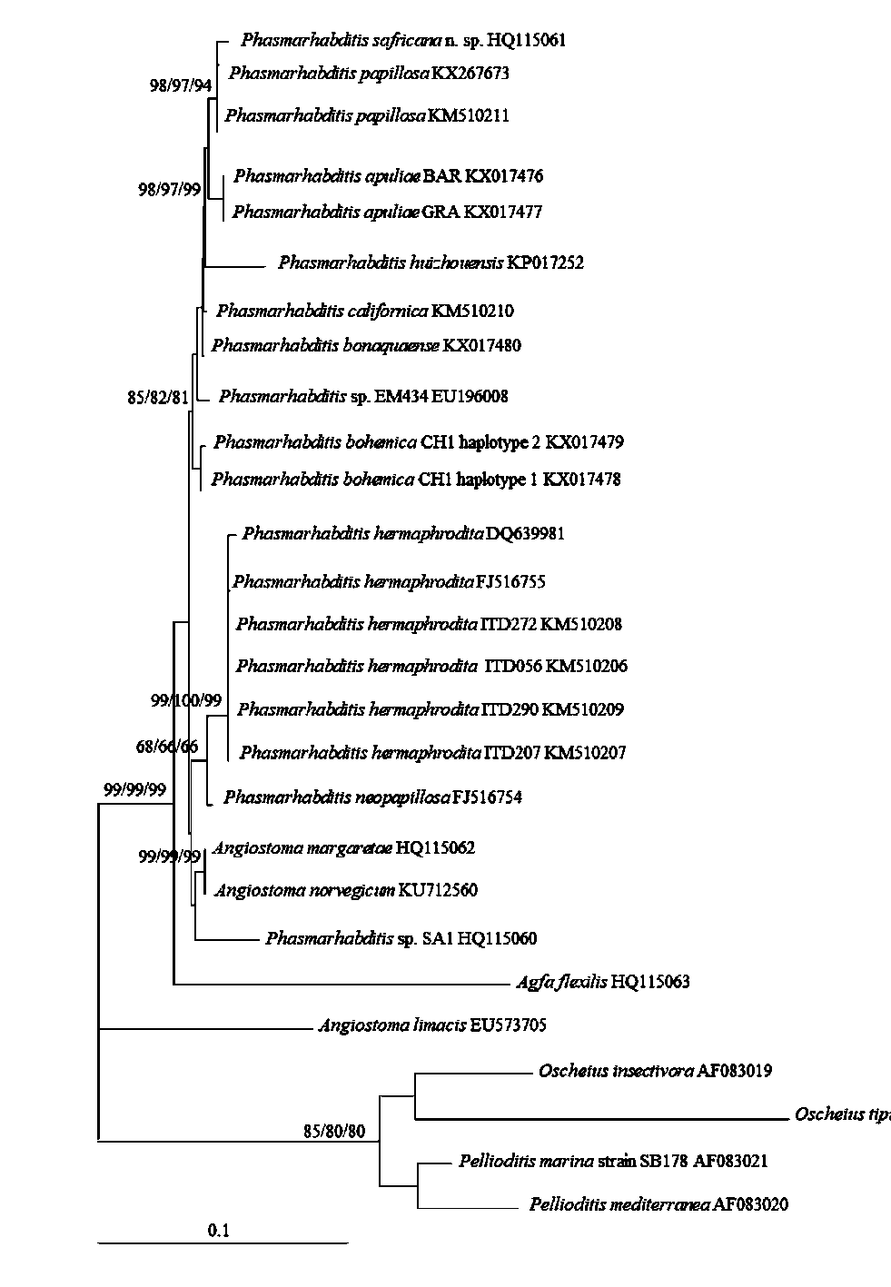
Phylogenetic analysis was conducted using the SSU and LSU rRNA genes of nematode taxa representing the genera: Agfa , Angiostoma , Phasmarhabditis , and Pellioditis . Oscheius tipulae and O. insectivora functioned as the outgroup. Although a representative ML tree is presented, bootstrap support is included for each method of analysis.
Phylogenetic analysis of the SSU placed P. safricana n. sp. with P. papillosa and Angiostoma dentiferum (Mengert) . The nematodes, along with P. apuliae , P. huizhouensis , P. bonaquaense , P. californica , Phasmarhabditis sp. EM434, and P. bohemica , created a sister group to P. hermaphrodita , P. neopapillosa , A. norvegicum Ross, Haukeland, Hatteland & Ivanova , A. margaretae Ross, Malan & Ivanova , and Phasmarhabditis sp. SA1. The aforementioned nematodes formed a sister group to Agfa flexilis (Dujardin) (99/99/99), with Angiostoma limacis Dujardin being in the basal position (100/100/100) ( Fig. 4 View FIGURE 4 ).
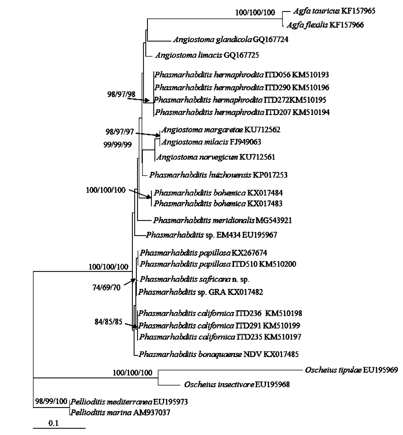
Phylogenetic analysis of the LSU grouped P. safricana n. sp. with P. papillosa and P. apuliae . The nematodes described, together with P. californica and P. bonaquaense , formed sister groups to Phasmarhabditis sp. EM434, P. meridionalis , P. bohemica , A. dentiferum , A. limacis , P. hermaphrodita , A. norvegicum , A. margaretae , A. milacis , P. huizhouensis , A. glandicola , A. tauricus , and A. flexilis (100/100/100) ( Fig. 5 View FIGURE 5 ).
Life cycle. Phasmarhabditis safricana n. sp. has been reared over the long term, under laboratory culture conditions, on freeze-killed slugs belonging to D. reticulatum , demonstrating the nematode’s ability to live on decaying organic material. However, like P. hermaphrodita , it is likely that the pathogenicity of P. safricana n. sp. is strongly influenced by the associated bacteria ( Wilson et al., 1995; Rae et al., 2010).
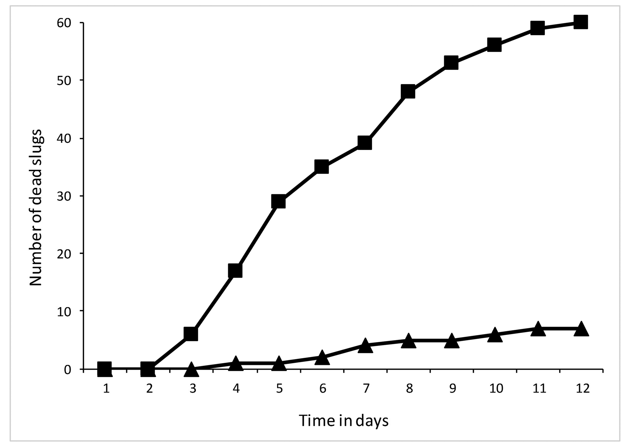
Virulence. Virulence tests demonstrated that P. safricana n. sp. caused mortality to the European invasive slug D. reticulatum , with the length of time and the type of treatment having a significant effect on mortality (P <0.001) ( Fig. 6 View FIGURE 6 ). After 5 days, half of the inoculated slugs were dead, with, by day 12, 100% mortality being recorded. Infection was confirmed by dissecting the dead slugs.

No known copyright restrictions apply. See Agosti, D., Egloff, W., 2009. Taxonomic information exchange and copyright: the Plazi approach. BMC Research Notes 2009, 2:53 for further explanation.
|
Kingdom |
|
|
Phylum |
|
|
Class |
|
|
Order |
|
|
Family |
|
|
Genus |
Phasmarhabditis safricana
| Ross, Jenna L., Pieterse, Annika, Malan, Antoinette P. & Ivanova, Elena 2018 |
Phasmarhabditis
| Ross & Pieterse & Malan & Ivanova 2018 |