
Thelepus hemeiensis, Hsueh, Pan-Wen & Li, Kuo-Rong, 2017
|
publication ID |
https://doi.org/ 10.11646/zootaxa.4244.3.10 |
|
publication LSID |
lsid:zoobank.org:pub:A36107A7-7E9F-4995-94A4-E62175719D91 |
|
DOI |
https://doi.org/10.5281/zenodo.6048904 |
|
persistent identifier |
https://treatment.plazi.org/id/0386D149-FFE7-9D7B-FF27-FD5FFDE8C6A3 |
|
treatment provided by |
Plazi |
|
scientific name |
Thelepus hemeiensis |
| status |
sp. nov. |
Thelepus hemeiensis View in CoL sp. nov.
Figs 1 View FIGURE 1 A–J, 2A–F
Material examined. Holotype ( NMNS 7743 - 1 View Materials ), Hemei (25°04´54˝N, 121°54´58˝E), New Taipei City, Taiwan, intertidal under rock on fine sand bottom, 17 October 2016.
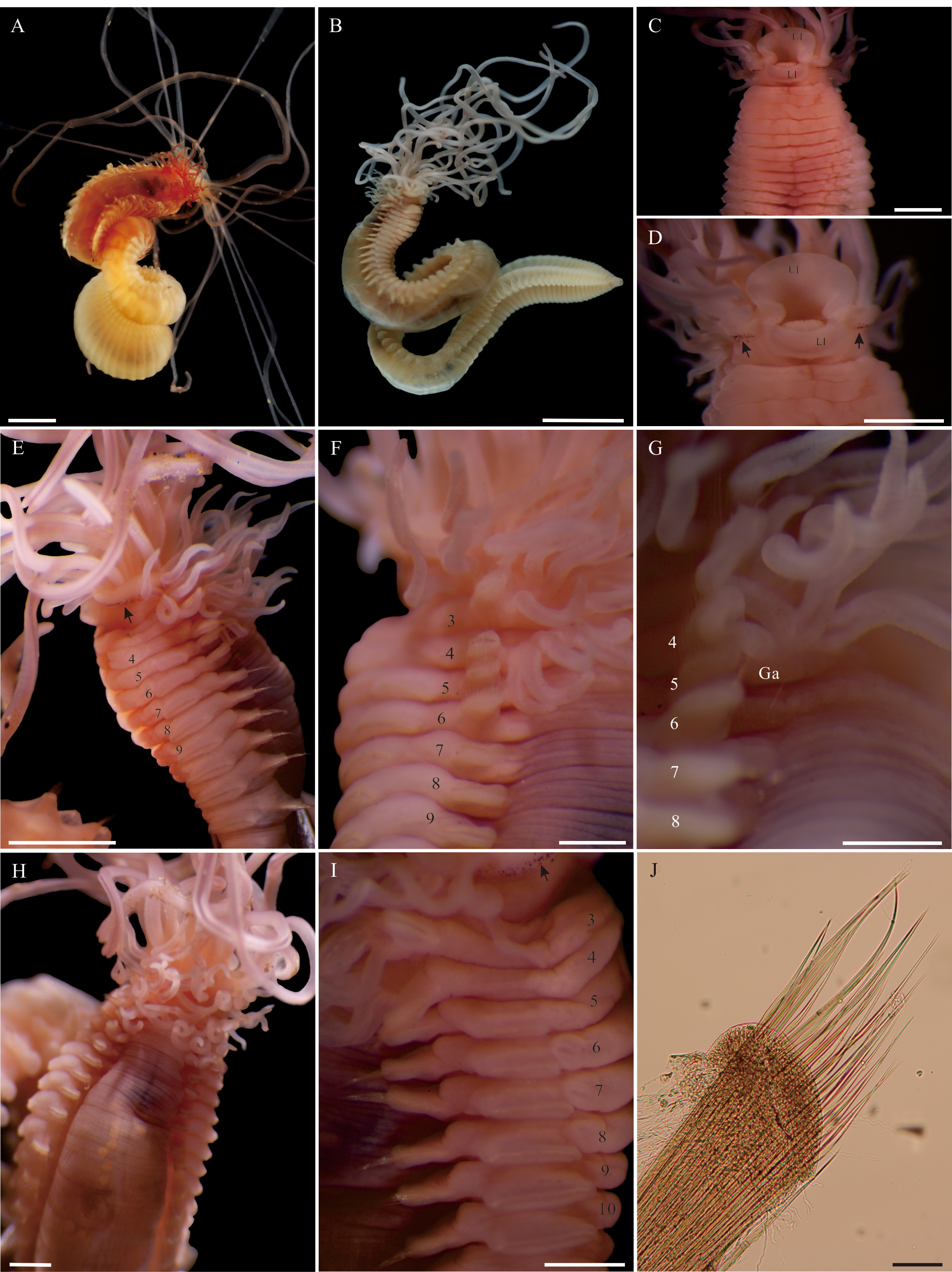
Description. Holotype, complete, ovigerous female; live specimen body uniformly orange to reddish with conspicuous blood vessels visible through body wall in anterior body, preserved body uniformly beige in alcohol, without distinct patterns of pigmentation ( Fig. 1 View FIGURE 1 A–B); body length 44.2 mm with 84 segments, maximum width 3.8 mm on segment 6.
Prostomium at base of upper lip; about twenty-two grooved buccal tentacles, thin, moderately elongate, arising along the distal part of prostomium, with long ones reaching to segments 41–44 ( Fig. 1 View FIGURE 1 C–D, H); eyespots present, forming a row at anterior base of prostomium, more concentrated latero-ventrally, becoming scattered at dorsal area of prostomial base ( Fig. 1 View FIGURE 1 C–E). Peristomium forming lips and continuing dorsally; upper lip large and thick, horseshoe-shaped; lower lip smooth, moderately large and thick ( Fig. 1 View FIGURE 1 C–D); pharyngeal organ exposed. Segment 1 with ventral lobe posterior to lower lip ( Fig. 1 View FIGURE 1 C–D). Ventral surface distinctly glandular, swollen, up to segment 10, more markedly glandular on segment 5 to 7 ( Fig. 1 View FIGURE 1 E).
Lateral lobe absent. Three pairs of branchiae, on segment 2 to 4, branchial filaments moderately thick and elongate, mostly not exceeding four body segments in length ( Fig. 1 View FIGURE 1 E–F); pairs arranged in transversal bands, arising from a glandular area, first pair lateral to the first pair of notopodia, following pairs placed progressively more dorsal, with wide medial gap between each pair, widest gap in the third pair ( Fig. 1 View FIGURE 1 E–H); pairs of segments 2–4 with 11–12, 5 and 4 simple branchial filaments on each side, respectively ( Fig. 1 View FIGURE 1 E–H).
Notopodia present from segment 3 to 39; first eight pairs similar in length, becoming longer from segment 11 to 21, thereafter shorter gradually to the last thoracic segment ( Fig. 1 View FIGURE 1 B, E), all notopodia aligned vertically ( Fig. 1 View FIGURE 1 H–I). Trapezoidal notopodia, with glandular forming pre-and postchaetal lobe from segment 3 to 11, notochaetae emerging from between lobes ( Fig. 1 View FIGURE 1 E, H–I), thereafter notopodia becoming less glandular. Notochaetae of all thoracic segments as two rows of winged chaetae, anterior row slightly shorter than posterior row, tapering to tips ( Fig. 1 View FIGURE 1 J).
Neuropodia present from segment 5 to 84, glandular and rectangular; neuropodia gradually wider in width up to segment 11 ( Fig. 1 View FIGURE 1 E, H), thereafter gradually narrower to posterior thoracic segment; abdominal neuropodia cylindrical progressively smaller to last segment prior to pygidium, as fleshy pinnules slightly raised from the surface of the body ( Fig. 1 View FIGURE 1 B); anterior neuropodia with small gap between pairs, wider from mid-body. Uncini in straight row, but slightly curved at both distal ends on anterior to mid-thoracic segments ( Fig. 1 View FIGURE 1 I, 2A), becoming Cshaped on posterior thoracic segments to the last abdominal segment ( Fig. 2 View FIGURE 2 B); dental formula MF:2:1 throughout, uncini with pointed prow and slightly upturned subterminal dorsal button ( Fig. 2 View FIGURE 2 C–D).
Genital and nephridial papillae not observed. Pygidium rounded, with crenulated opening ( Fig. 1 View FIGURE 1 B).
Etymology. The name is derived from the name of nearby village where the worm was collected.
Type locality. Hemei , New Taipei City, Taiwan.
Distribution. Only known from the type locality.
Remarks. Of forty-eight valid species described for the genus, seventeen were reported from the East and Southeast Asia. These species are: Thelepus abyssorum Caullery, 1944 (type locality: Southeast China), Thelepus angustitoris Caullery, 1944 (Southeast Asia), Thelepus binakayensis Pillai, 1965 ( Philippines) , Thelepus dubius Caullery, 1944 (Malay Archipelago) , Thelepus japonicus Marenzeller, 1884 ( Japan) , Thelepus leptoplocamus (Grube, 1878) ( Philippines) , Thelepus longtongensis Hsueh & Li, 2016 (Taiwan) , Thelepus malayensis ( Caullery, 1944) ( Malaysia) , Thelepus marenzelleri McIntosh, 1885 (Southern Japan), Thelepus microbranchiatus Caullery, 1944 ( Southeast Asia ), Thelepus opimus Hutchings, 1990 ( Hong Kong), Thelepus parcus (Grube, 1878) ( Philippines) , Thelepus paucibranchis (Grube, 1878) ( Philippines) , Thelepus pulvinus Hutchings, 1990 ( Hong Kong), Thelepus robustus (Grube, 1878) ( Philippines) , Thelepus taiwanensis Hsueh & Li, 2016 (Taiwan) , and Thelepus toyamaensis Okuda, 1936 ( Japan) ( Hsueh & Li 2016: 519, Table 1).
Among these species, only T. dubius , T. opimus , and T. toyamaensis have similar ranges of branchial filaments and the presence of eyespots ( Hsueh & Li 2016: 519, Table 1) as in Thelepus hemeiensis sp. nov.. However, T. dubius has only 29 pairs of notopodia and 60 abdominal segments ( Caullery 1944: 170–171), whereas Thelepus hemeiensis sp. nov. has 37 pairs of notopodia and 80 abdominal segments. Thelepus opimus differs from T. hemeiensis sp. nov. by having many more pairs of notopodia (>55 vs. 37) and different uncini dental formula on anterior and posterior tori (MF:2:2–3:Ɗ/MF:2–3:1–7:0–5 vs. MF:2:1 throughout) ( Hsueh & Li 2016: 519, Table 1; present study). Thelepus toyamaensis can be distinguished from T. hemeiensis sp. nov. by having very short branchial filaments (vs. moderate elongate branchial filaments), dorsal body surface with longitudinal furrows (vs. horizontally corrugated), about 30 segments with glandular ventral surface (vs. about 10 segments with glandular ventral surface), and one row of secondary teeth above main fang (vs. two rows of secondary teeth above main fang) ( Okuda 1936; present study).

| NMNS |
National Museum of Natural Science |
No known copyright restrictions apply. See Agosti, D., Egloff, W., 2009. Taxonomic information exchange and copyright: the Plazi approach. BMC Research Notes 2009, 2:53 for further explanation.