
Pseudoendorchis megalonemi, Alves & Chambrier & Scholz, 2021
|
publication ID |
https://doi.org/ 10.1007/s11230-021-09994-0 |
|
DOI |
https://doi.org/10.5281/zenodo.5645909 |
|
persistent identifier |
https://treatment.plazi.org/id/03878783-6065-FFB2-FD02-FAE6FD54FAB3 |
|
treatment provided by |
Felipe |
|
scientific name |
Pseudoendorchis megalonemi |
| status |
sp. nov. |
Pseudoendorchis megalonemi View in CoL n. sp.
Syns. Monticellia santafesina sensu de Chambrier et al. (2015) View in CoL , nec Arredondo and Gil de Pertierra (2010); Pseudoendorchis sp. 3 sensu Alves et al. (2021a)
Type-host: Megalonema platycephalum Eigenmann ( Siluriformes : Pimelodidae ).
Type-locality: Upper Amazon River (3°47 0 S, 73°20 0 W, Amazon River basin), near Iquitos, Region of Loreto, Peru.
Type-material: Holotype – complete specimen from M. platycephalum (PI 550g; MHNG-PLAT-0137396 ); paratype – complete specimen without scolex (on the same slide as the holotype (PI 550g; MHNG-PLAT-0137399 ); paratype – complete specimen mounted on two slides (PI 550b; IPCAS C-895 ). All specimens were collected by A. de Chambrier and R. Kuchta on 9 October 2009.
Voucher-material: Fragments of pregravid proglottids (PI 550a; MHNG-PLAT-69452 ); one specimen with scolex (on the same slide of the IPCAS paratype) and two pieces of strobila (on another slide = hologenophores) (PI 550b; IPCAS C-895 ) ; two fragments with immature and pregravid proglottids, and nine slides with serial sections (PI 550c; MHNG-PLAT-0120692 ); fragment of immature proglottids (PI 550d; MHNG-PLAT-0120693 ) ; one specimen without scolex (PI 550e; MHNG-PLAT-0121209 ); seven slides with serial sections (PI 550f; MHNG-PLAT-0121210 ).
Site of infection: Anterior intestine.
Infection rate: Only a single host individual was available; it was infected with seven, mostly fragmented tapeworms (see de Chambrier et al., 2015).
Representative DNA sequences and phylogenetic relationships: Two identical partial sequences of lsr DNA (length 1,430 bp) of two isolates ( PI 550b; MW 244616 View Materials , MW 244617 View Materials ) and complete sequence of COI (length 1,617 bp) of one isolate ( PI 550b; MW 244057 View Materials ). Phylogenetic analyses by Alves et al. (2021a) using both single (lsr DNA) and concatenated (lsr DNA + COI) dataset revealed the new species (= Pseudoendorchis sp. 3 in their analyses) to form a sister lineage to P. cristata (figs. 3 and 4 in Alves et al., 2021a). All but one species discovery and validation analyses suggested the validity of the new taxon. Only the Automatic Barcode Gap Discovery ( ABGD) distance-method indicated the conspecificity of Pseudoendorchis sp. 3 and P. cristata (see Alves et al., 2021a), but both species conspicuously differ from each other in their morphology and occur in distantly related fish hosts ( Pimelodidae versus Heptapteridae ). Therefore, they are considered to represent separate species.
ZooBank registration: To comply with the regulations set out in article 8.5 of the amended 2012 version of the International Code of Zoological Nomenclature ( ICZN, 2012), details of the new species have been submitted to ZooBank. The Life Science Identifier (LSID) for Pseudoendorchis megalonemi n. sp. is urn:lsid:zoobank.org:act:4E323CDF-4BCE-42E1-B24A-107DDEB08621
Etymology: The species is named after the generic name of the type host, and is formed as a noun in the genitive case, masculine.
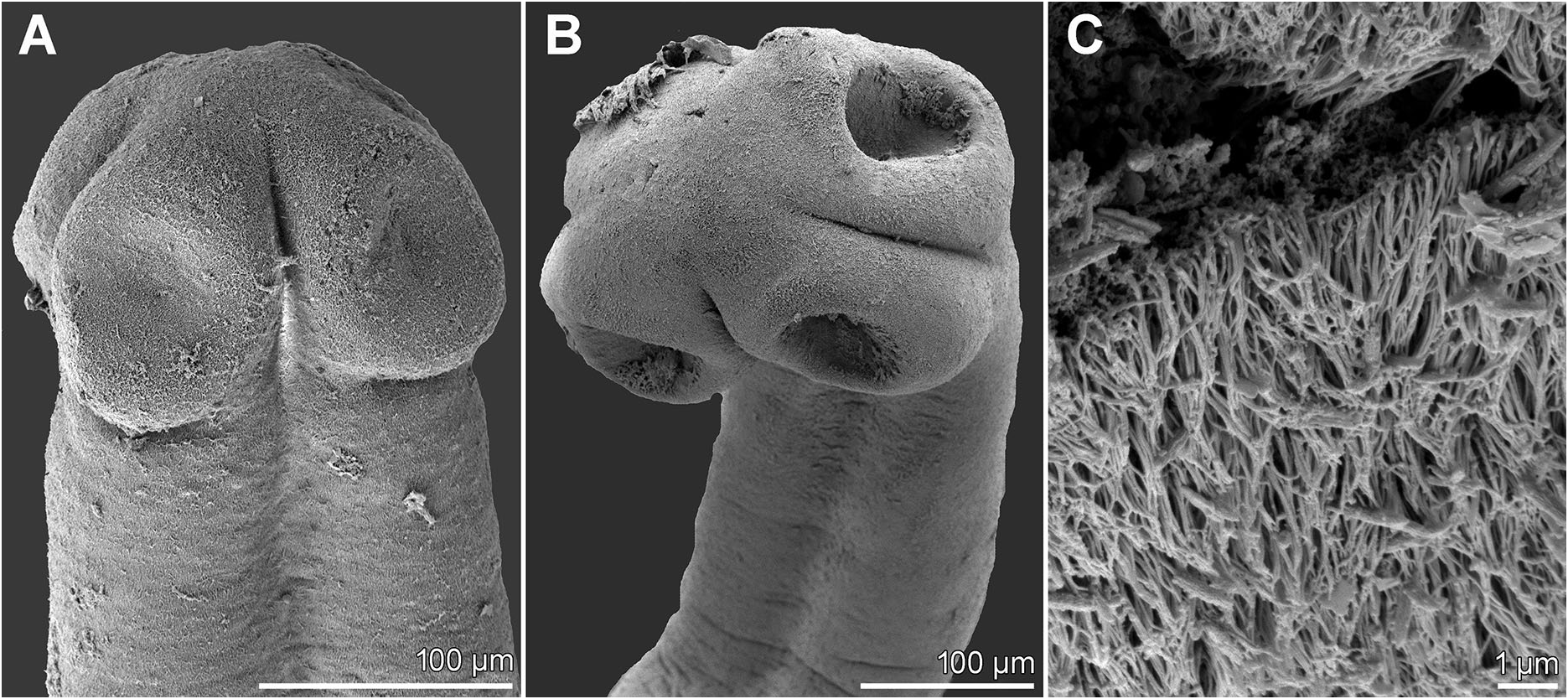
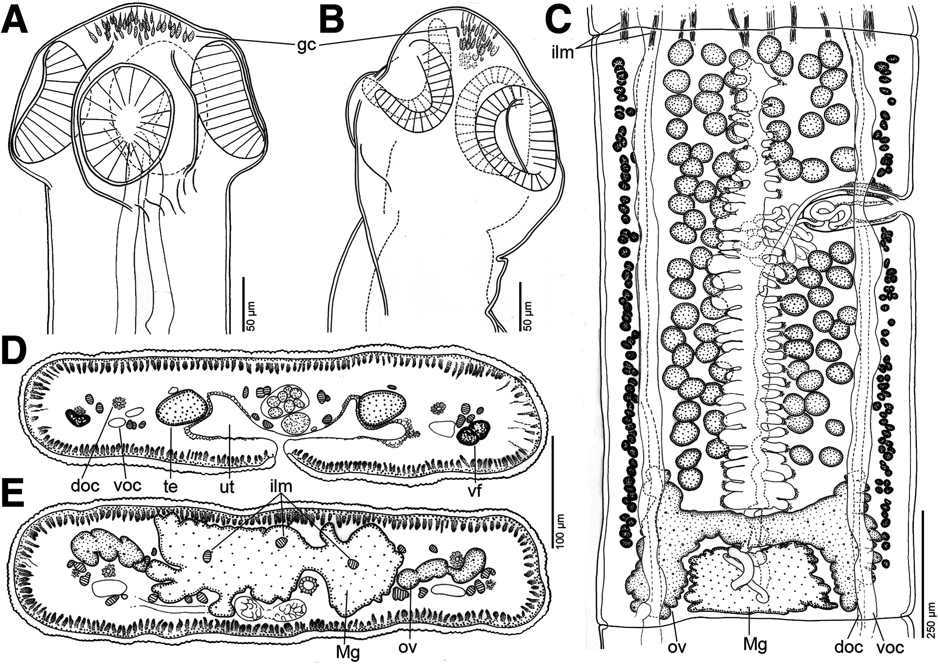
Description ( Figs. 1–3A View Fig View Fig View Fig , 5A View Fig )
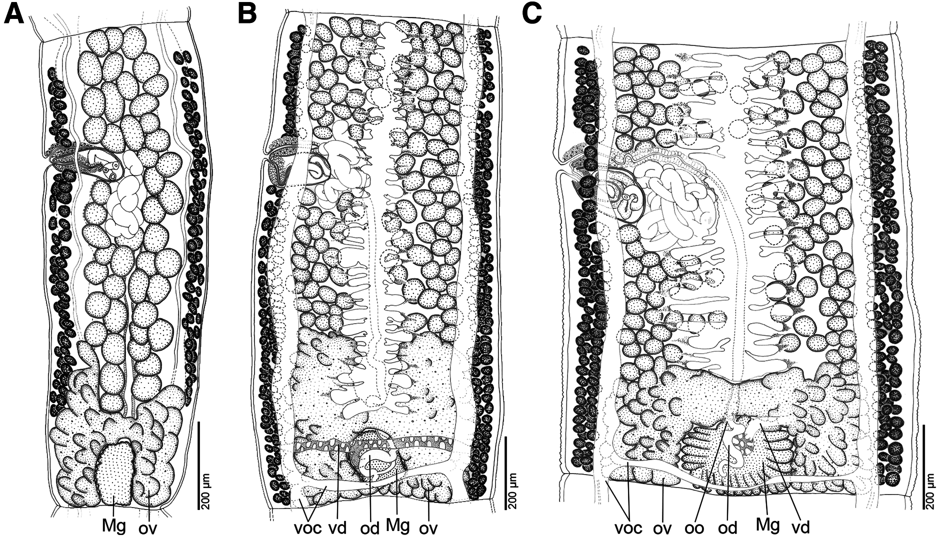
[Based on one complete specimen, four specimens without scoleces and parts of another two worms, all whole-mounted, and two scoleces observed using scanning electron microscopy – Fig. 1 View Fig ]. Proteocephalidae . Small-sized worm (total body length 29–36 mm), maximum width up to 640. Strobila acraspedote, anapolytic. Immature proglottids slightly wider than long to longer than wide (length: width ratio 0.90–2.95), mature and pregravid proglottids longer than wide (length: width ratio 1.87–4.10), gravid proglottids much longer than wide (length: width ratio 1.78–4.68).
Scolex 160–240 long and 230–240 wide, slightly wider than proliferative zone, 160–175 wide. Scolex bearing four oval, uniloculate suckers 105–120 in diameter ( Figs. 1A, B View Fig , 2A, B View Fig ), and dome-shaped apex without apical organ, but containing granular material ( Figs. 1B View Fig , 2A, B View Fig ). Strobila covered with numerous capilliform filitriches, interspersed with few gladiate spinitriches ( Fig. 1C View Fig ).
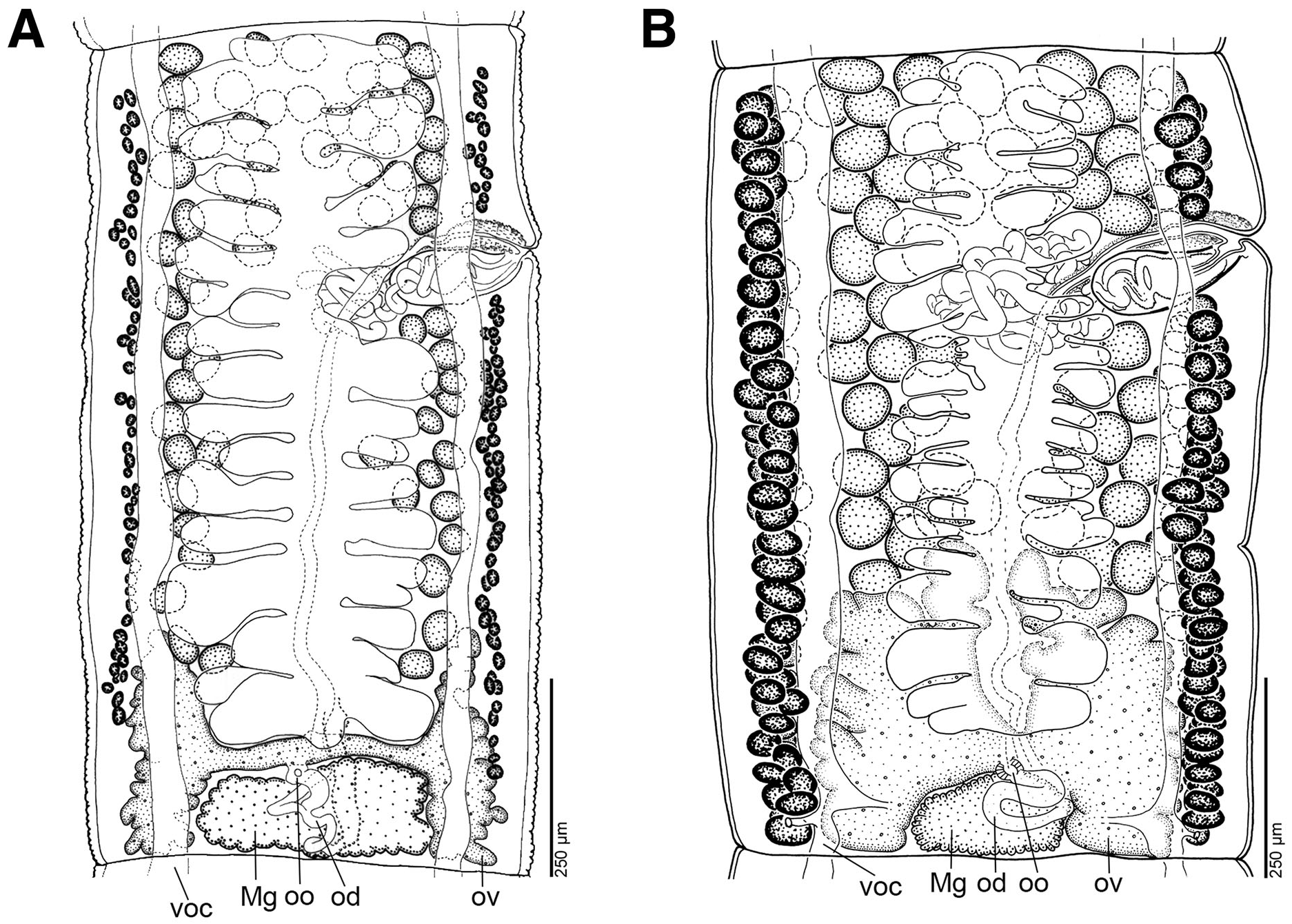
Inner longitudinal musculature formed by a few (about 4–5 dorsal and 4–5 ventral), wide, anastomosed bundles of sparsely distributed muscle fibres ( Fig. 2C– E View Fig ). Two pairs of osmoregulatory canals almost straight, situated at same level, between testes and vitelline follicles, partially overlapping lateralmost testes and sometimes medianmost vitelline follicles. Ventral canals thin-walled, wide, wider than dorsal one, 35–55 wide; ventral canals with anastomoses and sometimes connected posterior to ovary with secondary canals ending on ventral side of proglottid surface; dorsal canals thick-walled, surrounded by muscles fibres, 15–20 wide ( Fig. 2D, E View Fig ).
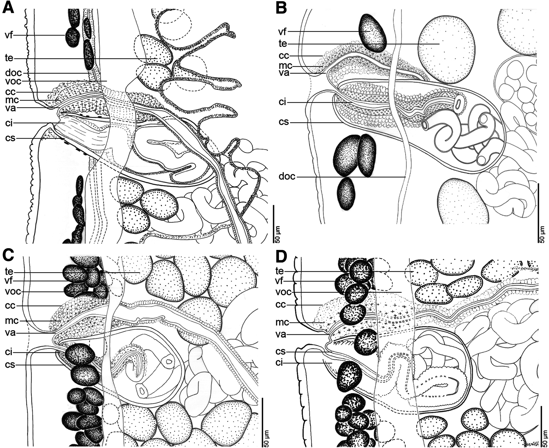
Testes medullary, oval to spherical, 50–75 long and 35–50 wide, in one layer, forming one field between anterior margin of proglottids and anterior lobes of ovary, 69–106 in number (x = 85, n = 15), never overlapping vas deferens and cirrus-sac, and less numerous at level of uterine stem ( Figs. 2C View Fig , 3A View Fig ). Testes present also in gravid proglottids. Vas deferens strongly coiled, with loops forming large oval field reaching to median line of proglottid or slightly crossing it ( Figs. 2C View Fig , 3A View Fig ). Cirrus-sac elongated, pyriform, 150–210 long and 50–85 wide (n = 22), its length representing 28–39% (x = 32%, n = 22) of proglottid width; cirrus sinuous, long, reaching up to 62% of cirrus-sac width ( Figs. 2C View Fig , 3A View Fig , 5A View Fig ). Genital pores alternating irregularly, pre-equatorial, situated at 24–35% (x = 30%, n = 20) of proglottid length from anterior margin ( Figs. 2C View Fig , 3A View Fig ); common genital atrium narrow, deep ( Figs. 2C View Fig , 3A View Fig ).
Ovary medullary, butterfly-shaped, with lateral lobules penetrating to cortex, occupying 76–83% (x = 80%, n = 17) of proglottid width; its length represents 25–35% (x = 28%, n = 20) of proglottid length ( Figs. 2C, E View Fig , 3A View Fig ). Relative ovarian size, i.e., ratio of surface of ovary to surface of proglottid (see de Chambrier et al., 2012), 8–9%. Mehlis’ gland unusually large, 240–360 in diameter, representing 41–50% of proglottid width (x = 46%, n = 18) ( Figs. 2C, E View Fig , 3A View Fig ). Vaginal canal slightly sinuous, with terminal part (pars copulatrix vaginae) surrounded by conspicuous, 90–110 long (51–62% of cirrus-sac length) concentration of chromophilic and muscular cells, but lacking ring-like vaginal sphincter ( Fig. 5A View Fig ); vagina always anterior to cirrus-sac (n = 34) ( Figs. 2C View Fig , 3A View Fig , 5A View Fig ).
Vitelline follicles cortical, arranged in lateral rows on each side, not reaching anterior and posterior margins of proglottids; follicles absent on ventral side at level of cirrus-sac. Length of bands represents 80–92% and 79–92% of proglottid length on poral and aporal side, respectively (n = 13) ( Figs. 2C View Fig , 3A View Fig ).
Uterus cortical, with 15–29 (x = 21, n = 18) lateral diverticula on each side penetrating to medulla, occupying up to 62% of proglottid width ( Figs. 2C, D View Fig , 3A View Fig ); development of type 2 according to de Chambrier et al. (2004) ( Fig. 2C View Fig ). Intrauterine eggs spherical, with embryophore 18–23 in diameter; oncosphere spherical, 12–13 in diameter (measured on mounted slides); hooks 5–7 long.
Remarks
The new species is placed in Pseudoendorchis because it possesses a remarkably large Mehlis’ gland (more than 1/5 of the proglottid width), the vagina is always anterior to the cirrus-sac and its terminal part (pars copulatrix vaginae) is surrounded by conspicuous, elongate concentration of chromophilic and muscular cells, but is devoid of a ring-like vaginal sphincter, the testes and ovary are in the medulla, whereas vitelline follicles are cortical, arranged in lateral rows on each side of the proglottid, and genital pores are preequatorial ( Alves et al., 2021a). The first two characteristics distinguish Pseudoendorchis from any other proteocephalid genera ( Alves et al., 2021a).
Tapeworms found in M. platycephalum in Peru, identified as M. santafesina by de Chambrier et al. (2015), actually belong to P. megalonemi n. sp. because the former species has a much smaller Mehlis’ gland, the diameter of which represents only c. 10% of the proglottid width, and the vagina can be anterior or posterior to the cirrus-sac ( Arredondo & Gil de Pertierra, 2010).
Pseudoendorchis megalonemi n. sp. differs from its congeners by possessing uniloculate suckers rather than biloculate, and by a smaller relative size of the ovary (see Table 1 View Table 1 ). In addition, the new species can be distinguished from all nominal species but P. souzalimae by a larger Mehlis’ gland in relation to the proglottid width, and all but P. cristata by shorter longitudinal bands of vitelline follicles ( Table 1 View Table 1 ; compare Figs. 2C View Fig , 3A View Fig of the new species with Figs. 3B View Fig , 4 View Fig of the congeners). The new species also differs from P. auchenipteri by a smaller scolex, more posterior position of the genital pore and a wider ovary in relation to the proglottid width ( Table 1 View Table 1 ).
Pseudoendorchis sp. 2 from P. maculatus in Brazil (Paraná River), which was not described formally as a new species due to shortage of material, is the second species in the genus with uniloculate suckers ( Alves et al., 2021a). Interestingly, this species and P. megalonemi n. sp. are not closely related (see figs. 2, 3 in Alves et al., 2021a) and differ from each other also by the morphology of the scolex apex (dome-shaped in P. megalonemi n. sp. versus conical in Pseudoendorchis sp. 2 ), shape of the suckers (more elongated in the undescribed species), and relative size of Mehlis’ gland (41–50% in the new species versus 28–36% in Pseudoendorchis sp. 2 ) (see Alves et al., 2021a).
| PI |
Paleontological Institute |
| MW |
Museum Wasmann |
| COI |
University of Coimbra Botany Department |
No known copyright restrictions apply. See Agosti, D., Egloff, W., 2009. Taxonomic information exchange and copyright: the Plazi approach. BMC Research Notes 2009, 2:53 for further explanation.
|
Kingdom |
|
|
Phylum |
|
|
Class |
|
|
Order |
|
|
Family |
|
|
Genus |
Pseudoendorchis megalonemi
| Alves, Philippe Vieira, Chambrier, Alain de & Scholz, Tomáš 2021 |
Monticellia santafesina sensu
| de Chambrier 2015 |