
Platydoris angustipes (Mörch, 1863), Morch, 1863
|
publication ID |
https://doi.org/ 10.11646/zootaxa.3745.2.2 |
|
publication LSID |
lsid:zoobank.org:pub:D87FBB64-5DE2-4D19-9338-6E9BE212FAEF |
|
DOI |
https://doi.org/10.5281/zenodo.6146314 |
|
persistent identifier |
https://treatment.plazi.org/id/0387C073-FF83-6333-FF22-0C6CB0F95C01 |
|
treatment provided by |
Plazi |
|
scientific name |
Platydoris angustipes (Mörch, 1863) |
| status |
|
Platydoris angustipes (Mörch, 1863) View in CoL
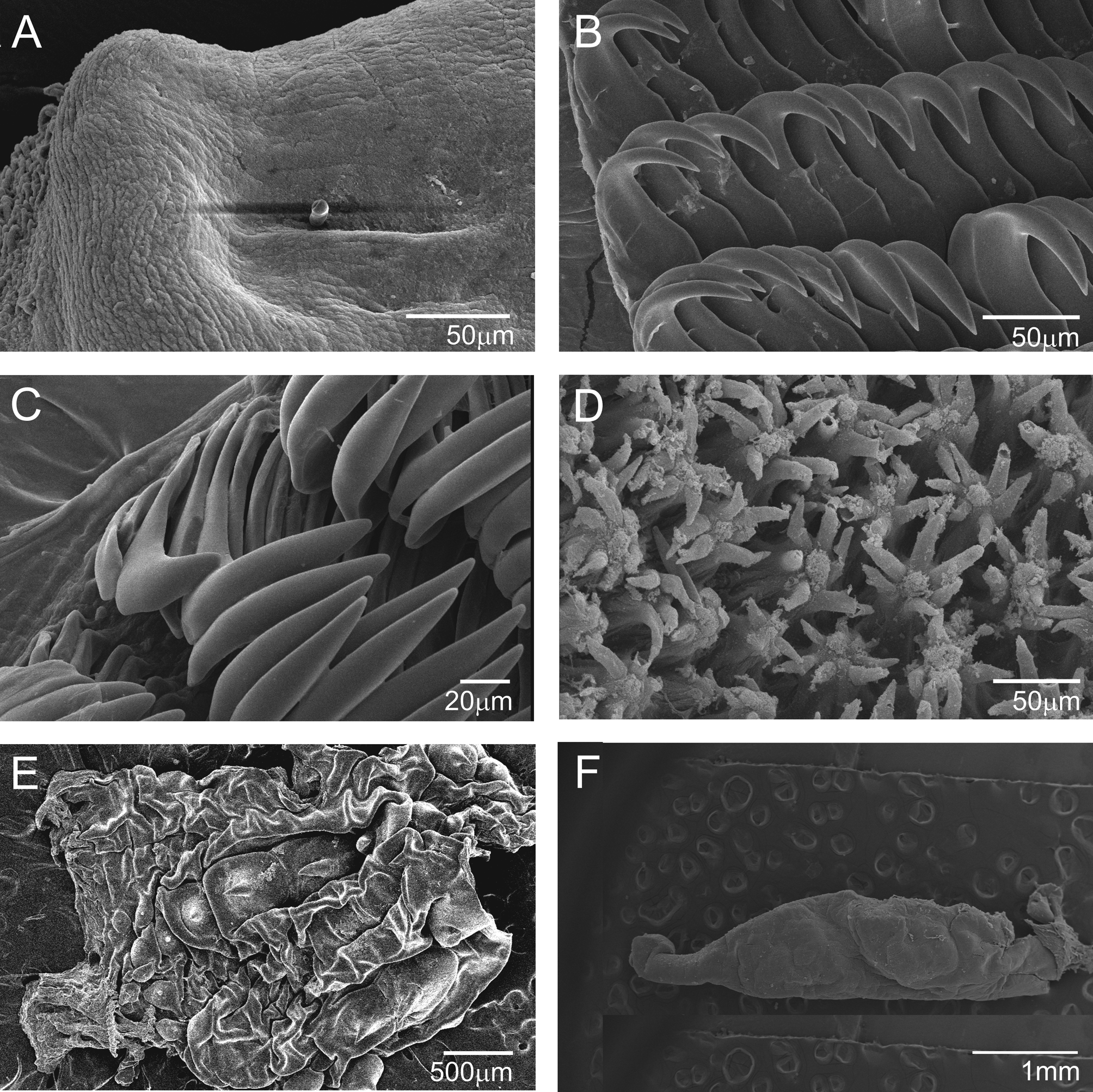
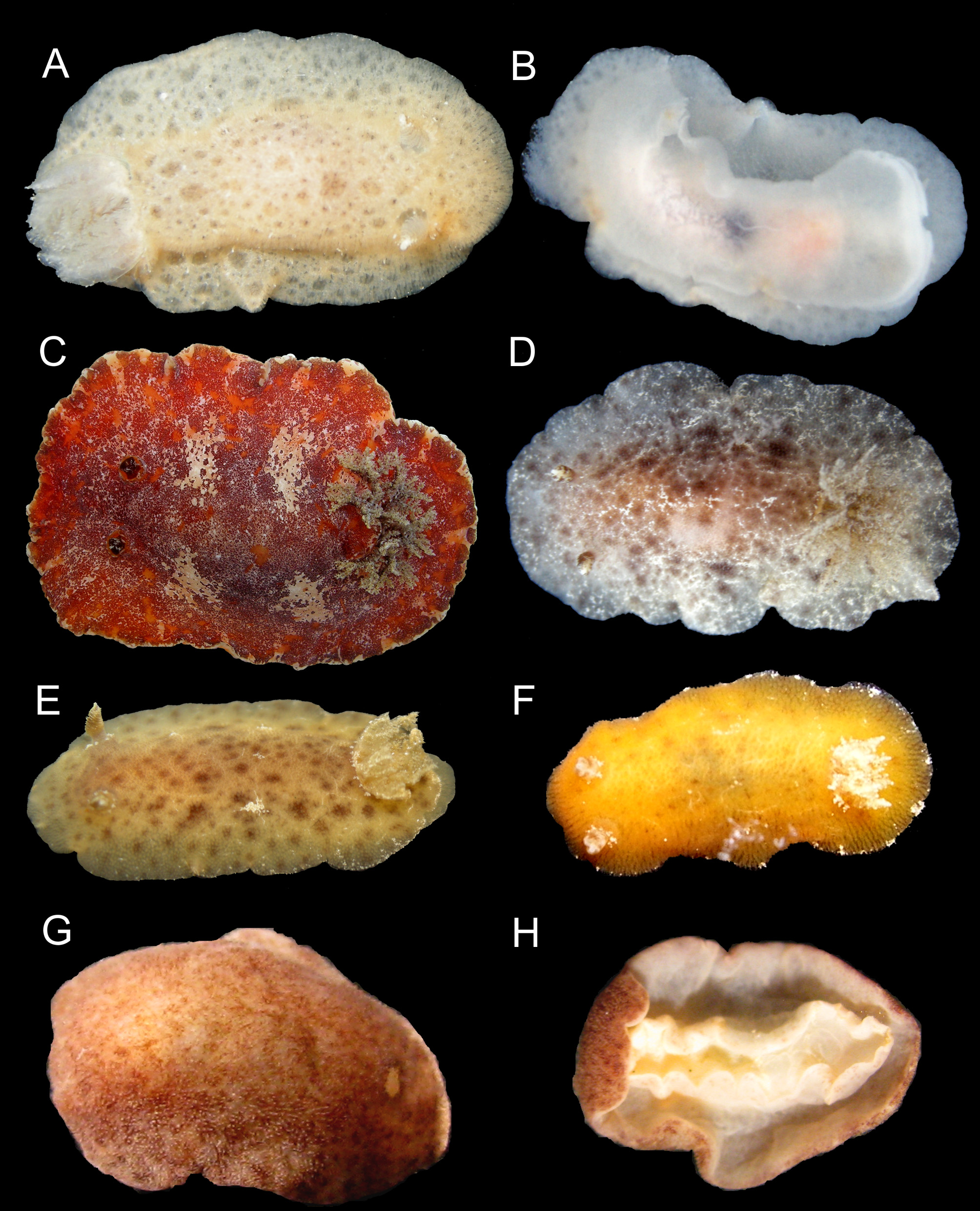
( Figures 2 View FIGURE 2. A – B C; 21–22)
Doris (Argus) angustipes Mörch, 1863: 32
Platydoris angustipes var. alaleta Bergh, 1877: 505 –506, plt. 58, figs. 13–18.
Platydoris rubra White, 1952: 118 , fig. 17, plt. 6, fig. 6.
Platydoris angustipes : Er. Marcus (1957: 422, fig. 81–89); Ev. Marcus & Er. Marcus (1967a: 93, fig. 112); Er. Marcus & Ev. Marcus (1970: 67, fig. 121); Meyer (1977: 301); Humann (1992: 243); Dorgan et al. (2002: 282, figs. 1B, 2B, 11–13); Espinosa et al. (2005); Valdés et al. (2006: 182); Debelius & Kuiter (2007: 245); García et al. (2008: 148); Rios (2009: 428); Padula et al. (2012: 3).
Type material. Unkown. Holotype ( Platydoris angustipes var. alaleta ) ZMUC-GAS 2020, St. Croix, Virgin Islands, up to 25mm.
Type locality. St. Thomas.
Material examined. Honduras: Gulf of de Honduras: MZSP 75996, 16 /vii/1969, Marcus [1]; Brazil: Pernambuco state: Baía dos Porcos: MZSP 30925, 21 /vii/1999, L. R. Simone & Souza Jr. colls. [2]; Praia do Meio: MZSP 31050, 22 /vii/1999, L. R. Simone & Souza Jr. colls. [1]; Bahia state: Itapoã: MZSP 37947 [1]; Ilha de Itaparica: MZSP 58647, i/2000, A. R. R. Neto coll. [1; one dissected]; Rio de Janeiro state: Cabo Frio: Ilha Comprida: MNRJ 13189, 29 /iii/2008, V. Padula coll. [1; one dissected]; Arraial do Cabo: Prainha: MNRJ 13177, 03 /iii/2007, P. M. S. Costa coll. [1; one dissected]; Without locality data: MZSP 75762 [1] (ex-Marcus collection).
Geographical distribution. Florida, Mexico, Honduras, Costa Rica, Panama, Cayman Islands, Dry Tortugas, Cuba, Jamaica, Aruba, Puerto Rico, Virgin Islands, St. Maarten / St. Martin, St. Bartholomew, Turks & Caicos, Antigua, St. Lucia, Martinique, Barbuda, St. Vincent and the Grenadines, Grenada, Trinidad & Tobago, Brazil (Valdés et al., 2006): Pernambuco state: Fernando de Noronha; Alagoas state: Saco da Pedra (Padula et al., 2012); Bahia state: Praia de Itapoã (García et al.: 2008); Rio de Janeiro state: Cabo Frio: Ilha Comprida; Arraial do Cabo: Prainha (present study).
Description. External morphology ( Figures 2 View FIGURE 2. A – B C; 21D): body elliptical, slightly depressed, up to 46.0 mm long alive; with 1.5 times greater length than width. Mantle hardened, densely covered by rounded caryophyllidia irregularly positioned and approximately equidistant with different diameters (21 µm to 36 µm) and uniform height; caryophyllidia with ciliary tuft and with five to 7 spicules that protrude outside. Rhinophoral sheaths prominent and irregular, covered by caryophyllidia. Rhinophores long, with cylindrical apex and 19 to 25 diagonal perfoliations. Branchial sheath prominent with caryophyllidia. Gill with six retractile, tripinnate branchial leaves, symmetrically positioned along longitudinal axis of body; high anal cone, located between two most posterior branchial leaves. Foot narrower than mantle; anteriorly bilabiate and notched on upper “lip”. Oral tentacles conical. Color of living specimens predominantly red-orange, with several white blotches of irregular sizes and arrangement on dorsum; mantle edge darker with purplish and white pigment; ventrally, orange, with some red spots on ventral part of mantle; rhinophores dark red, with some white spots on perfoliation and cylindrical apical part; branchial leaves brown with dark spots.
Labial cuticle and radula ( Figures 21 View FIGURE 21 A–C): Labial cuticle smooth. Radula formula 62 x 2.78.0.78. 2 in specimen measuring 46.0 mm in length; lateral teeth hook-shaped and smooth larger and more developed in center of rows; two marginal teeth spatulate.
Reproductive system ( Figures 21 View FIGURE 21 E–F; 22): hermaphrodite duct connected to convolute ampulla. Postampullary gonoduct short, connecting to oviduct and prostate. Prostate granular and rounded, divided into two parts; bigger and less dense proximal part and smaller and denser distal part. Vas deferens elongated, wide, folding on bursa copulatrix, becoming thinner near prostate. Penis elongated. Accessory gland elongated, attached at joint atrium of deferent duct and vagina. Vagina elongated, slightly wider than deferent duct, narrowing abruptly near opening of bursa copulatrix. Vagina, near gonopore, bearing two rows of spines; row with larger spines (up to 625 µm) in middle of vagina, subsequent rows composed by small spines (up to 14 µm), located on extremities.
Rounded/spherical bursa copulatrix. Bursa serially arranged, convoluted vaginal duct connecting to seminal receptacle; seminal receptacle with ¼ to 1/5 diameter of bursa copulatrix. Very short uterine duct.
Remarks. Dorgan et al. (2002) reviewed the genus Platydoris , adding micrographs of the penis, vagina and radula to the description of Platydoris angustipes , which until that point had been illustrated only in drawings. Our examination of several specimens allowed the recognition of some variation in the rhinophores and gill of P. angustipes . The re-description by Dorgan et al. (2002) reported 25 perfoliations in the rhinophore, whereas we observed 19 to 25 perfoliations; the specimens from Brazil always have beige branchial leaves with numerous tiny dark spots, in contrast to the white to beige gills described by Dorgan et al. (2002). The radula examined here is wider compared to those recorded by Er. Marcus (1957). The two last lateral teeth are described as hook shaped (Er. Marcus, 1957), whereas in the specimens studied here they are spatulate ( Fig. 21 View FIGURE 21 C). In this paper, we also add micrographs of the caryophyllidia ( Fig. 21 View FIGURE 21 D).
No known copyright restrictions apply. See Agosti, D., Egloff, W., 2009. Taxonomic information exchange and copyright: the Plazi approach. BMC Research Notes 2009, 2:53 for further explanation.