
Taringa iemanja, Alvim, Juliana & Pimenta, Alexandre Dias, 2013
|
publication ID |
https://doi.org/ 10.11646/zootaxa.3745.2.2 |
|
publication LSID |
lsid:zoobank.org:pub:D87FBB64-5DE2-4D19-9338-6E9BE212FAEF |
|
DOI |
https://doi.org/10.5281/zenodo.6146320 |
|
persistent identifier |
https://treatment.plazi.org/id/0387C073-FF89-633A-FF22-0F30B7605C24 |
|
treatment provided by |
Plazi |
|
scientific name |
Taringa iemanja |
| status |
sp. nov. |
Taringa iemanja View in CoL sp. nov.
(Figures: 2G–H; 25–26)
Type material. Holotype: MNRJ 13180, P.M. S. Costa leg., 15.0 mm preserved length [dissected].
Type locality. Bacia de Campos (22°06'9875"S–40°10'8204"W), Rio de Janeiro state, Brazil, 150 m depth.
Etymology. Taringa is derived from an Aboriginal word meaning “strong”. " Iemanjá " (Yoruba expression "Yèyé omo ejá", which means mother whose children are fish) is an African deity who represents the queen of the sea.
Diagnosis. Radula with innermost lateral teeth having three denticles on their outer side, first denticle is tiny. Deferent duct long, narrow and convoluted, near opening presenting a ring-shaped and smooth cuticle. Bursa copulatrix large and sherical, not flattened, covering practically entire reproductive system.
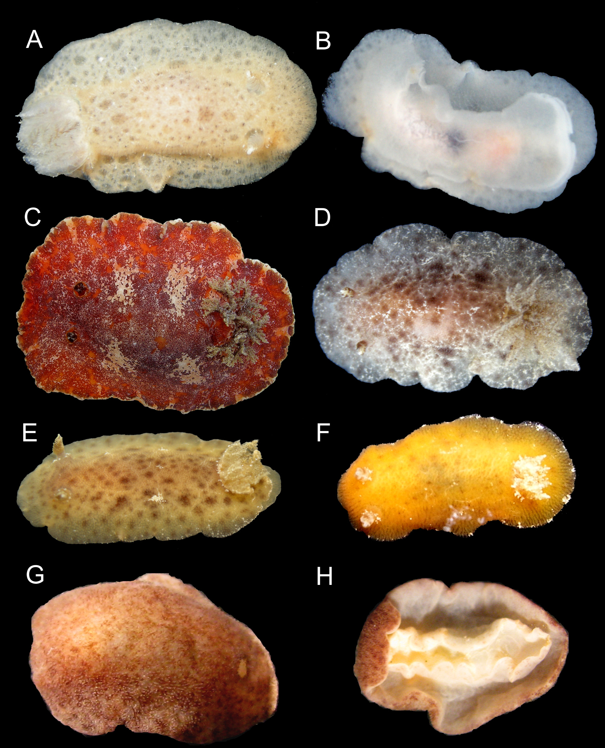
Description. External morphology ( Figures 2 View FIGURE 2. A – B G–H; 25E): body oval, slightly depressed, 15.0 mm long in a preserved specimen, with two times greater length than width. Mantle densely covered by equidistant caryophyllidia; caryophyllidia lower at mantle edge and in center of mantle (48 µm–50 µm) than those on sides of mantle (100 µm–110 µm), with variable number of spicules protruding around tubercle; tubercle apex with a ciliary tuft, elongated and oval. Rhinophoral sheaths moderately high and irregular covered by caryophyllidia of same size of those in centre of mantle. Rhinophores long with cylindrical apex, 12 diagonal perfoliations. Branchial sheath low with lobulated edge. Gill with six retractile, tripinnate branchial leaves, arranged symmetrically at longitudinal axis of body; anal cone high, located between two most posterior branchial leaves. Foot narrower than mantle, anteriorly bilabiated and notched on two lips. Oral tentacles triangular. Preserved specimen presenting mantle coloration orange-brown in edges and center, and with predominance of reddish brown on sides of mantle; mantle edge with white blotches; rhinophores with chestnut-colored perfoliations with white blotches and white apex; branchial leaves in proximal part translucent yellow and distal part brownish; ventrally body is yellowish, side of foot with brownish circular blotches.
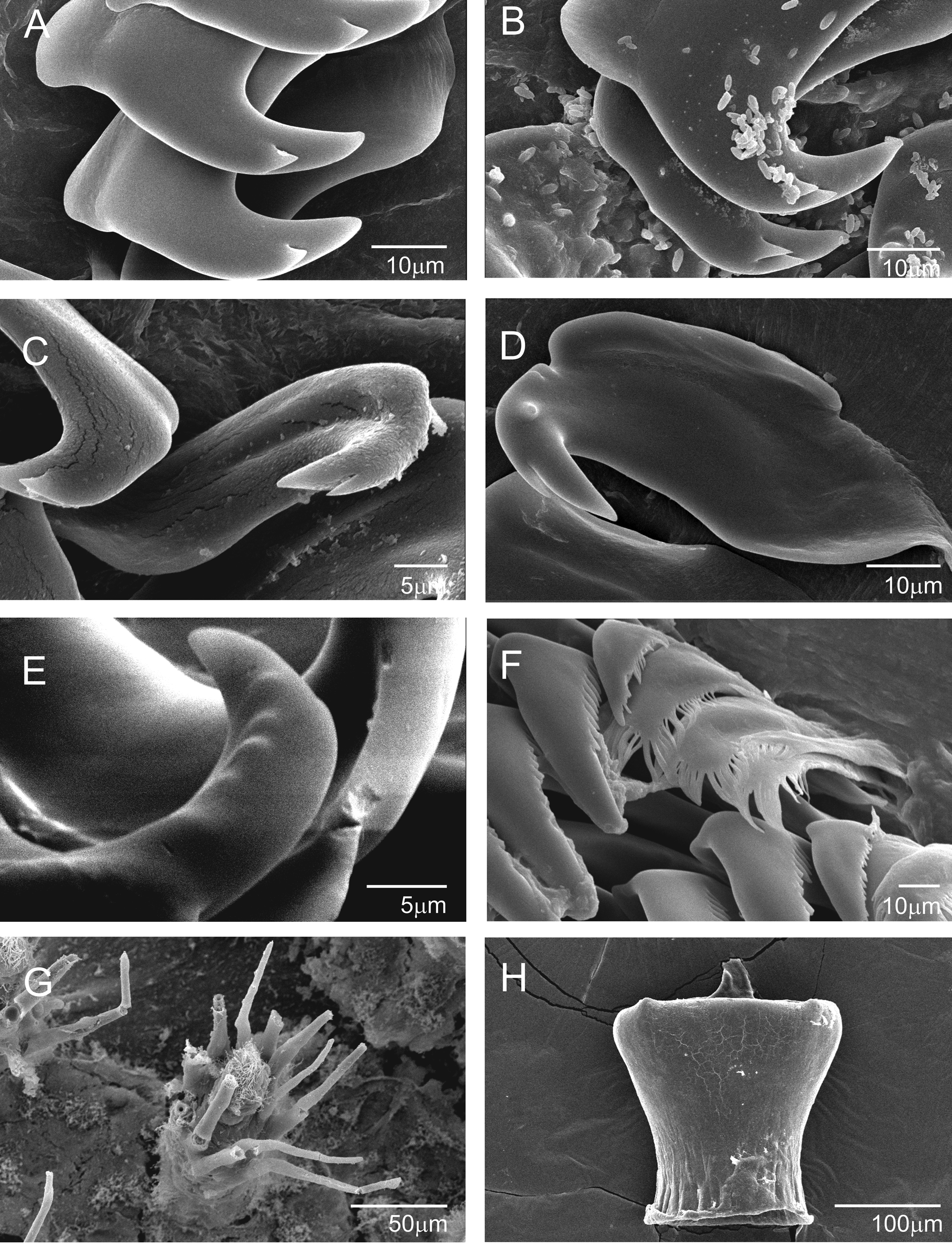
Labial cuticle and radula ( Figures 25 View FIGURE 25 A–D): labial cuticle smooth. Radula formula 29 x 3 –4.34-35.0.34-35.3– 4 in preserved specimen measuring 15.0 mm in length; innermost lateral teeth hook-shaped with three denticles on outer surface, first denticle smaller than others, and with one protuberance on inner surface; subsequent lateral teeth hook-shaped with only external denticles increasing in number outwards (up to 7); 3–4 marginal teeth spatulate and non-pectinate.
Reproductive system ( Figures 25 View FIGURE 25 F–26): hermaphrodite duct connecting to long and slightly convoluted ampulla. Postampullary gonoduct short, connecting to oviduct and prostate. Prostate granular and rounded, divided into two parts; proximal part smaller, whitish, less dense, and distal part larger, yellowish, denser. Vas deferens long, narrow and convoluted, opening into common atrium with vagina; vas deferens near gonopore presenting a ring-shaped and smooth cuticle. Vagina elongate, narrow and opening into bursa copulatrix. Bursa copulatrix large and rounded/spherical, covering almost entire reproductive system. Bursa serially arranged, convoluted vaginal duct connecting to small-stalked seminal receptacle. Uterine duct very short. Seminal receptacle with 1/5 of diameter of bursa copulatrix.
Remarks. The single specimen studied here fits perfectly in the genus Taringa , presenting all the diagnostic features of this genus (Er. Marcus, 1955), including the dorsum covered by elongated caryophyllidia ( Fig. 25 View FIGURE 25 E); tripinnate gill; smooth labial armature ( Fig. 25 View FIGURE 25 A); radula without rachidian teeth and with hook-shaped lateral plates bearing denticles on outer side ( Figs. 25 View FIGURE 25 B–D); prostate divided into two parts (Fig. 26), and a penis with cuticular structure ( Fig. 25 View FIGURE 25 F).
According to McDonald (2009), and considering Taringa telopia and Taringa disa as distinct species (see remarks relating to Taringa telopia ), there are 14 valid species in the genus Taringa , of which three are reported from the Western Atlantic: T. telopia , T. disa and Taringa tritorquis Ortea, Peréz & Llera, 1982 , the latter from Costa Rica, Cuba and the Azores. As indicated by the table presented by García-Gómez et al. (1993), T. iemanja sp. nov. differs from Taringa tritorquis in that the latter species possesses smooth rhinophoral sheaths with 6–8 lobes, whereas the sheaths in T.iemanja sp. nov. are irregularly covered by caryophyllia. The cuticular structure of the penis was not described for T. tritorquis , therefore we could not compare this feature.
T. telopia and T. disa are clearly distinct from T. iemanja sp. nov. in that the cuticular structure of the penis in these species is cylindrical, whereas in T. iemanja sp. nov. it is ring-shaped. Taringa telopia is the most similar species to T. iemanja sp. nov., but differs in its almost complete lack of pigmentation in preserved specimens; T. iemanja sp. nov., on the other hand, has maintained coloration of the orange-brown mantle with white blotches on the mantle edge in preserved specimens ( Fig. 2 View FIGURE 2. A – B G). Furthermore, the color of the rhinophores and gill of T. iemanja sp. nov. was found to be different from that of T. telopia . In live specimens of T. telopia , the color of the rhinophores and gill is uniformly beige, whereas in the preserved specimen of T. iemanja sp. nov. the rhinophores are chestnut with white blotches and a white apex; furthermore, the branchial leaves in the proximal part are translucent yellow and those in the distal part are brown. The external face of the innermost lateral tooth of T.iemanja sp. nov. presents three denticles, one of which is tiny ( Fig. 25 View FIGURE 25 B), whereas T. telopia presents only one or two denticles on the outside of the innermost lateral tooth ( Figs. 23 View FIGURE 23 A–B). We cannot assess the diversity of the inner surfaces of the innermost lateral teeth of T.iemanja sp. nov. relative to that of T. telopia ( Figs. 23 View FIGURE 23 C–E), because we obtained only one specimen of the former species. The outermost lateral tooth of T.iemanja sp. nov. is spatulated and non-pectinate ( Fig. 25 View FIGURE 25 D), whereas in T. telopia it is spatulated and pectinate ( Fig. 23 View FIGURE 23 F). Additionally, the deferent duct is convoluted in T.iemanja sp. nov. (Fig. 26); however, in T. telopia it is short and straight ( Fig. 24 View FIGURE 24 ). Finally, T.iemanja sp. nov. presents a bursa copulatrix that is proportionately longer and larger in volume (Fig. 26), covering practically the entire reproductive system, than that of T. telopia ( Fig.24 View FIGURE 24 ).


No known copyright restrictions apply. See Agosti, D., Egloff, W., 2009. Taxonomic information exchange and copyright: the Plazi approach. BMC Research Notes 2009, 2:53 for further explanation.
|
Kingdom |
|
|
Phylum |
|
|
Class |
|
|
Order |
|
|
Family |
|
|
Genus |