
Thordisa diuda
|
publication ID |
https://doi.org/ 10.11646/zootaxa.3745.2.2 |
|
publication LSID |
lsid:zoobank.org:pub:D87FBB64-5DE2-4D19-9338-6E9BE212FAEF |
|
DOI |
https://doi.org/10.5281/zenodo.6146274 |
|
persistent identifier |
https://treatment.plazi.org/id/0387C073-FFA2-6317-FF22-0DB1B07A582E |
|
treatment provided by |
Plazi |
|
scientific name |
Thordisa diuda |
| status |
|
Thordisa diuda View in CoL Er. Marcus, 1955
( Figures 1 View FIGURE 1 A; 3–4)
Thordisa diuda Er. Marcus, 1955: 140, figs. 133–140; Ev. Marcus (1971: 943); Thompson (1980: 89, figs. 7; 10); Valdés et al.
(2006: 176); Debelius & Kuiter (2007: 246); Rios (2009: 428, in part).
Type material. Probably lost. Not located in MZSP.
Type locality. Ilha de São Sebastião, São Paulo state, Brazil.
Material examined. Brazil: Rio de Janeiro state: Cabo Frio, Praia das Conchas: MNRJ 13194, 28 /x/2007, coll. V. Padula [1; one dissected]; MNRJ 13195, 15 /viii/2007, coll. J. Bahia [2; two dissected]; Arraial do Cabo, Praia do Forno: MNRJ 15015, vii/2007, coll. J. Alvim [1]; Búzios, Praia da Tartaruga: MNRJ 15014, 25 /v/2009, coll. J. Alvim [1; one dissected]. Without locality data: MZSP 75298, Ev. Marcus det. [2; one dissected]
Geographical distribution. Bahamas, Jamaica (Valdés et al., 2006); Brazil: Rio Grande do Norte state (Ev. Marcus, 1971); Rio de Janeiro state: Búzios (Debelius & Kuiter, 2007), Praia da Tartaruga (present study); Cabo Frio, Praia das Conchas (present study); Arraial do Cabo, Praia do Forno (present study); São Paulo state: Ilha de São Sebastião (Er. Marcus, 1955).
Description. External morphology ( Figures 1 View FIGURE 1 A; 3F): body oval to elliptical, slightly depressed, up to 9.0 mm long alive, with 1.5 to 3 times greater length than width. Mantle covered by conical to rounded, simple tubercles, irregularly disposed and approximately equidistant; spicules protruding from all sides of tubercles, except from apex; tubercles larger in central part of mantle than along mantle edge or on rhinophoral and branchial sheaths. Low rhinophoral and branchial sheaths covered by tubercles. Rhinophores with 12 diagonal perfoliations and cylindrical apex. Gill with 6 retractile, unipinnate branchial leaves, arranged to form a closed circle around anal cone. Foot narrower than mantle; anterior foot border notched on upper “lip”. Oral tentacles tiny and conical. Color of living specimens uniformly yellow to orange-brown; few specimens with dark blotches on dorsum; rhinophores and branchial leaves with basal part darker than apical part (this differentiation more evident in adults); ventrally, yellowish.
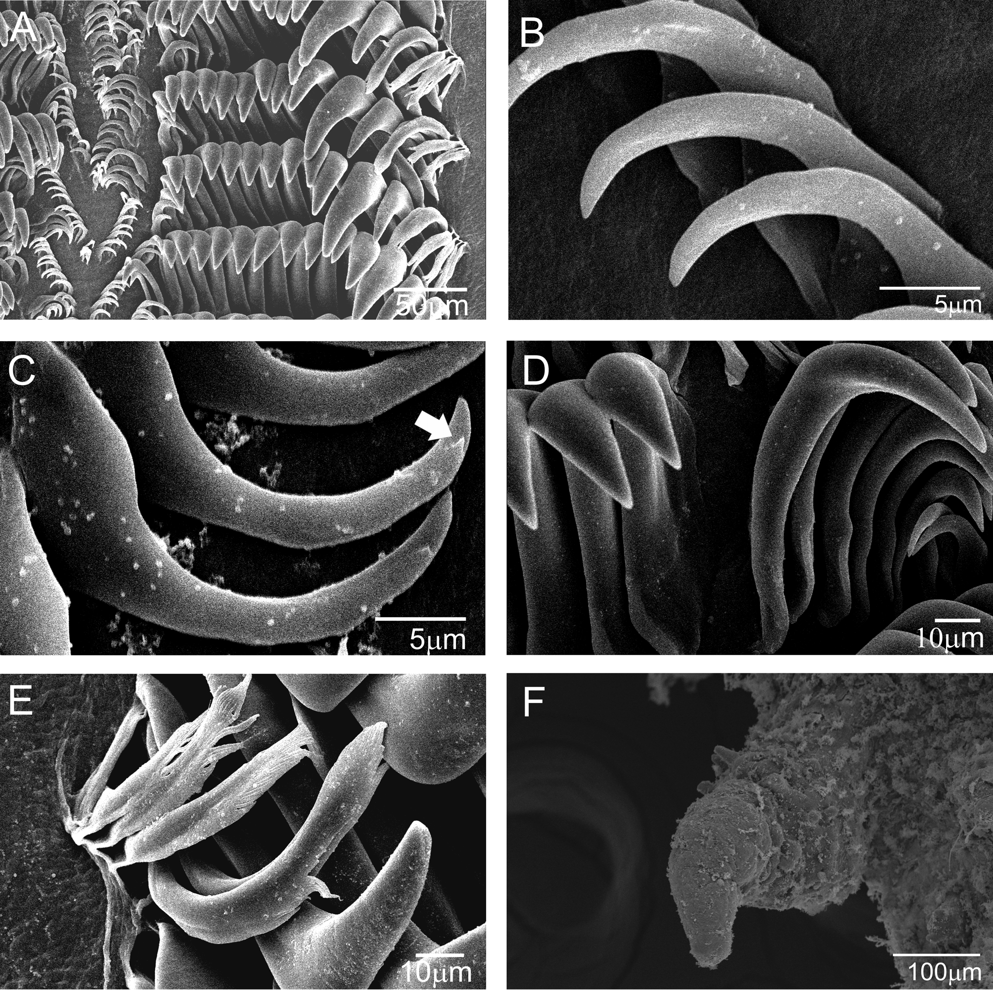
Labial cuticle and radula ( Figures 3 View FIGURE 3 A–E): Labial cuticle rigid and smooth. Radular formula 25 x 24.0. 24 in preserved specimen measuring 3.0 mm in length and 29 x 30.0. 30 in preserved specimen measuring 5.0 mm in length; lateral plates smooth and hook-shaped, larger and more developed in center of rows; lateral teeth divided in two size classes: first, smaller, comprising first to sixteenth tooth, these smaller teeth presenting one denticle at outer side of cusp; second, larger, without outer denticles. Five marginal teeth spatulate, smooth or pectinate.
Reproductive system ( Figure 4 View FIGURE 4 ): hermaphrodite duct connecting to short and slightly convoluted ampulla. Postampullary gonoduct very short that connects to oviduct and prostate. Prostate granular, divided into two parts, less dense part proximal, denser distal part. Distal portion of vas deferens thin, about same diameter of vagina, elongated, opening in common atrium with vagina. Vagina elongated and non-convoluted, with single accessory gland attached to its base; gland not visible in small animals (3.0 mm in length; Fig. 4 View FIGURE 4 B). Vagina opening into rounded bursa copulatrix, completely covered by prostate and situated above female gland mass. Bursa serially arranged, vaginal duct folding twice and connecting to rounded to bean-shaped, short-stalked seminal receptacle; uterine duct short. Bursa and receptacle of similar size.
Remarks. Thordisa diuda fits perfectly in the genus Thordisa , because this species presents all the diagnostic features of the genus, including the dorsum covered with soft, elongate tubercles ( Figs. 1 View FIGURE 1 A–3F); labial cuticle smooth; radula composed of simple, hamate teeth ( Figs. 3 View FIGURE 3 A–D); outermost lateral teeth multidenticulate ( Fig. 3 View FIGURE 3 F); reproductive system with a flattened, granular prostate having two well differentiated regions ( Figs. 4 View FIGURE 4 A–B); and there is one accessory gland ( Fig. 4 View FIGURE 4 A) (Valdés, 2002).
Thordisa diuda was described by Er. Marcus (1955) based on a single specimen from the coast of São Paulo state, Brazil, and subsequent publications expanded the known geographic distribution but did not add data on internal anatomy (Ev. Marcus, 1971; Thompson, 1980). Almost all of the major characteristics of specimens examined herein agree with the description made by Er. Marcus (1955), with the exception of the inner lateral teeth. Examining the radula of some specimens of Thordisa diuda , we observed one denticle on the outer surface of the first until the sixteenth lateral teeth ( Fig. 3 View FIGURE 3 C). This was not mentioned by Er. Marcus (1955) and Thompson (1980), who described all the lateral teeth as smooth. The denticles might have been overlooked initially because of their diminutive size (approximately 0.75 µm) ( Fig. 3 View FIGURE 3 C). The remaining teeth, which are larger in size, do not exhibit this denticle on the external surface ( Fig. 3 View FIGURE 3 D).
Chan & Gosliner (2007) mentioned that Thordisa oliva Chan & Gosliner, 2007 appears to be sister to Thordisa diuda because these two species share a unique apomorphy of having a pair of circular pits on the sides of the mouth. Thus, we would like to emphasize that the round button on a disc beside the mouth, as described by Er. Marcus (1955) and Chan & Gosliner (2007) were found in the specimens analyzed in this study.
Thordisa azmani Cervera & García-Gomes, 1989 from the Iberian Peninsula was distinguished from T. diuda by the presence of two accessory glands at the base of the vagina, in contrast to T. diuda , which has a single accessory gland (Er. Marcus, 1955). According to Ortea and Martínez (1990), who proposed synonymy between T. diuda and T. azmani , the single gland described by Er. Marcus (1955) was a misinterpretation. Ortea and Martínez (1990) suggested that the two glands appear united, giving the impression of being a single bilobed gland. Ortea and Cabrera (1999) reported the identification of T. diuda in Cape Verde, based on the presence of two accessory glands near the vagina of the specimens they examined.
The two sexually mature specimens dissected here are consistent with the description by Er. Marcus (1955) in having only one non-bilobed accessory gland near the vaginal opening ( Fig. 4 View FIGURE 4 A). Furthermore, there are other differences between T. diuda and T. azmani , for example in the number and color of branchial leaves, presence or absence of the denticle on the outer surface of the smaller class of lateral teeth, and rhinophoral and branchial coloration. Thus, we consider that T. diuda occurs in Brazil, Bahamas, and Jamaica (Valdés et al. 2006), but not in northern Spain (Ortea & Martínez, 1990), Iberian Peninsula (Cervera & García-Gomez, 1989), and Cape Verde (Ortea & Cabrera, 1999).
Thordisa lurca (Ev. Marcus & Er. Marcus, 1967b), originally described in the genus Nuvuca (Ev. Marcus & Er. Marcus, 1967b), was recorded from Brazil by Valdés et al. (2006) without specifying the exact location in Brazil or state in which collection this specimen was deposited. Thus, it is impossible compare with this material. Thordisa lurca only was described in its original description and according to that some different features to separate these two species were found. First, Ev. Marcus and Er. Marcus (1967b) do not metion the existence of a round button beside the mouth in T. lurca , that it is present in T. diuda ; T. lurca presents the tubercles of the notum with different sizes, larger and smaller ones mixed, while T. diuda presents tubercles larger in the central part of the mantle than along the mantle edge or on rhinophoral and branchial sheaths; T. lurca has the anterior border of the foot grooved and entire, not notched, while in T. diuda it is notched on the upper “lip”; T. lurca presents the inner lateral teeth smooth, while T. diuda presents the lateral teeth divided in two size classes: first, smaller, comprising first to sixteenth tooth, these smaller teeth presenting one denticle at the outer side of the cusp, while the second class of size is larger and teeth are smooth.


No known copyright restrictions apply. See Agosti, D., Egloff, W., 2009. Taxonomic information exchange and copyright: the Plazi approach. BMC Research Notes 2009, 2:53 for further explanation.