
Microporella genisii ( Audouin & Savigny, 1826 )
|
publication ID |
https://doi.org/ 10.5281/zenodo.207232 |
|
DOI |
https://doi.org/10.5281/zenodo.6184656 |
|
persistent identifier |
https://treatment.plazi.org/id/0387F320-FFE1-FFFE-D4AF-F94BFE38F90B |
|
treatment provided by |
Plazi |
|
scientific name |
Microporella genisii ( Audouin & Savigny, 1826 ) |
| status |
|
Microporella genisii ( Audouin & Savigny, 1826)
( Fig. 7 View FIGURE 7. M A–F, Table 4 View TABLE 4 )
Flustra View in CoL ? genisii Audouin, 1826 , p. 67 (1828); Savigny, 1817, pl. 9, fig. 5.1–2. Microporella ciliata: d’Hondt 2006 View in CoL , p. 42.
Microporella intermedia Aristegui, 1984 View in CoL , p. 321, fig. 67a–c, pl. 24, figs 1–3. Microporella orientalis View in CoL :? Tilbrook et al. 2001, p. 87, figs 19 C–D;? Sternhell et al. 2002, p. 229, fig. 4b. Not M. intermedia Livingstone, 1929 View in CoL : Gordon 1984, p. 102, pl. 38 C–D.
Material examined. Neotype: 2010-0005-0001 DPUV; large colony (ca 10 x 11 mm) including many ovicells, from South Sinai, Ras Mohammed, ‘Yolanda’ wreck, 18 m, 15 May 1983. Additional material examined: Lebanon: (1) Tripoli, Ramkine Island, 12 m, one colony on pottery debris, 22 October 1999; (2) Batroun, “Phoenician wall, 9 m, two colonies on serpulid tubes, 16 October 1999; (3) Jonnieh Bay, Aquamarina, 20–30 m, two colonies on stone, 10 July 2003; (4) Beirut, Harf el Kalb, 34 m, one colony on Spondylus shell, 21 October 1999; (5) Tyre (Sour), El Kasmieh, 36–42 m, two colonies on shell, 25 October 1999. Red Sea: (1) South Sinai, Ras Mohammed, ‘Yolanda’ wreck, 18 m, 10 colonies on aluminium plates, 15 May 1983; (2) Safaga Bay, transect B2, depth 42 m, one colony on Cymodocea sp.; (3) Safaga Bay, west part of Safaga Is., transect A5, 1– 2 m, 6 colonies on Cymodocea sp., September 1992.
Description. Colony small or medium-sized, unilaminar. Autozooids relatively small, mainly hexagonal or oval, longer than broad (in average, L/W = 1.3–1.4); frontal shield coarsely granular, perforated with 18–30 irregularly scattered pseudopores; marginal elongate areolae occasionally visible. Ascopore separated from proximal border of orifice by a distance shorter than orifice length, oval or slightly compressed distally; median process not very prominent in most cases or even absent, with 8–15 short denticles leaving a wide lumen; surrounded by a thick, prominent rim, frequently with a proximal bulge (particularly in Lebanese colonies and in Red Sea colonies collected on sea-grasses). Primary orifice wider than long; distolateral edge (anter) smooth; proximal border straight and crenulate with 8–16 low, flat or rounded beads, and without condyles at the corners. Oral spines slen- der, 4 in most cases, sometimes 3 or 5 (6 in zone of astogenetic change). Avicularium single, on right or left, sometimes missing, lateral or proximolateral to ascopore, orientated distolaterally or laterally; opesia moderate-sized; rostrum short, with narrow truncated tip; mandible elongated, lanceolate, shorter than autozooid width, guttershaped on lower side when dry, bearing 2 pointed lateral processes curved basally, which lean against rostrum tip. Ovicells often numerous and appearing early among post-ancestrular zooids ( Fig. 7 View FIGURE 7. M E), ooecium double-walled with membranous ectooecium visible in non-cleaned colonies; entooecium thickly calcified, globular, granular, perforated with tiny ‘pseudopores’, which may be filled in with calcification; proximal edge bordered by smooth rim. Maternal zooids with personate peristome formed by arched collar rising vertically close to ascopore and sometimes hiding it partially, fused side by side with proximal rim of ooecium; 1–2 oral spines often visible at corners where ooecium and proximal collar meet. In some colonies from Safaga Bay, Red Sea, ovicells with either fully or partially developed or even missing proximal collar were co-occurring. Ancestrula tatiform with 11 or 12 spines and narrow cryptocyst, budding 2 distal autozooids.
Remarks. The beautiful Savigny drawing (pl. 9, fig. 5.2) of the Red Sea Microporella species named Flustra ? Genisii by Audouin (1826) clearly shows personate ovicells, three to five oral spines, a single avicularium placed proximolaterally to ascopore and at a variable distance to it, directed distolaterally with long and narrow mandible, persistent and well visible; a round or oval ascopore, sometimes with median distal process and peripheral ring; conspicuous frontal nodules (or pseudopores?), round ovicell, slightly broader than long, maternal zooid with a personate collar adjacent to ascopore, vertical and relatively narrow, not completely fused with the ooecium rim but leaving a narrow suture. The close agreement in most respects between Flustra ? Genisii and the Microporella specimens described here from Lebanon and the Red Sea argues for considering the former as a valid Microporella species and for referring the latter to it. A neotype has thus been selected among the Red Sea material. The only differences concern the crenulation of the proximal edge of the orifice and the paired spines at the proximal corners of the ooecium, which are missing in Savigny’s drawing. However, these tiny morphological characters were not observable with the optical means available in the early 19th century.
Flustra View in CoL ? Genisii was synonymized by Harmer (1957) with Microporella ciliata (Pallas, 1766) View in CoL , the type species of Microporella View in CoL , without comment. This opinion was already expressed by Hastings (1927, p. 242) and followed by d’Hondt (2006) in his review of the bryozoan species illustrated by Savigny. Microporella View in CoL species with a single avicularium proximolateral to the ascopore and having a setiform mandible have often been ascribed to M. ciliata View in CoL , to which a cosmopolitan distribution was often wrongly attributed. Recently, a neotype of M. ciliata View in CoL was chosen by Kuklinsky and Taylor (2008) among specimens collected at Naples. The features of M. ciliata View in CoL , now fixed by this neotype designation, differ from those shown by specimens of M. ciliata View in CoL commonly present in the Mediterranean and Adriatic (e.g. Gautier 1962; Hayward & McKinney 2002) or in British waters (e.g. Hayward & Ryland 1999). It also clearly differs from M. genisii , particularly in having orifices with the proximal edge finely serrated between distinct condyles and bordered by 1–4 oral spines.
The Sinai and Safaga Bay specimens of M. genisii differ from the Lebanese specimens in having a larger cystid size (mean AzL and AzW are respectively 24% and 33% larger in Sinai, and 14% and 11% larger in Safaga Bay) and a higher frequency of occurrence of one or two spines at the proximal corners of the ovicell. The other features, such as the number of spines, the ornamentation of the proximal side of the orifice, the avicularium, the personate ovicell and the ancestrula, are similar.
Aristegui (1984) described in his unpublished thesis a new species of Microporella View in CoL from Tenerife, Canary Islands — M. intermedia View in CoL sp. nov. — that he considered intermediate between M. ciliata View in CoL and M. orientalis ( Harmer, 1957) View in CoL . This species name is a junior primary homonym of M. intermedia Livinstone, 1929 View in CoL , from New Zealand, which clearly differs (cf. Gordon 1984) from the Canarian species in many characters. Thus, according to the Principle of Priority, M. intermedia Aristegui, 1984 View in CoL is invalid. We consider that this Canarian species is conspecific with M. genisii from Lebanon, South Sinai and Safaga Bay redescribed here. The identity of the specimens from the Canaries with the Lebanese and Red Sea specimens is attested by the following characters: four oral spines, one or two remaining visible in the ovicell corners; ascopore circular, oval or reniform, surrounded by a bulging ring; avicularia single, located below the ascopore, orientated distolaterally with an angle <45, sometimes missing (cf. Aristegui 1984: pl. 24-2), with a narrow-lanceolate, gutter-shaped mandible moderately long, bearing two lateral processes on the lower side ( Aristegui 1984: fig. 67-c); ovicells personate ( Aristegui 1984, fig. 67a, pl. 24-1–2), ancestrula tatiform with 11 spines and narrow cryptocyst. Aristegui did not notice any crenulation of the proximal edge of the primary orifice. However, this apparent difference between the Lebanese and Red Sea specimens may not be real as these beads are low and not easily visible.
It is likely that M. genisii has been confused with other personate Microporella View in CoL species with a single avicularium, particularly M. orientalis Harmer, 1957 View in CoL . The species growing on artificial substrata at Eilat (Red Sea, Gulf of Aqaba) recorded as M. orientalis View in CoL by Sternhell et al. (2002) presents the general aspect of M. genisii based on the following characters: appearance of the cryptocystal walls; shape and position of the single avicularium; and shape and proportions of personate collar and ovicell, including a pair of spines visible at the proximal corners of each ovicell. Unfortunately, the number of oral spines of non-ovicellate zooids and the ornamentation of the proximal border of the orifice are not visible in the SEM photo of this specimen. Also, both photos and description of specimens from Vanuatu Islands ascribed to M. orientalis View in CoL by Tilbrook et al. (2001) strongly suggest that they belong to M. genisii .
The description of M. epihalimeda Tilbrook, 2006 View in CoL from the Solomon Islands indicates that it is closely related to M. genisii . Both species have 3–4 spines, similarly shaped avicularia and maternal zooids, a tatiform ancestrula with 11 spines and a primary orifice without condyles. In M. epihalimeda View in CoL the primary orifice is described as smooth but the SEM illustration ( Tilbrook 2006, pl. 46D) shows a rather uneven proximal edge, resembling that of M. genisii although less rippled. Another similarity is the oral spines seen at the proximal corners of the personate ovicells — although “none visible in ovicellate zooids” is mentioned in Tilbrook’s description (p. 213). The only apparent difference is that the setiform avicularian mandible has a length equal to zooidal width and no basal processes in M. epihalimeda View in CoL .
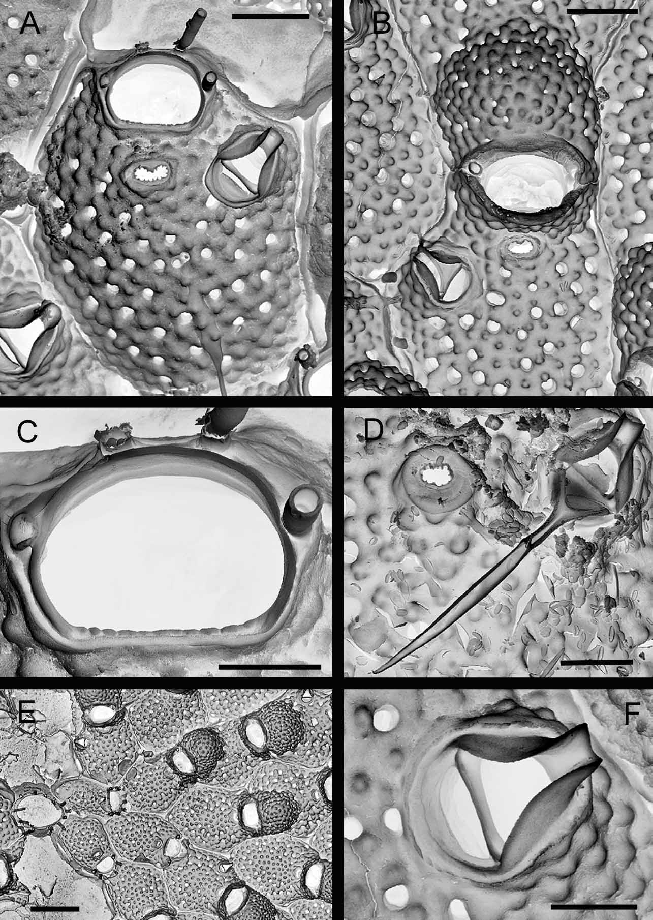
TABLE 4. Morphometrics (in µm) of specimens of M. genisii from Lebanon (5 localities), Ras Mohammed and Safaga Bay. Length (L) and width (W) of autozooid (Az), ovicell (Ov), primary orifice (Or) and avicularium mandible (Md). Mean standard deviation, range and number of measurements (in brackets).
| Lebanon | S Sinai | Safaga |
|---|---|---|
| AzL 383.5 ±31.6 | 475.8± 33.9 | 436.7 ±25.3 |
| - 320–440 (23) | 440–530 (18) | 380–490 (27) |
| AzW 280.0 ±34.7 | 372.6 ±51.9 | 309.6± 28.2 |
| - 245–400 (23) | 295–485 (18) | 250–380 (27) |
| OvL 186.3 ±12.5 | 182.2 ±9.5 | 192.2± 11.4 |
| - 170–205 (17) | 170–200 (16) | 170–220 (38) |
| OvW 225.0 ±21.6 | 210.9 ±12.7 | 217.1± 11.4 |
| - 195–265 (17) | 180–230 (16) | 200–250 (38) |
| OrL 58.8 ±4.4 | 70.6 ±2.7 | 61.3± 3.0 |
| - 50–65 (17) | 65–72 (7) | 60–70 (15) |
| OrW 91.1 ±7.8 | 111.5 ±5.3 | 94.3± 5.6 |
| - 80–108 (17) | 105–120 (14) | 90–105 (15) |
| MdL 155.8± 13.2 | 181.8± 17.7 | 200.4 ±10.7 |
| - 145–180 (6) | 160–220 (16) | 190–220 (13) |
No known copyright restrictions apply. See Agosti, D., Egloff, W., 2009. Taxonomic information exchange and copyright: the Plazi approach. BMC Research Notes 2009, 2:53 for further explanation.
|
Kingdom |
|
|
Phylum |
|
|
Class |
|
|
Order |
|
|
Family |
|
|
Genus |
Microporella genisii ( Audouin & Savigny, 1826 )
| Harmelin, Jean-Georges, Ostrovsky, Andrew N., Cáceres-Chamizo, Julia P. & Sanner, Joann 2011 |
Microporella ciliata: d’Hondt 2006
| d'Hondt 2006 |
M. epihalimeda
| Tilbrook 2006 |
Microporella intermedia
| Aristegui 1984 |
M. intermedia
| Aristegui 1984 |
M. orientalis (
| Harmer 1957 |
M. orientalis
| Harmer 1957 |
M. intermedia
| Livingstone 1929 |
M. intermedia
| Livinstone 1929 |
genisii
| Audouin 1826 |
Microporella ciliata
| Pallas 1766 |