
Microporella coronata ( Audouin & Savigny, 1826 )
|
publication ID |
https://doi.org/ 10.5281/zenodo.207232 |
|
DOI |
https://doi.org/10.5281/zenodo.6184654 |
|
persistent identifier |
https://treatment.plazi.org/id/0387F320-FFE5-FFFD-D4AF-FE53FD99FD1B |
|
treatment provided by |
Plazi |
|
scientific name |
Microporella coronata ( Audouin & Savigny, 1826 ) |
| status |
|
Microporella coronata ( Audouin & Savigny, 1826) View in CoL
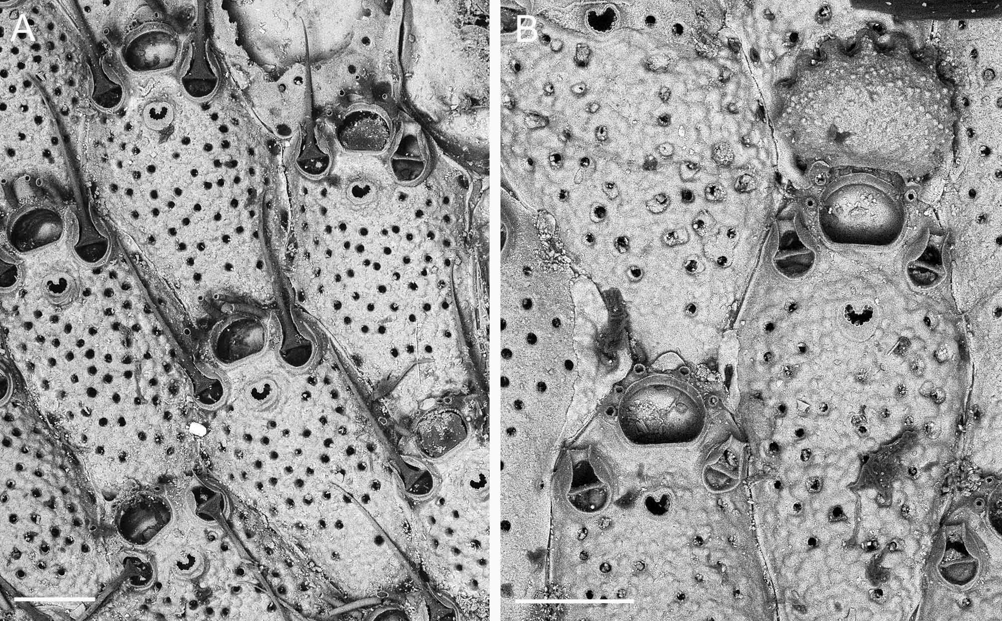
( Figs 4 View FIGURE 4. M A–E, 5, Table 3)
Flustra coronata Audouin, 1826 , p. 67 (1828); Savigny, 1817, pl. 9, fig. 6.
Flustra umbracula Audouin, 1826 , p. 67 (1828); Savigny, 1817, pl. 9, fig. 7.
Microporella coronata: Balavoine 1959 View in CoL , p. 274, pl. 3, fig. 7-8; d’Hondt 2006, p. 44;? Norman 1909, p. 297, pl. 39, fig. 4;? Gautier 1962, p. 173;? Zabala 1986, p. 513–514, fig. 180.
Microporella ciliata View in CoL var. coronata :? Hastings 1930, p. 727.
Microporella umbracula: Harmer 1957 View in CoL , p. 964;? Di Geronimo et al. 1998, p. 250 ( Tab. 1 View TABLE 1 );? Koçak et al. 2002, p. 236 ( Tab. 1 View TABLE 1 );? Morri et al. 1999, p. 733 ( Tab. 1 View TABLE 1 );? Nicoletti et al. 1995, p. 398 ( Tab. 1 View TABLE 1 ).
Not Microporella coronata: Waters 1909 View in CoL , p. 142, pl. 12, figs 6–9 (= Microporella View in CoL n. sp., see below); Osburn 1952, p. 386 (= Microporelloides coronula Soule, Chaney & Morris, 2003 View in CoL ).
Not Microporella ciliata View in CoL var. coronata: Hastings 1927 , p. 340, figs 83–84 (= Microporella View in CoL n. sp., see below).
Not Microporella umbracula: Balavoine 1959 View in CoL , p. 274, pl. 5, fig. 3 (= Predanophora longiuscula View in CoL ).
Not Microporella umbracula: Winston 1982 View in CoL , p. 150, fig. 83; Winston 1986, p. 21, fig. 49; Winston & Håkansson 1986, p. 29, figs 68–69; Aristegui 1984, p. 331, fig. 68a,b, pl. 25, figs 3–4 (= Microporella View in CoL n. sp., see below).
Material examined. Neotype: 2010-0004-0001 DPUV; part of an uncleaned colony comprising ca. 26 zooids with 4 ovicells. Lebanon, Kafar Abida, overhang, 7–8 m, 30 May 2000. Other material examined: Lebanon: (1) Tripoli, Ramkine Island, cave wall, 5–7 m, 14 July 2003; (2) Anfey, pebbles, 14 m, 26 October 1999; (3) Chak El Hatab, cave wall, 12 m, 21 September 2002; (4) Kafar Abida, overhangs, on scleractinians ( Phyllangia , Polycyathus ), 9 m, 6 August 1995; (5) Kafar Abida, overhangs, 7–8 m, 30 May 2000; (6) Tyre (Sour), pebbles, 14 m, 26 October 1999. Balavoine Collection: Red Sea, Gulf of Suez, MNHN n 7758, Al Sayad Stn XI, labelled Microporella coronata ( Audouin, 1826) ; MNHN n 7808, Al Sayad Stn X, labelled Microporella umbracula ; MNHN n 7803, Al Sayad Stn X, labelled Microporella umbracula .
Description. Colony small, unilaminar. Autozooids hexagonal or less frequently quadrangular, longer than broad. Frontal shield moderately convex, evenly covered with rounded nodules and 50–60 small frontal pseudopores; 1–3 marginal larger pores occasionally visible. Ascopore relatively close to proximal edge of orifice, i.e. at a distance about half orifice length, crescentic, with rounded median process and C-shaped lumen, both spurred with small spines, and surrounded by low rim with bulge developing proximally. Primary orifice broader than long, with smooth, rounded distal edge and proximal edge slightly concave, smooth, with low step-shaped condyles at corners. Oral spines 7 in most cases (59%), less frequently 6 (22%), rarely 8 (2%). Avicularia paired in most zooids, sometimes single, particularly in periancestrular zooids, placed symmetrically on both sides of zooid at level of ascopore or a little more distally, directed distally or distolaterally with a small angle; rostrum small, truncated, cross-bar complete; proximal area rounded, moderately broad; mandible setoid, slender, as long as zooid length or a little shorter, with lower side gutter-shaped and bearing a pair of triangular pointed processes corresponding to the rostrum tip. Ovicell prominent, rounded, slightly broader than long (L/W = 0.85 in average), nodular and ‘pseudoporous’ like the frontal shield; proximal edge of ectooecium calcified, forming a thick visor overhanging proximal, smooth edge of entooecium; a raised peristomial collar including ascopore fused laterally to ooecium proximal corners and presenting two lateral flaps that can join in midline when particularly developed, forming bridge over orifice, or can be reduced to a lower prominence; 2 oral spines occasionally visible against junction between vizor and collar. Ancestrula tatiform, oval, with proximal gymnocyst moderately developed, the large oval opesia bordered with 10 spines (5 distal, 2 median, 3 proximal); cryptocyst narrow, developed both proximally and distally. Two autozooids distally budded by ancestrula, the smaller with 8 spines, the larger with 7 spines, both with single avicularium placed proximolateral to ascopore.
AzL: 582.1± 67.2, 460–730 (17)
AzW: 403.5± 60.5, 315–535 (17)
OvL: 256.4± 20.3, 220–290 (11)
OvW: 303.5 ±17.3, 280–340 (11)
OrL: 97.7± 10.5, 85–112 (11)
OrW: 126.2 ±11.1, 110–140 (11)
MdL: 403.7± 54.0, 315–510 (15)
Remarks. Savigny’s drawings of two specimens from the Red Sea, one without ovicells ( Savigny 1817, plate 9, fig. 6.1-2) and the other with many ovicells ( Savigny 1817, plate 9, fig. 7.1-4), undoubtedly show the same species ( Fig. 5 View FIGURE 5 ). Audouin (1826), however, gave two different species names to these specimens, coronata and umbracula for Savigny’s figures 6 and 7 respectively. As stated by d’Hondt (2006), the name coronata , cited before umbracula in Audouin’s text, has priority and must be used. The specific name umbracula has often been preferred to coronata (e.g. Harmer, 1957) because of the illustration of ovicells. The non-ovicellate zooids of both specimens illustrated by Savigny clearly show zooids with six oral spines and paired avicularia with long setoid mandibles while the ovicellate zooids present the same avicularia and personate ovicells. The occurrence in this species of a particular peristomial structure in maternal zooids was noticed by Busk (1854, p. 74) when describing Lepralia personata : “The form of the ovicell in F. (L.) umbracula ... is very similar apparently to that of L. personata ....”. Personate ovicells with a collar presenting lateral flaps that can fuse into a bridge is also a typical feature of M. pontifica Osburn, 1952 , redescribed by Soule et al. (2003). This particular structure can occasionally be found in British and Dutch specimens attributed to M. ciliata by Hayward and Ryland (1999, fig. 136D) and De Blauwe (2009, p. 384, Foto 414). It also characterizes a Canarian species, M. cooki , described by Aristegui (1984) in his unpublished thesis, which differs from M. coronata in having a personate collar that does not surround the ascopore and avicularia that are generally single and in a more proximal position.
In the present collection, specimens of M. coronata were recorded at six localities along the coast of Lebanon, from the north (Tripoli) to the south (Tyre). The ascription of these Lebanese specimens to M. coronata is based on the occurrence of paired avicularia with long setoid mandibles directed distally and placed in the distal quarter of the autozooid, orifices with 6–7 spines, smooth distal and proximal borders, and personate ovicells with lateral flaps that can fuse into a bridge over the orifice and the ascopore.
Considering that the collection illustrated by Savigny (1817) was most likely lost (d’Hondt 2006) and that both coronata and umbracula species names have been improperly attributed in many cases, leading to wrong information concerning geographic and stratigraphic distribution, a neotype of M. coronata has been chosen from the present Lebanese collection. The choice of a Lebanese specimen for neotype designation, despite the asserted Red Sea origin of the specimens illustrated by Savigny (see Audouin 1828, p. 67) is justified by good morphological congruence and the existence of faunal exchanges between the two basins (see below).
Microporella coronata View in CoL was listed several times as M. umbracula View in CoL in faunal inventories from warm regions of the Mediterranean ( Turkey: Nicoletti et al. 1995; Ionian Sea: Di Geronimo et al. 1998; Milos Is.: Morri et al. 1999; Cyprus: Koçak et al. 2002). According to Hastings (1927), Waters (MS) had also recorded M. coronata View in CoL from the Mediterranean. However, although this is very probable, the distribution of M. coronata View in CoL or of another species with paired avicularia in the eastern Mediterranean cannot be ascertained from these records without comment or figures. Similarly, the fossil records of M. coronata View in CoL from Miocene to Pleistocene assemblages around the Mediterranean (e.g. Rosso 1987; Moissette 1988) are not easy to interpret and may correspond to other species ( Berning 2006), such as that recorded by Hastings (1927) (see below). Zabala (1986, pp. 513–514, fig. 180) recorded M. coronata View in CoL from the western Mediterranean but without personal collected material or a precise Mediterranean reference. His description and figure (copied from Osburn 1952, pl. 45, fig, 1, Bay of California) indicate 5–6 oral spines, paired avicularia with long setoid mandibles directed distolaterally and non-personate ovicells with a few ‘pores’ identical to the pseudopores of the zooidal frontal shield.
Microporella coronata View in CoL was recorded from Madeira by Norman (1909, p. 297) and M. umbracula View in CoL from the Canary Isles by Aristegui (1984, p. 331). Norman’s description and illustration of M. coronata View in CoL depict a species with six oral spines and paired avicularia with long setoid mandibles directed distally. These characters may correspond to Audouin’s species, but curiously the proximal area of the avicularia was illustrated as having the same pointed shape as the rostrum. Also the ovicell presents no lateral flaps. Canu and Bassler (1928, p. 112, pl. 34, fig. 5), without justification that the Madeiran material differed from M. coronata View in CoL , erected a new species, M. normani View in CoL . Paradoxically they illustrated it with a Pliocene specimen from Panama bearing single avicularia. This species group also includes species from Florida and Jamaica ( Winston 1982, 1986; Winston & Håkansson 1986) recorded as M. umbracula View in CoL , which have non-personate ovicells, a smaller number of oral spines and zooids commonly with a single avicularium. Microporella lunifera (Haswell) View in CoL , redescribed by Hayward and Ryland (1995) from the Great Barrier Reef, may belong to the same species group having paired avicularia with long, setiform mandibles, placed distally and directed distolaterally, and non-personate ovicells. The record of M. coronata View in CoL from Marmar (Red Sea, south of Jeddah) by Gautier (1962) is disputable as the description without illustration mentions 5–6 oral spines with a dark base and does not indicate a peristome associated with the ovicell. In Balavoine’s collection from the Gulf of Suez, specimen MNHN n 7803 labelled Microporella umbracula View in CoL is M. harmeri View in CoL . Another specimen labelled Microporella umbracula View in CoL in the same collection (MNHN n 7808, Al Sayad Stn X, 28–62 m), recorded as Microporella umbracula View in CoL and figured by Balavoine (1959, p. 274, pl. 5, fig. 3), is not a Microporella View in CoL but Predanophora longiuscula ( Harmer, 1957) View in CoL .
The Albatross collections of bryozoans from the Philippine Archipelago (Canu & Bassler 1929), kept at the USNM, Washington, include an uncleaned specimen (Albatross Stn 5141) labelled Microporella View in CoL that resembles M. coronata View in CoL . SEM photos of this specimen show paired avicularia with robust setiform mandibles, six oral spines, a crescentic, spinous ascopore with rounded median process placed close to the proximal border of the orifice and encircled by a broad plate-shaped rim with a smooth surface and the edges raised distally. This Microporella View in CoL , in which the ovicells are incompletely developed, differs from M. coronata View in CoL at least in having not step-shaped condyles at the corners of the primary orifice.
Specimens collected in the Suez Canal near its Red Sea entrance (Km. 157), identified by Hastings (1927) as M. ciliata View in CoL var. coronata , clearly belong to another species despite the occurrence of paired distal avicularia with setoid mandibles. SEM photos (courtesy of M. Spencer Jones and J. Scholz) of these specimens ( Fig. 6 View FIGURE 6. M ), in the Natural History Museum, London (n 1926.9.6.238), show that they differ from M. coronata View in CoL in the following characters: four oral spines in most cases (range 3–6) in non-ovicellate zooids, two (sometimes three) remaining free at the proximal corners of the ovicells; a primary orifice with smooth edges but without step-like condyles; a frontal shield with large pseudopores; paired avicularia with truncate rostra and proximal area clearly broader than in M.
coronata View in CoL , with robust setoid mandibles; and ovicells with finely nodular calcified entooecium without ‘pseudopores’, encircled by 9–14 notches corresponding to marginal pores. The peculiarity of the ovicells with large marginal pores in these specimens was already noticed by Berning (2006, p. 104). The same combination of typical features occurs in a specimen from Suez docks figured by Waters (1909, pl. 12, figs 6–7) and recorded as M. coronata View in CoL : four oral spines in non-ovicellate zooids, absence of personate collar, paired avicularia with robust mandibles, ovicell with indented border and a pair of oral spines at the proximal corners. The same type of ovicell was present in a specimen from the Gulf of Suez (MNHN 7758, Al Sayad Stn XI) recorded as Microporella coronata View in CoL by Balavoine (1959, p. 274, pl. 3, figs 7–8). Similarly, specimens from the Canary Islands ascribed to M. umbracula by Aristegui (1984, pp. 331–332, figs 68a–b, pl. 25, figs 3–4) are also characterized by 4–6 oral spines in non-ovicellate zooids and ovicells without personate structure and ‘pseudopores’ and encircled by particularly large marginal pores.
The particular features of these specimens justify their placement in a new species within the M. coronata View in CoL species group. We propose to name it Microporella hastingsae n. sp., in honour of Anna B. Hastings, with specimen NHM n 1926.9.6.238 as holotype.


| MNHN |
Museum National d'Histoire Naturelle |
No known copyright restrictions apply. See Agosti, D., Egloff, W., 2009. Taxonomic information exchange and copyright: the Plazi approach. BMC Research Notes 2009, 2:53 for further explanation.
|
Kingdom |
|
|
Phylum |
|
|
Class |
|
|
Order |
|
|
Family |
|
|
Genus |
Microporella coronata ( Audouin & Savigny, 1826 )
| Harmelin, Jean-Georges, Ostrovsky, Andrew N., Cáceres-Chamizo, Julia P. & Sanner, Joann 2011 |
Microporelloides coronula
| Soule, Chaney & Morris 2003 |
Microporella umbracula:
| Winston 1982 |
Microporella coronata:
| Balavoine 1959 |
Microporella umbracula:
| Balavoine 1959 |
Microporella umbracula:
| Harmer 1957 |
Predanophora longiuscula (
| Harmer 1957 |
var. coronata:
| Hastings 1927 |
Microporella coronata:
| Waters 1909 |
Flustra coronata
| Audouin 1826 |
Flustra umbracula
| Audouin 1826 |