
Microcambeva jucuensis, Costa & Katz & Mattos & Rangel-Pereira, 2019
|
publication ID |
https://doi.org/10.1080/00222933.2019.1669729 |
|
DOI |
https://doi.org/10.5281/zenodo.3664997 |
|
persistent identifier |
https://treatment.plazi.org/id/03887322-FFEF-FFBE-FE66-D4D9DBE6FB77 |
|
treatment provided by |
Valdenar |
|
scientific name |
Microcambeva jucuensis |
| status |
sp. nov. |
Microcambeva jucuensis sp. nov.
( Figures 5–6 View Figure 5 View Figure 6 )
Holotype
UFRJ 12124, 27.2 mm SL; Brazil: Estado do Espírito Santo: Município de Viana: stream close to Nova Campo Grande village , Rio Jucu basin, 20.337°S, 40.449°W, altitude about 25 m asl; A. Katz, F. Pereira and J.L. Mattos, 15 July 2016. GoogleMaps
Paratypes
UFRJ 11083, 18 ex., 19.3–27.1 mm SL ; UFRJ 11011 , 4 ex., 19.1–22.3 mm SL ; UFRJ 11840 , 4 ex., 21.1–22.0 mm SL (C&S) ; CICCA 03524 , 2 ex., 19.2–20.9 mm SL ; all collected along with holotype.
Diagnosis
Microcambeva jucuensis differs from all other congeners, except M. draco and M. mucuriensis , by the presence of a distal widening on the posterior process of the autopalatine (vs absence) and a rudimentary anterior autopalatine ossification, consisting of a minute thin ossification, with its length about one-fourth of the width of the anterior cartilage head of autopalatine (vs ossification well developed in M. barbata , its length nearly equal to the width of the anterior cartilage head of autopalatine; autopalatine ossification absent in M. ribeirae ). Microcambeva jucuensis differs from M. draco and M. mucuriensis in having more opercular odontodes (13–15 vs 9–12) and by the presence of a robust interopercle, with a rudimentary anterior process (vs a thin interopercle, with a well-developed anterior process).
Description
Morphometric data appear in Table 2. Body slender, subcylindrical and slightly depressed anteriorly, compressed posteriorly. Greatest body depth at vertical line just in front of pelvic-fin base. Dorsal and ventral profiles of head and trunk slightly convex, approximately straight on caudal peduncle. Anus and urogenital papilla in vertical line through posterior third of dorsal-fin base. Head narrow, sub-triangular in dorsal view. Anterior profile of snout convex in dorsal view. Eye elliptical, dorsally positioned in head. Posterior naris slightly nearer to anterior naris than to anterior margin of orbit. Tip of maxillary barbel reaching base of opercular patch of odontodes; tip of rictal barbel reaching middle part of interopercular patch of odontodes; tip of nasal barbel reaching between anterior margin of orbit and eye centre. Mouth subterminal. Chin region with paired finger-like projections. Jaw teeth conical, arranged in two series: 19 or 20 teeth on premaxilla, 13–15 on dentary. Branchial membrane attached to isthmus only at its anterior point. Opercular odontodes 13–15, interopercular odontodes 8 or 9. Dorsal surface of neurocranium with broad lozenge-shaped fontanelle between frontals and anterior portion of sesamoid supraorbital. Branchiostegal rays 6.
Dorsal and anal fins subtriangular; dorsal-fin rays ii + 6 + 1i, anal-fin rays ii + 4 + I; rudimentary unsegmented ray immediately posterior to both dorsal- and anal-fin origin; anal-fin origin in vertical line posterior to dorsal-fin base. Caudal fin slightly forked, ventral portion slightly longer than dorsal portion; principal caudal-fin rays 13 (i + 11 + i), dorsal procurrent rays v–vii, ventral procurrent rays iv–vi. Pelvic fin slightly pointed, its tip reaching vertical line through middle of dorsal-fin base, pelvic-fin bases medially separated by interspace about three-quarters pelvic-fin base width; pelvic-fin rays i + 4. Pectoral fin subtriangular in dorsal view, first pectoral-fin ray terminating in short filament reaching about 5% of pectoral-fin length without filament; pectoral-fin rays i + 6. Dorsalfin origin on vertical line through vertebra 15 or 16, anal-fin origin on vertical line through vertebra 21, pelvic-fin insertion on vertical line through vertebra 12 or 13. Vertebrae 34. Ribs 3–4.
Mesethmoidal region and adjacent structures ( Figure 6 View Figure 6 (a)). Anterior margin of mesethmoid nearly straight, mesethmoid cornua narrow, rod-like. Antorbital and sesamoid supraorbital rod-like, sesamoid supraorbital slightly longer than antorbital. Premaxilla slender, subtriangular, bearing prominent sharp lateral process. Maxilla slen- der, laterally terminating in long and sharp process. Autopalatine elongate, lateral margin nearly straight, medial margin concave; width of middle portion about three-quarters of autopalatine cartilage width; latero-posterior process of autopalatine slender and long, about half autopalatine length without latero-posterior process, with subterminal widening creating arrow shape in dorsal view; cartilaginous head of autopalatine prominent, its length about one-third of autopalatine length without latero-posterior process; anterior autopalatine ossification rudimentary, scale-like.
Suspensorium and opercular apparatus ( Figure 6 View Figure 6 (b)). Metapterygoid minute, subtriangular, its greatest length about half length of antero-dorsal portion of quadrate. Quadrate slender, its length about 60% length of hyomandibula without anterior process, its depth about one-quarter total length of quadrate; dorsoposterior process rudimentary or absent. Hyomandibula with narrow, pointed anteriorly directed process, its length about 70% hyomandibula longitudinal length excluding process, its tip anteriorly reaching vertical line through anterior fifth of quadrate length. Opercle robust, odontode patch width about three-quarters of width of dorsal portion of hyomandibula. Interopercle compact, width of distal portion of odontode patch about two-thirds of width of dorsal portion of hyomandibula; anterior process rudimentary.
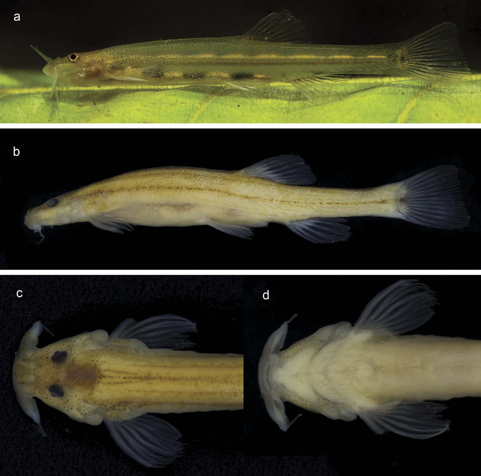
Colouration in life ( Figure 5 View Figure 5 (a)). Trunk and head almost translucent, with minute dark orange chromatophores on superficial layer of skin. Small superficial melanophores arranged in longitudinal zones, along mid-lateral, dorso-lateral and mid-dorsal portions of trunk, and scattered on post-orbital area and central portion of dorsal surface of head. Internal dermal layer of trunk with row of alternating elongate dark grey and pale orangish-yellow spots along mid-lateral line of body; another similarly coloured row between pectoral-fin base and anus. Dark grey stripe between nasal barbel and orbit. Iris pale yellow, with small superficial melanophores. Fins hyaline, with dark chromatophores concentrated on basal portion of dorsal and caudal fins forming small black spots.
Colouration in alcohol. After fixation, specimens became opaque light yellowish grey. Superficial chromatophores still visible.
Distribution and habitat notes
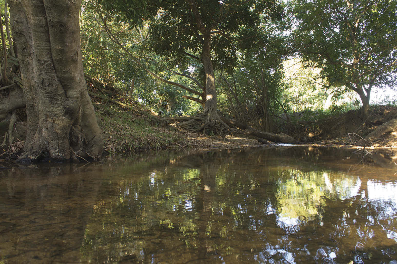
Microcambeva jucuensis was collected in a single locality (20°20 ʹ 12 ʺ S, 40°26 ʹ 55 ʺ W; Figure 3 View Figure 3 ), in a small clearwater stream, about 5 m wide at the collection site. The stream is a sub-tributary of the Rio Jucu, which with a course of about 180 km forms an isolated basin in eastern Brazil. The type locality area is in a highly deforested area close to a vast urban area and about 15 km from the sea. At the collection site, the river margin was protected by some remnant trees ( Figure 7 View Figure 7 ). The water was clear, and it was possible to see specimens of M. jucuensis swimming over the sandy substrate in sunlight, at a maximum depth of 50 cm.
Etymology
The name jucuensis is an allusion to the occurrence of the new species in the Rio Jucu basin.


No known copyright restrictions apply. See Agosti, D., Egloff, W., 2009. Taxonomic information exchange and copyright: the Plazi approach. BMC Research Notes 2009, 2:53 for further explanation.
|
Kingdom |
|
|
Phylum |
|
|
Class |
|
|
Order |
|
|
Family |
|
|
Genus |