
Craspodema reflectans Gerlach, 1964
|
publication ID |
https://doi.org/ 10.11646/zootaxa.3972.3.6 |
|
publication LSID |
lsid:zoobank.org:pub:A49A0FD5-FBBB-459F-AFF4-27CB8ADFA945 |
|
DOI |
https://doi.org/10.5281/zenodo.6112534 |
|
persistent identifier |
https://treatment.plazi.org/id/03888797-7906-6A62-61A7-FDA3F31BFEE6 |
|
treatment provided by |
Plazi |
|
scientific name |
Craspodema reflectans Gerlach, 1964 |
| status |
|
Craspodema reflectans Gerlach, 1964
( Figs 1–5 View FIGURE 1 View FIGURE 2 View FIGURE 3 View FIGURE 4 View FIGURE 5 ; Table 1)
Material examined. One adult male, one adult female and a juvenile. The specimens were collected by Giuseppe Baldelli on March 2009 during an ecological survey in the inner lagoon of the Suvadiva atoll. In detail, the male was collected at Dhevadhoo, the female and juvenile at Funandhoo. Overall, the sites were medium-coarse sands characterized by the following sediment fractions: gravel from 4.6 to 2.5%; sand from 92.7 to 93.8% and pelite from 2.7 to 3.7%. Both sites were at 61 m depth.
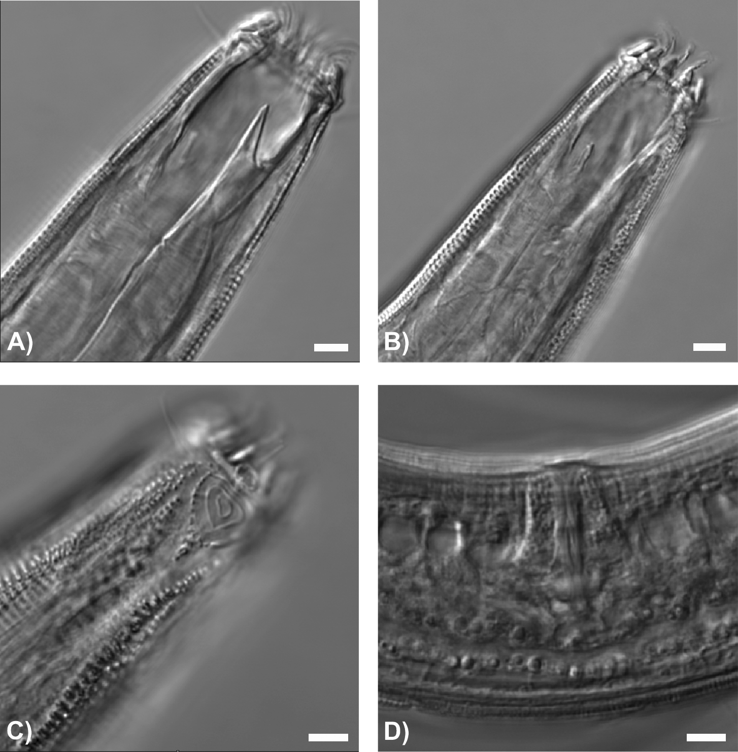
Description. Male. Body slender and regularly attenuated toward the tail. Cuticle annulated, with transverse rows of puntactions. Lateral differentiation begins at level of the amphideal fovea and consists of two prominent rows of enlarged punctations. Additional lateral fields (8 in total) arise between them at about 45 µm from anterior body end and extend to the tail region. Cephalic setae arranged in two circles. Six labial setae (respectively, the sub-median and lateral setae are 3.5 and 8 µm long) and ten (6+4) cephalic setae, 12 µm long. Amphideal fovea based on a plaque, anterior border of fovea located at 9 µm from anterior body end. Amphideal fovea spiral (3.5 turns), hand-mirror shaped, occupying about 60% of corresponding body diameter. Buccal cavity deep (about 32 µm in length). Cheilostoma with twelve conspicuous and well-distinct cheilorhabdia. Pharyngostoma with a welldeveloped dorsal tooth (24 µm long) and two smaller and equal subventral teeth (11 µm long). Pharynx cylindrical, ca 268 µm long. Nerve ring at ca 41% of the PL. Male with two testes situated on opposite sides of the intestine. Spicules arcuate (41 µm long, 1.4 abd). Gubernaculum characterized by having two parts: the proximal part paired and connected to the median piece which is knee-shaped proximally and bent like a hook distally ( Fig. 1 View FIGURE 1 E). Seventeen ventral pre-anal supplementary organs; each consisting of a stout cylindrical body having a flat cover with a central opening. The supplementary organs may or may not be elevated above the body cuticle depending on the body curvature. Pre-anal papilla present (5 µm long). Tail conical, 119 µm long, one terminal seta observed (6 µm long).
Female. General appearance similar to that of male. Lateral differentiation of body cuticle prominent as longitudinal rows of enlarged punctations, arising at level of the amphideal fovea. Broad lateral fields arise 40 µm from the anterior end and extend to the tail region. Cephalic setae arranged in two circles. Six labial setae (respectively, the sub-median and lateral setae are 2 and 7 µm long) and ten (6+4) cephalic setae, 9 µm long. Amphideal fovea based on a plaque, anterior border of fovea located at 9 µm from anterior body end. Amphideal fovea spiral (2.5 turns), hand-mirror shaped, occupying ca 51% of corresponding body diameter. Buccal cavity ca 37 µm long, with twelve conspicuous cheilorhabdia, one well-developed dorsal tooth (24 µm long) and two smaller subventral teeth (15 µm long). Pharynx cylindrical, ca 243 µm long. Nerve ring at about 47% of the PL. Genital system didelphic-amphidelphic with anterior and posterior antidromously reflexed gonads. Vulva located at 55% of the body length. Vagina surrounded by constrictor muscles, granular vaginal glands present. Tail 101 µm long, no terminal setae observed.
Juveniles. Similar to adults in most morphological aspects.
Remarks. The general morphology of the specimens from the inner lagoon of Suvadiva fits the original description of Gerlach (1964) and, in particular, the structure of the male copulatory apparatus. The De Man ratios of the Suvadiva male appear higher (a =48 vs 33; b =5.7 vs 4.3; c =12.9 vs 11.8) than in the original description, while the female shows higher a and lower b and c ratios (a =33 vs 29; b =4.4 vs 5.2; c=10.5 vs 12.8). However, these may be intraspecific variations.
The most relevant differences of the Suvadiva specimens are in the cephalic region. In particular, the morphology of the labial region is more complex than that reported by Gerlach (1964) ( Figs 1–5 View FIGURE 1 View FIGURE 2 View FIGURE 3 View FIGURE 4 View FIGURE 5 ) with the cheilostoma walls armoured with 12 stout rugae protruding outside the opened mouth. This is clearly visible in the 3D-reconstruction obtained by CLSM in which the rugae, drawn by Gerlach as inside the buccal cavity, form a sort of ring around the mouth opening ( Fig. 5 View FIGURE 5 A). The insertion of the rugae in the labial region is also clearly visible in Fig. 5 View FIGURE 5 B and further supports the ability of these cuticularized structures to protrude.
TABLE 1. Morphometric measurements for Craspodema reflectans Gerlach, 1964 (n/a: not available; all measurements in µm, except ratios).
The amphideal fovea was described by Gerlach as a spiral (3 and 2 turns in the male and female, respectively), hand-mirror shaped, and based on a plate. However, the morphology of this sense organ as seen here appears slightly different from the original description, being more triangular both in male and female. Further, Gerlach reported a more oval shape of this organ in female than in male, but it appears an erroneous interpretation. No such difference is discernible in the recently collected specimens, but the spiral appears closed in the female and open in the male ( Figs 1 View FIGURE 1 C, 2B, 3A, 4C, 5C,D).
The structure of the gubernaculum, consisting of two elements, is as outlined in the original description. CLSM supports Gerlach’s observation, and provides some additional evidence. Gerlach described the gubernaculum as having paired dorsal pieces joined with a median piece. The latter appeared knee-shaped in its proximal part and bent like a hook in the distal one. The 3D-reconstruction of the spicule apparatus confirms the gubernaculum subdivision into two parts, and shows that they are also characterized by different auto-fluorescence emissions. The only difference with Gerlach’s drawing is the presence of a long, thin protrusion on the ventral side of the median piece of the gubernaculum ( Figs 1 View FIGURE 1 E, 5F).


No known copyright restrictions apply. See Agosti, D., Egloff, W., 2009. Taxonomic information exchange and copyright: the Plazi approach. BMC Research Notes 2009, 2:53 for further explanation.