
Sardiniella guizhouensis Y.Y. Chen & Jian K. Liu., 2021
|
publication ID |
https://doi.org/ 10.11646/phytotaxa.508.2.7 |
|
DOI |
https://doi.org/10.5281/zenodo.5485151 |
|
persistent identifier |
https://treatment.plazi.org/id/038887AC-FFB5-646A-FF79-FC25FE2DD98A |
|
treatment provided by |
Marcus |
|
scientific name |
Sardiniella guizhouensis Y.Y. Chen & Jian K. Liu. |
| status |
sp. nov. |
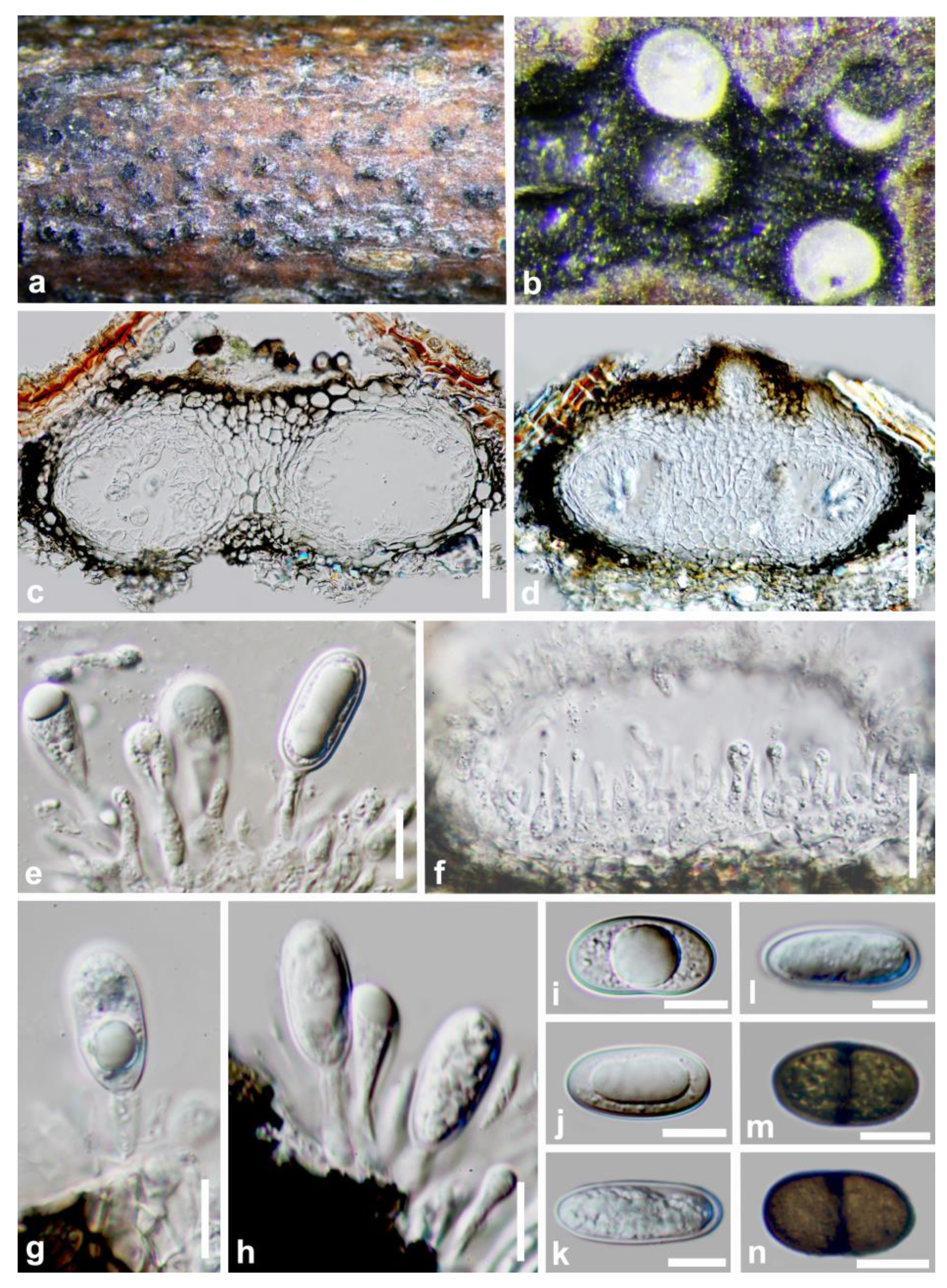
Sardiniella guizhouensis Y.Y. Chen & Jian K. Liu. View in CoL , sp. nov ( FIGURES 2 View FIGURE 2 , 3 View FIGURE 3 )
Index Fungorum number: IF558352, Facesoffungi number: FoF09647.
Etymology:—Name refers the location where the fungus was collected, Guizhou province, China.
Saprobic on decaying aerial stem. Sexual morph: Ascostromata 175–250 µm high × 220–285 µm diam. (x = 190 × 265 µm, n = 20), black, pseudothecial, solitary, initially immersed in host, erumpent at maturity, uniloculate. Peridium 50–80 μm thick, outer layer composed of heavily pigmented thick-walled cells of textura angularis, inner layer composed of hyaline thin-walled cells of textura angularis. Pseudoparaphyses up to 3−4 µm wide, hyphaelike, septate, slightly constricted at septum. Asci 78–92 × 23–27 µm (x = 85 × 24 µm, n = 25), 2–4(–6)-spored, bitunicate, fissitunicate, clavate, pedicellate, with a well-developed ocular chamber, arising from base of the ascoma. Ascospores 26−31 × 14−16 µm (x = 29 × 15 µm, n = 40), uniseriate or irregularly biseriate, initially hyaline and aseptate, becoming pigmented brown and 1-septate, ovate to subclavate, constricted at septum, thick-walled. Asexual morph: Conidiomata 95–151 µm high × 114–294 µm diam. (x = 129 × 187 µm, n = 20), immersed, arranged singly or in small groups within the bark, globose to subglobose, dark brown to black, solitary or gregarious. Ostiole central. Peridium 22–27 µm thick, outer layer composed of pigmented thick-walled cells of textura angularis, inner layer composed of hyaline thin-walled cells of textura angularis (3–5-layered). Conidiogenous cells lining the inner surface of the conidioma, hyaline, short obpyriform to subcylindrical. Conidia 16–28 × 9–12 µm (x = 26 × 10, n = 40), ellipsoid to obovoid, immature conidia hyaline, mature conidia becoming medium to dark brown.
Culture characteristics: —Ascospores germinating on WA within 18 h and producing germ tubes from the septum. Colonies growing on PDA, reaching a diam. of 4 cm after 3d at 25°C, effuse, velvety, with entire to slightly undulate edge. Colonies initially white and later turning green.
Materials examined: — China, Guizhou province, Guiyang District, Xiaochehe wetland park, on decaying aerial stem, 20 May 2017, Y. Y . Chen, 18-53 ( HKAS 113023 View Materials , holotype; GZAAS 19-1809 , isotype); ex-type living culture CGMCC 3.19222 View Materials ( GZCC 19-0090 ); ibid, Huaxi wetland park, 16 April 2017, 19-62 ( GZAAS 19-1935 , paratype), living culture GZCC 19-0216 ; ibid, Libo District, Maolan nature reserve , 19 July 2017, 18-60 ( GZAAS 19-1813 , paratype), living culture GZCC 19-0094 .
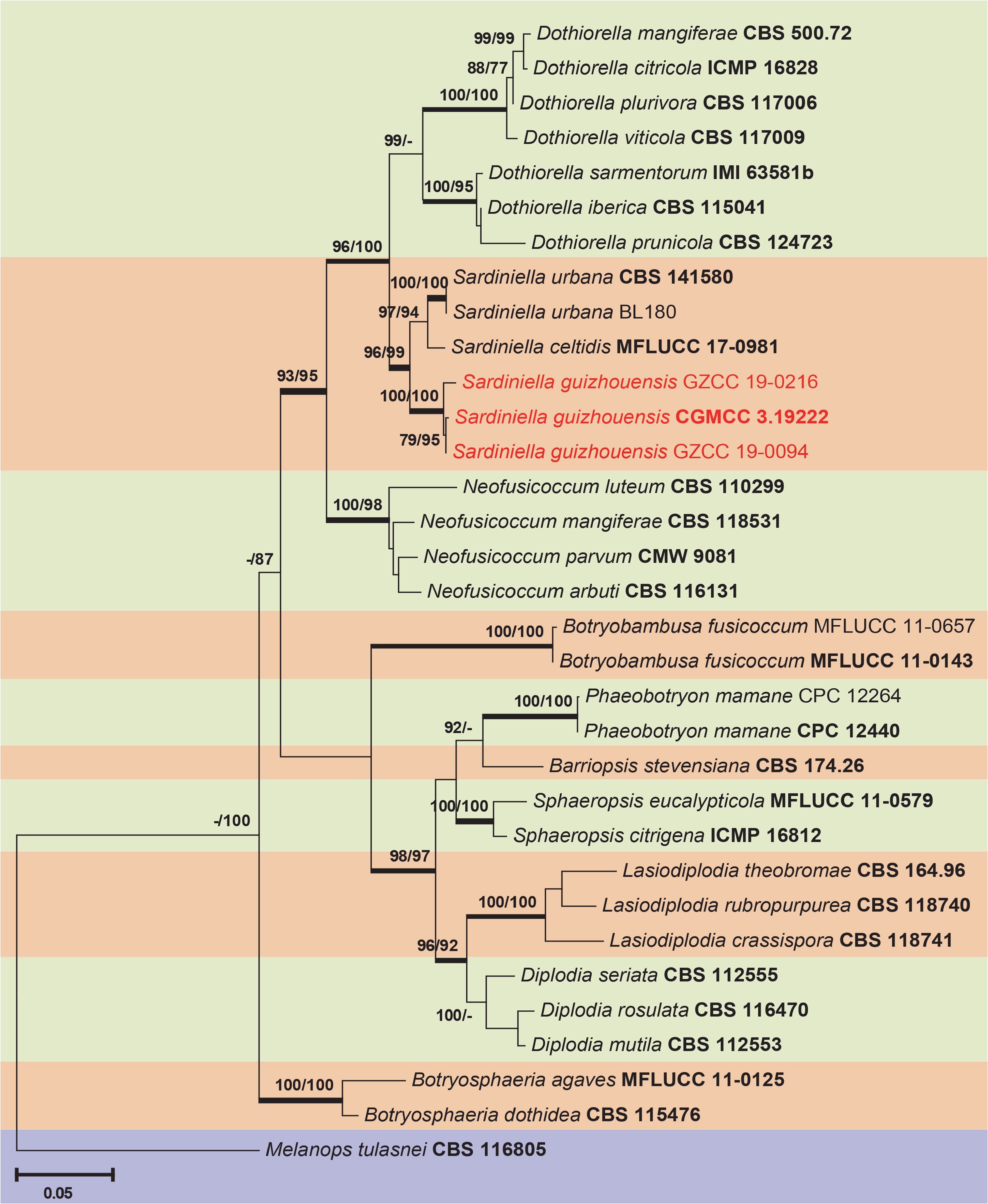
Notes: —The phylogenetic results showed that six taxa clustered together and formed a well-supported clade ( ML / MP / BI = 96/99/1.0) representing the genus Sardiniella ( FIG. 1 View FIGURE 1 ). Sardiniella guizhouensis can be distinguished from S. celtidis (9/ 631 in ITS and 24/ 364 in tef1), and from S. urbana (5/ 839 in LSU, 6/ 631 in ITS and 25/ 364 in tef1). In addition, Sardiniella guizhouensis differs from other two known Sardiniella species in having multiloculate conidiomata ( FIG. 2 c,d View FIGURE 2 ). The sexual morph is known only in Sardiniella guizhouensis and thus no comparisons between species can be made.

| Y |
Yale University |
| ML |
Musee de Lectoure |
| MP |
Mohonk Preserve, Inc. |
| BI |
Istituto Ortobotanico |
| LSU |
Louisiana State University - Herbarium |
No known copyright restrictions apply. See Agosti, D., Egloff, W., 2009. Taxonomic information exchange and copyright: the Plazi approach. BMC Research Notes 2009, 2:53 for further explanation.