
Ceratomyxa herouardi Georgévitch, 1916
|
publication ID |
https://doi.org/ 10.11646/zootaxa.3887.2.3 |
|
publication LSID |
lsid:zoobank.org:pub:B697D6BA-836B-44E2-A8D9-07661554FE59 |
|
DOI |
https://doi.org/10.5281/zenodo.5626244 |
|
persistent identifier |
https://treatment.plazi.org/id/038887C4-FFC3-FFD3-B3BB-FF1580F042E5 |
|
treatment provided by |
Plazi |
|
scientific name |
Ceratomyxa herouardi Georgévitch, 1916 |
| status |
|
Ceratomyxa herouardi Georgévitch, 1916
Type host: Sarpa salpa Linnaeus, 1758 goldline sea bream ( Perciformes : Sparidae )
Type localities: Mediterranean off Tunisia: Location 1: Gulf of Tunis (36°45’N, 10°15’E); Location 2: Bay of Bizerte (37°20’ N, 9°53’ E).
Site of infection: Within gall bladder
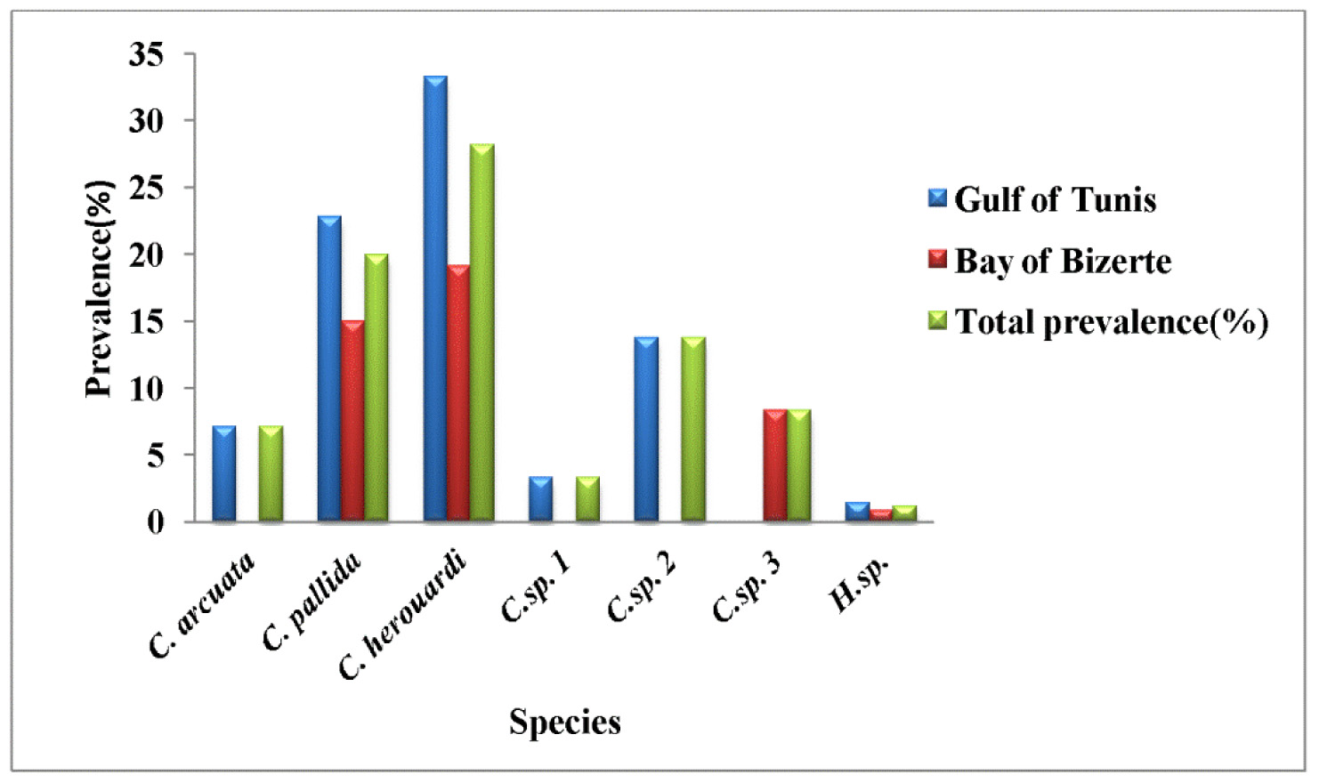
Prevalence: The overall prevalence is 28.2% (93/330) ( Fig. 9 View FIGURE 9 ). At location 1, the prevalence of infection was 33.3% (70/210) distributed as following, 03/2012: 36.7% (11/30); 04/2012: 30% (9/30); 05/2012: 40% (12/30); 06/ 2012: 46.7% (14/30); 07/2012: 23.3% (7/30); 08/2012: 30% (9/30); 05/2013: 25% (5/20); 06/2013: 30% (3/10). At location 2, the prevalence of infection is 19.2% (23/120) distributed as following, 03/2013: 13.3% (4/30); 04/2013: 13.3% (4/30); 05/2013: 26.7% (8/30); 06/2013: 23.3% (7/30) (see Table 4).
Mean intensity: 211.2 ± 50.6 spores/infected fish (++++++) ( Fig. 9 View FIGURE 9 ) (see Table 4).
Type-material: Digitized photos of syntype spores were deposited in the parasitological collection of the Museum National d’Histoire Naturelle ( MNHN), Paris, Coll. No. ZS 117.
Description
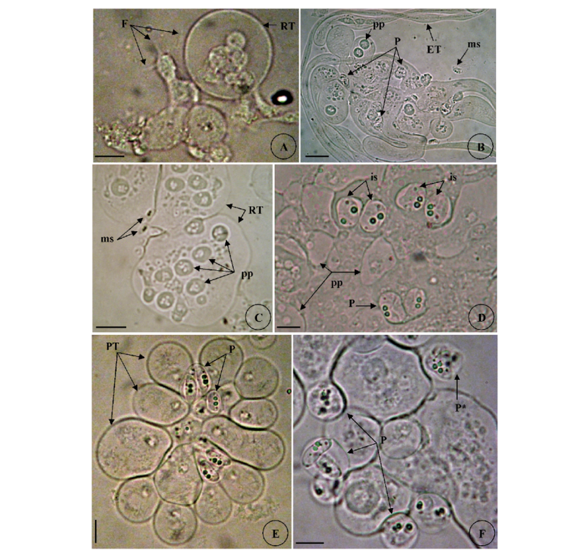
Vegetative stages. Trophozoites (n = 100) were freely floating in bile of the gallbladder in different stages of maturation and some seen attached to gall bladder epithelium. Polymorphous with great variety of shape and size, elongated with same breadth or tapering to one end or club-shaped with roundish enlargements ( Fig. 3B View FIGURE 3 A – F ). Young trophozoïtes were spherical or pyriform ( Figs. 3E–F View FIGURE 3 A – F ). Trophozoïtes were attached to each other by their pseudopodia while other possessed a long pseudopodia-like filopodia probably used for their motility ( Fig. 3A View FIGURE 3 A – F ). Protoplasm homogeneous with fine refractile granules and inner generative cells. Subspherical to spherical plasmodia measuring 30.23 ± 5.5 (22–40) µm in length and 31.8 ± 5.1 (27–44.5) µm in width (n = 50). Disporous, each plasmodium contained ordinarily two identical spores ( Figs.3C–E View FIGURE 3 A – F ), or polysporous, with formation of numerous disporic plasmodia attached to each other within the trophozoïte mother ( Figs. 3C–D View FIGURE 3 A – F ).
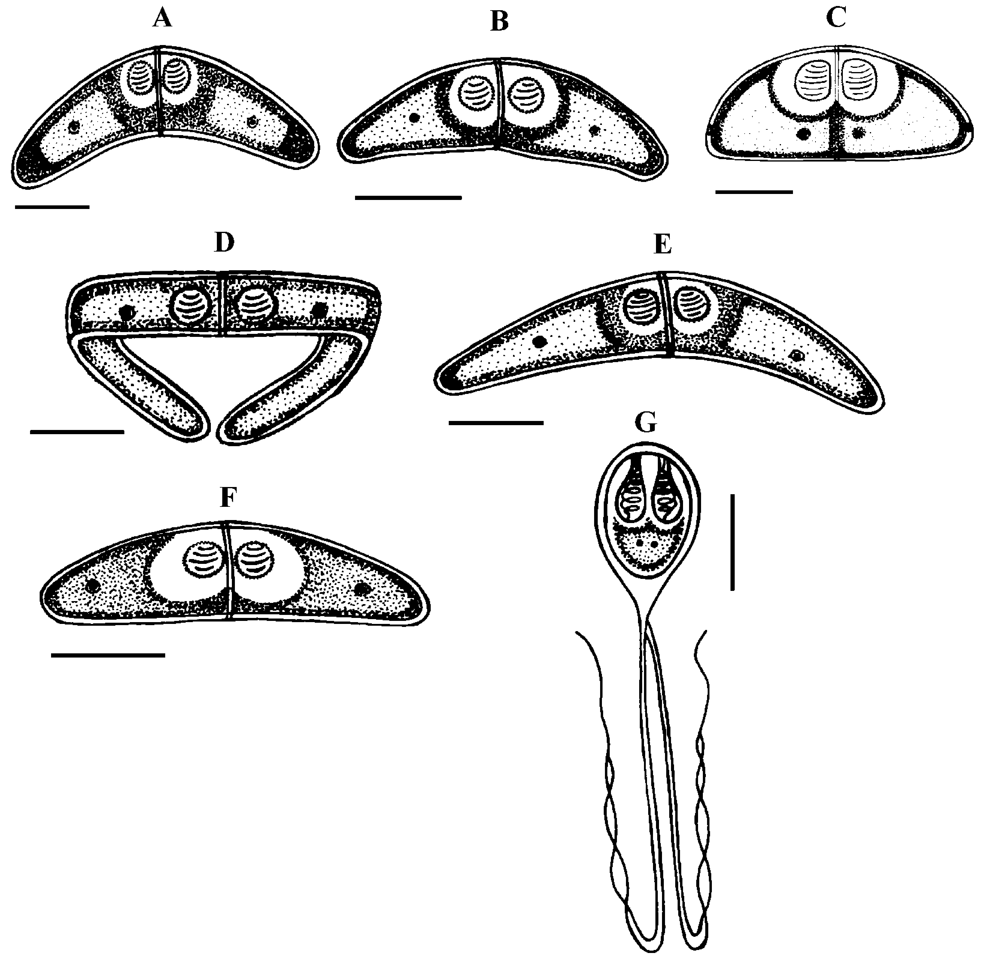
Spores (n = 100 fresh spores). Mature spores were elliptic to hemispherical broadly with round ends in sutural view ( Figs. 3I –K,M View FIGURE 3 A – F View FIGURE 3 G – N , 8 View FIGURE 8 C), measuring 10.5 ± 1.2 (8–12) Μm in length and 21.6 ± 1.6 (20–24) Μm in thickness. Posterior angle was flattened to straight 172.5 ± 6.8 (165–180°). The suture line divided the spore in two equal valves smoothly ovoid in lateral view ( Fig. 3J View FIGURE 3 A – F View FIGURE 3 G – N ). Polar capsules were nearly spherical to sub-spherical 3.91 ± 0.25 (3.5–4.5) Μm in length and 3.89 ± 0.27 (3.5–4.5) Μm in width (n = 100). The polar filament formed five to six turns arranged along the longitudinal axis of the capsule. A binucleate sporoplasm almost filled the spore cavity and was generally distributed symmetrically. Occasionally, aberrant spores with 3 polar capsules and 3 valves were observed ( Figs. 3F View FIGURE 3 A – F , L View FIGURE 3 G – N ).
Remarks
The first description of C. herouardi was illustrated by Georgévitch (1916a, 1916b, 1916c, 1917) from S. salpa captured in Mediterranean coast of Monaco, France. The description was focused on the huge variety of the vegetative forms. Georgévitch (1916) declared that C. herouardi has been especially deserved the most attention from all the Ceratomyxa spp. identified in that time by its extraordinary vegetative forms that were very diverse ( Figs. 3A–F View FIGURE 3 A – F ): round, elliptical, pyriform, elongated and very unequal in size and by its mature spores that could show dissimilar shape in different views. In result of that, Jameson (1913) mentioned the same forms and variety of trophozoïtes in the same host, organ and locality ( Monaco) without recognized the spores. He couldn’t determined the species and he declared in his study that the form of parasite found in the gall bladder of S. salpa has something of the appearance of a Lepthoteca ( Kudo 1920). Georgévitch (1917) believed that the variety of the trophozoites and the complexity of it developmental cycle according to the process of endogenous or exogenous budding, were an important characteristics to identify C. herouardi from the other species of the same genus. This author made a traditional demonstration about the life cycle of C. herouardi based to observations in light microscopy and some erroneous hypothesis that attached to myxosporeans described many years ago. In recent paper, the different vegetative forms of C. herouardi are recognized and the first measurements of the sporogonic stages and as well as mature spore are given (see Table 5).
Ecological notes
During the sampling period, the overall rate of infection is 28.2% and this myxosporean has a parasitic status as less frequent species even it has a great mean of intensity with 211.2 ± 50.6 spores per individual infected host ( Fig. 10 View FIGURE 10 ). Infection by C. herouardi was detected during the whole period of investigation. In Gulf of Tunis, the infection started from March to August with maximum prevalence in June 46.7% while the infection commenced from March to June in Bay of Bizerte and maximum prevalence was recorded in May 26.7%. During examination of goldline sea bream’s gall bladder, the intensity of infection was important and showed no significant variation with months (see Table 4).


| MNHN |
Museum National d'Histoire Naturelle |
No known copyright restrictions apply. See Agosti, D., Egloff, W., 2009. Taxonomic information exchange and copyright: the Plazi approach. BMC Research Notes 2009, 2:53 for further explanation.