
Brunetorhynchus deconincki, Schockaert, Ernest R., Martens, Paul M., Revis, Nathalie, Janssen, Toon, Willems, Wim & Artois, Tom J., 2014
|
publication ID |
https://doi.org/ 10.11646/zootaxa.3755.3.4 |
|
publication LSID |
lsid:zoobank.org:pub:C60EB7B9-77F2-488E-8583-CAD0C364575E |
|
DOI |
https://doi.org/10.5281/zenodo.6135446 |
|
persistent identifier |
https://treatment.plazi.org/id/038887EB-FF99-FFBE-FF07-FF7628DCF8E8 |
|
treatment provided by |
Plazi |
|
scientific name |
Brunetorhynchus deconincki |
| status |
sp. nov. |
Brunetorhynchus deconincki View in CoL n. sp. Schockaert, Martens & Artois
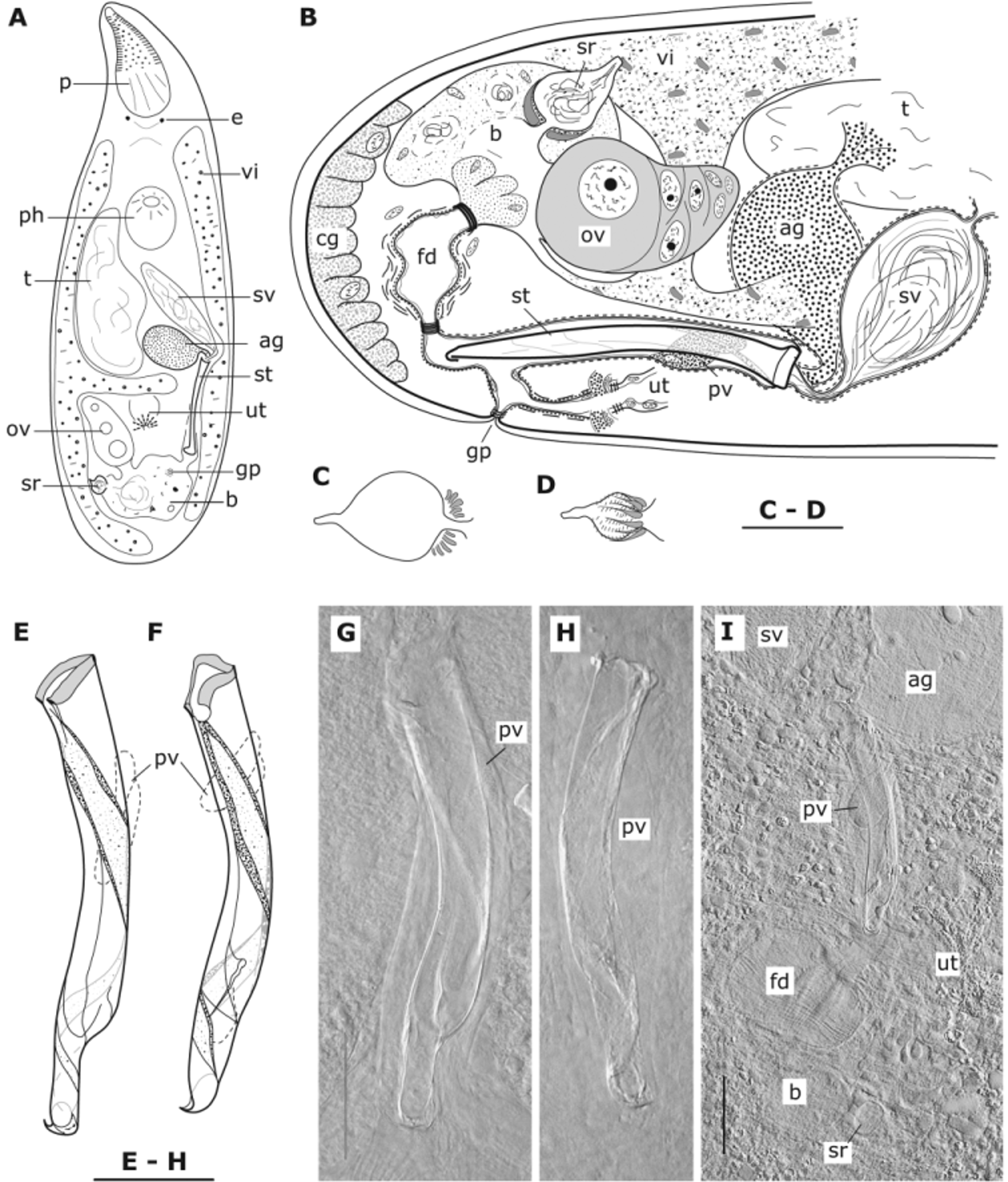
( Fig. 1 View FIGURE 1 )
Holotype. A whole mount ( SMNH 7836), Bay of Marseille, France, “La Pierre Joseph” near the Isle Plane, 17 m deep, fine-grained sand (type locality)(11 May 1967, leg M. Brunet).
Paratype. One serially-sectioned specimen from the type locality (HU, no 541).
Other material and localities. One whole mount and two serially-sectioned specimens (HU, nos VI.1.47–49), Bay of Marseille, France, west from the northern tip of Isle Pomègues, muddy sand with small pebbles, 35 m deep (26 July 1967). A whole mount and one serially-sectioned specimen (HU, nos VI.1.50–VI.2.1), Bay of Marseille, France, Cap Canaille, near Cassis, 16 m deep, fine-grained sand (26 January 1967). One whole mount (HU, no VI.2.2), Corsica, Bay of Calvi, France, fine-grained sand with some detritus and silt, 39 m deep (11 June 1982). Two whole mounts and one serially-sectioned specimen (HU, nos VI.2.3–VI.2.5), harbour of the Station de Recherches Sous-marines et Océanographiques (STARESO), Pointe Revellata, Corsica, Bay of Calvi, France, coarse-grained sand with some silt, 4 m deep (7 April 1983).
Etymology. Species epithet in honour of the late Prof. Dr. Lucien De Coninck, Ghent University, Belgium, teacher of the first author (ERS).
Diagnosis. Species of Brunetorhynchus with a slightly bent, tubular stylet 75–100 µm long and 8–10 µm broad, narrower in the distal quarter, with a thickened proximal rim, open at one side from where two spirallyrunning ridges depart and run counter clock-wise over the stylet. Prostate vesicle (type III) rudimentary, in approximately 1/3 of the proximal part of the male atrium.
Description. Colourless to light brownish-pink animals, 0.8–1 mm long, with eyes far apart from each other (i.e. closer to the lateral rim than to each other). The proboscis is about 1/4th to 1/5th of the body length (in sections) with parallel cone retractors and a thick internal circular muscle layer. There are four proboscis retractors and a pair of ventral integument retractors. There are no nuclei at the junction but there are four nuclei about half way along the proboscis sheath where the epithelium is slightly thicker and where four sheath dilatators insert. The syncytial epidermis with few lobate nuclei is 2.5–3 µm thick with a slightly thicker basement membrane and cilia 2.5–3 µm long. The epidermis contains densely-packed small granules under its surface. The pharynx with the usual polycystidid construction is at about 30–40%.
The common genital pore is at about 80% in the living animal but closer to the caudal end in sections. The genital atrium receives the uterus (with the normal polycystidid construction) at its anterior side and continues in the dorsal direction, where it receives the male atrium frontally and the female duct dorsally. The atria are lined with a very low epithelium with depressed nuclei and are surrounded by a weak muscle layer.
The single testis is situated next to and just behind the pharynx at the left side. The male atrium, at the right side, is surrounded by a relatively weak, spirally running muscle layer. The large seminal vesicle is lined with a low epithelium and surrounded by thin, spirally running muscle fibres. Distally it narrows to a short seminal duct towards the stylet. Next and left to the seminal vesicle there is the large accessory vesicle (type II), filled with a coarse-grained, basophilic secretion and surrounded by a spiral muscle layer. Distally it forms a narrow duct that enters the stylet next to the seminal duct. The male atrium is somewhat broadened in its proximal third where it contains a small prostate vesicle (type III). Its slightly basophilic secretion is arranged in two or three fascicles, probably formed by one single cell, since only one nucleus can be found. The stylet (an accessory stylet type II) is 75–100 µm (m = 91 µm, n = 5) long and 8–10 µm broad over the proximal 4/5th, narrower in the last 5th. The proximal rim is thickened and open at one side from where two ridges run spirally, counter-clock wise, over the entire length of the stylet. The distal opening is subterminal.
The ovary is unpaired, at the left side; the paired vitellaria join close to the ovary to form the vitelloduct, which enters into a wide oviduct, next to the ovary. In its caudal part the oviduct is transformed into a resorbing bursa. Dorsally and close to the ovary a pear-shaped vesicle, the seminal receptacle, is attached to the oviduct. It is surrounded by thin inner circular muscles and outer longitudinal muscles, both layers gradually disappearing distally. At the side opposite to the ovary, the female duct type I departs. The opening towards the female duct is surrounded by eosinophilic glands and is guarded by a sphincter. The female duct is lined with a ruffled or low epithelium and surrounded by inner circular muscles and outer longitudinal muscles, arranged in bundles (see Fig. 1 View FIGURE 1 I). In several specimens (sectioned and in whole mounts, but not in all) the female duct has a spherical aspect, probably a fixation artefact.
| SMNH |
Saskatchewan Museum of Natural History |
No known copyright restrictions apply. See Agosti, D., Egloff, W., 2009. Taxonomic information exchange and copyright: the Plazi approach. BMC Research Notes 2009, 2:53 for further explanation.