
Cyproidea liodactyla Hirayama, 1978
|
publication ID |
https://doi.org/ 10.11646/zootaxa.4097.3.1 |
|
publication LSID |
lsid:zoobank.org:pub:7DC7B6D8-6C7B-423C-A85E-EBE555FA3CCD |
|
DOI |
https://doi.org/10.5281/zenodo.6056313 |
|
persistent identifier |
https://treatment.plazi.org/id/0388D03F-FFD8-FFE2-82DF-FCDBFDD5F9CD |
|
treatment provided by |
Plazi |
|
scientific name |
Cyproidea liodactyla Hirayama, 1978 |
| status |
|
Cyproidea liodactyla Hirayama, 1978 View in CoL
[Japanese name: Hosozume-hoteiyokoebi] ( Figs 2–6 View FIGURE 2 View FIGURE 3 View FIGURE 4 View FIGURE 5 View FIGURE 6 )
Cyproidea liodactyla Hirayama, 1978: 245 View in CoL , figs 1–3 (type locality: Tomioka Bay, Amakusa, Kumamoto Prefecture, Japan).— Hirayama, 1983: 124.— Barnard & Karaman, 1991: 259.— Ishimaru, 1994: 53.
Material examined. One male (OMNH-Ar-9940), 3.6 mm, off Jogashima Island, Miura City, Kanagawa Prefecture, 35°07'49"N, 139°33'57"E, 97.3–100 m in depth, sand and shell bottom, using dredge, 19 April 2012, coll. H. Kohtsuka; 1 female (OMNH-Ar-9941), 3.0 mm, Ariake Sea, 32°30'56"N, 130°16'58"E, 18 m in depth, sandy-muddy bottom, using dredge, 17 September 2005, coll. K. Mori; 1 male (OMNH-Ar-9942), 3.3 mm, Ariake Sea, 32°31'11"N, 130°20'02"E, 15 m in depth, sandy-muddy bottom, using Smith-McIntyre grab, 12 May 2006, coll. K. Mori; 1 female (OMNH-Ar-9943), 3.6 mm, Ariake Sea, 32°30'56"N, 130°16'58"E, 18 m in depth, sandymuddy bottom, using Smith-McIntyre grab, 17 September 2005, coll. K. Mori; 1 female and 2 juveniles (OMNH- Ar-9944–9946), 2.7, 2.0, 1.9 mm, Koganezaki-park beach, Nishi-izu Town, Shizuoka Prefecture, 34°50'37"N, 138°45'49"E, 10–12 m in depth, sandy bottom, using SCUBA, 17 July 2012, coll. M. Fujii; 3 males and 1 juvenile (OMNH-Ar-9947–9950), 2.5, 2.5, 2.4, 2.0 mm, off Moroiso Bay, Misaki City, Kanagawa Prefecture, 10 m in depth, sandy bottom, using dredge, 1 July 2014, coll. H. Kohtsuka. Paratypes 1–7 (not dissected), sex unknown, 3.0, 3.3, 3.8, 3.2, 2.2, 3.5, 3.5 mm, Tomioka Bay, Amakusa, Kumamoto Prefecture, 10 m in depth, sandy bottom, 15 July 1978.
Description. Male [based on 3.6 mm (OMNH-Ar-9940), and 3.3 mm (OMNH-Ar-9942) for lower lip and pleonal epimera]. Body ( Fig. 2 View FIGURE 2 ) oval; head shorter than pereonite 1, eyes large, diameter about 0.7 times as long as head; pereonites 1, 4 and 5 dorsally long, posteroventral corners of pereonites 5–7 protruded; coxa 5 far exceeding posteroventral corner of pereonite 6; pleonites 1–3 each with dorsolateral ridge extending to urosomite 1; urosome slender.
Antenna 1 ( Fig. 3 View FIGURE 3 A, A1) short, with ratio of lengths of peduncular articles 1–3 1:1.2:0.7, tooth of article 2 occupying 30% of whole length; accessory flagellum vestigial, with 4 setae; primary flagellum with 7 articles, articles 1–6 with 2, 2, 2, 2, 2, 1 aesthetascs at each distal end, respectively. Antenna 2 ( Fig. 3 View FIGURE 3 B) short, with ratio of lengths of peduncular articles 3–5 1:2.2:2.0; flagellum with 3 articles.
Upper lip ( Fig. 3 View FIGURE 3 C), ventrolateral margins with short setae. Mandible ( Fig. 3 View FIGURE 3 D, D1, E, E1), each incisor bearing 6 cusps, left lacinia mobilis fan-shaped, distal margin minutely serrated, right lacinia mobilis lacking, number of accessory blades 5 in left and 4 in right; molar conical; palp article length ratio 1:1.6: 1.4 in left and 1:1.4: 1.2 in right, article 2 with 2–3 short setae, tip of article 3 with 1 long seta, ventral surfaces of articles 2–3 covered with minute setae. Lower lip ( Fig. 3 View FIGURE 3 F), inner lobes indistinct; anteromedial corners of outer lobes each with 1 acute and 1 truncate projections, mandibular processes small. Maxilla 1 ( Fig. 3 View FIGURE 3 G, G1 – 2), inner plate roundish triangular, without setae; mediodistal corner of outer plate acutely projected, distal margin with 7 robust setae, medial margin bearing 3 robust setae; distal end of palp projected in middle, with 2 minutely-serrated robust setae and 4 robust setae. Maxilla 2 ( Fig. 3 View FIGURE 3 H), inner plate shorter than outer, terminal ends of both plates each with a few setae, dorsal surfaces of both plates setose. Maxilliped ( Fig. 3 View FIGURE 3 I, I1 – 2), inner plate truncate, mediodistal corner with 1 robust seta dorsally; outer plate with 2 distal and 2 ventral robust setae, mediodistal margin serrated; palp articles 1 – 2 with 1 and 4 setae, respectively, article 3 covered with short setae dorsally, with 1 robust seta on mediodistal corner, article 4 slender.
Gnathopod 1 ( Fig. 4 View FIGURE 4 A, A1), coxa quadrate; basis long, narrow; posterodistal corner of ischium not projected; merus short; carpus long, posterodistal corner slightly protruded, bearing several long setae, posteromedial surface covered with thin setae; propodus long, about 1.6 times as long as carpus, palm with many acute denticles, anteromedial surface covered with thin setae; dactylus very long, gradually curved, posteroproximal margin bearing 1 long and 6 short acute spines. Gnathopod 2 ( Fig. 4 View FIGURE 4 B, B1) stouter than gnathopod 1; coxa roundish quadrate; posterodistal corner of ischium projected, with several setae; merus short; posterodistal projection of carpus as long as carpus proper, bearing 4 medial setae, posteromedial surface covered with thin setae; distal half of propodus slightly swollen posteriorly, palm with many blunt denticles; dactylus strongly flexed at a point of one third, gradually narrower, posteroproximal margin bearing 8 acute denticles.
Pereopod 3 ( Fig. 4 View FIGURE 4 C), coxa triangular, anterior margin rounded, posterior margin slightly concave, posteroproximal corner with small projection, posteroventral corner with small medial hollow; basis and merus – propodus long. Pereopod 4 ( Fig. 4 View FIGURE 4 D), coxa extremely wide, swollen posteriorly, anterior margin slightly convex, with small projection on anteroventral corner, ventral margin also slightly convex, posteroproximal corner a little projected; basis and merus long. Pereopod 5 ( Fig. 4 View FIGURE 4 E), coxa lanceolate, width about 3.1 times of length, anterior margin with small hollow, posterior end pointed; basis and merus – propodus long. Pereopod 6 ( Fig. 4 View FIGURE 4 F) almost same length as pereopod 5; coxa roundish trapezoidal; basis and merus – propodus long. Pereopod 7 ( Fig. 4 View FIGURE 4 G), coxa roundish rectangular; basis and merus long.
Pleonal epimera ( Fig. 5 View FIGURE 5 A), epimera 1 – 2 projected bluntly on posteroventral corner, each with oblique lateral ridge, epimeron 3 rounded posteroventrally, all ventral margins bare. Pleopods ( Fig. 5 View FIGURE 5 B – D) almost same length; inner rami with 6 articles, outer rami with 8, 8, 7 articles in pleopods 1 – 3, respectively. Uropods ( Fig. 5 View FIGURE 5 E – G), peduncle and both rami without robust setae, but bearing minute denticles on edges of dorsal surfaces; peduncle of uropod 1 long, both rami about 0.7 times as long as peduncle; uropod 2 about 0.8 times length of uropod 1, both rami 93% as long as peduncle; uropod 3 short, 55% length of uropod 1, peduncle relatively short, inner ramus about 1.3 times as long as peduncle, outer ramus almost same length as peduncle. Telson ( Fig. 5 View FIGURE 5 G) ovoid, breadth about 0.6 times of length, without setae.
Female [3.6 mm (OMNH-Ar-9943)]. Generally similar to male except for antenna 1 and oostegites. Antenna 1, peduncular article 2 cuspidate, flagellum 6-articulated. Gnathopods 1–2 ( Fig. 5 View FIGURE 5 H–I) almost same as those in male except for oostegite on coxa 2, but carpi–dactyli shorter.
Paratypes-1–7 (sex unknown, 2.2 – 3.8 mm). Antenna 1 ( Fig. 5 View FIGURE 5 J), peduncular articles 2 in all paratypes cuspidate, flagellum in paratype-2 (3.3 mm) with 6 articles.
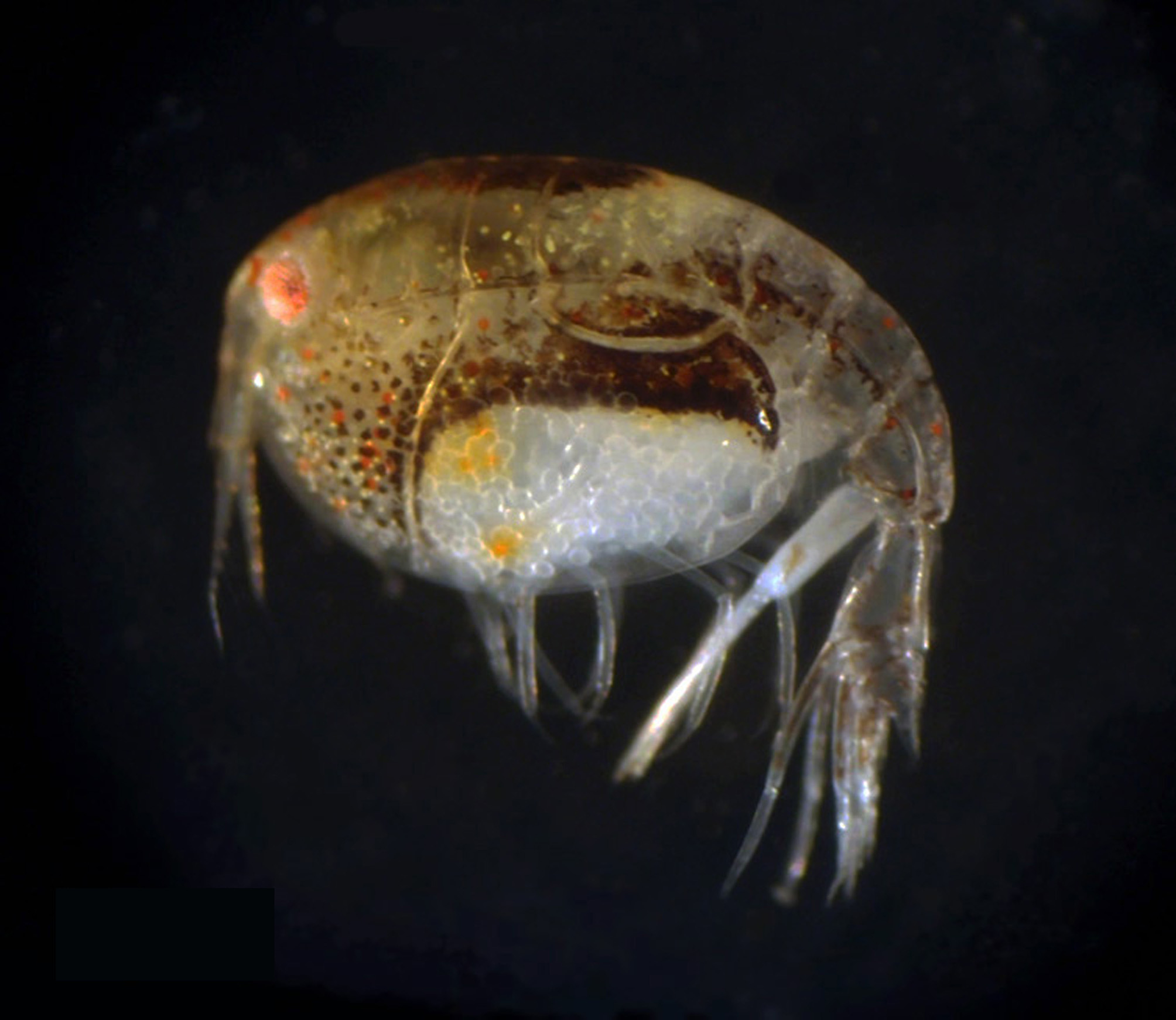
Coloration in fixed specimen ( Fig. 6 View FIGURE 6 ). Eyes red; dorsal surfaces from head to pereonite 5 dark brown, lateral surfaces of them translucent, lateral surfaces from pereonite 6 to pleonite 2 partly dark brown; proximal–middle part of coxa 3 dark brown, with several red pigments; proximal part of coxa 4 dark brown, middle–distal part translucent, with a few orange pigments; coxa 5 dark brown; from posterior part of urosomite 1 to peduncles of uropods also dark brown.
Remarks. Morphological characters of the examined specimens almost agree with the original description and figures of Cyproidea liodactyla , except for the shape of antenna 1. Although Hirayama (1978) stated on the antenna 1 “distal margin of peduncle 2 produced into two pointed and triangular processes” and “flagellum four segmented”, the examined specimens including the paratypes have a 1-pointed large acute projection similar to the other Cyproidea species, and a 6–7-articulated flagellum. However, the examined specimens can be identified as C. liodactyla because of the following reasons: (1) the morphological characters except for the antenna 1 are similar to the original description, (2) the shape of antenna 1 is the same in all the specimens including the paratypes, and (3) the distribution of brown pigments resembles well Hirayama’s Fig. 1 View FIGURE 1 . The antenna 1 in the holotype is considered to be abnormal caused by omission and regeneration.
Distribution ( Fig. 1 View FIGURE 1 ). Japan: Miura City in Kanagawa Prefecture, Nishi-izu Town in Shizuoka Prefecture, Ariake Sea (present study); Amakusa in Kumamoto Prefecture ( Hirayama 1978).
Habitat. Sandy or sandy-muddy bottom, 10–100 m in depth. This species occurs together with Terepeltopes dolichorhunia in Nishi-izu Town.

No known copyright restrictions apply. See Agosti, D., Egloff, W., 2009. Taxonomic information exchange and copyright: the Plazi approach. BMC Research Notes 2009, 2:53 for further explanation.
|
Kingdom |
|
|
Phylum |
|
|
Class |
|
|
Order |
|
|
Family |
|
|
Genus |
Cyproidea liodactyla Hirayama, 1978
| Ariyama, Hiroyuki 2016 |
Cyproidea liodactyla
| Ishimaru 1994: 53 |
| Barnard 1991: 259 |
| Hirayama 1983: 124 |
| Hirayama 1978: 245 |