
Rhamphostomella pacifica ( O’Donoghue, 1925 )
|
publication ID |
https://doi.org/10.11646/zootaxa.5131.1.1 |
|
publication LSID |
lsid:zoobank.org:pub:CF550031-D6A9-48A3-A953-A1BD40C72F5E |
|
DOI |
https://doi.org/10.5281/zenodo.6520683 |
|
persistent identifier |
https://treatment.plazi.org/id/03892374-0B28-331F-FF73-ACA61C14FB9D |
|
treatment provided by |
Plazi |
|
scientific name |
Rhamphostomella pacifica ( O’Donoghue, 1925 ) |
| status |
|
Rhamphostomella pacifica ( O’Donoghue, 1925)
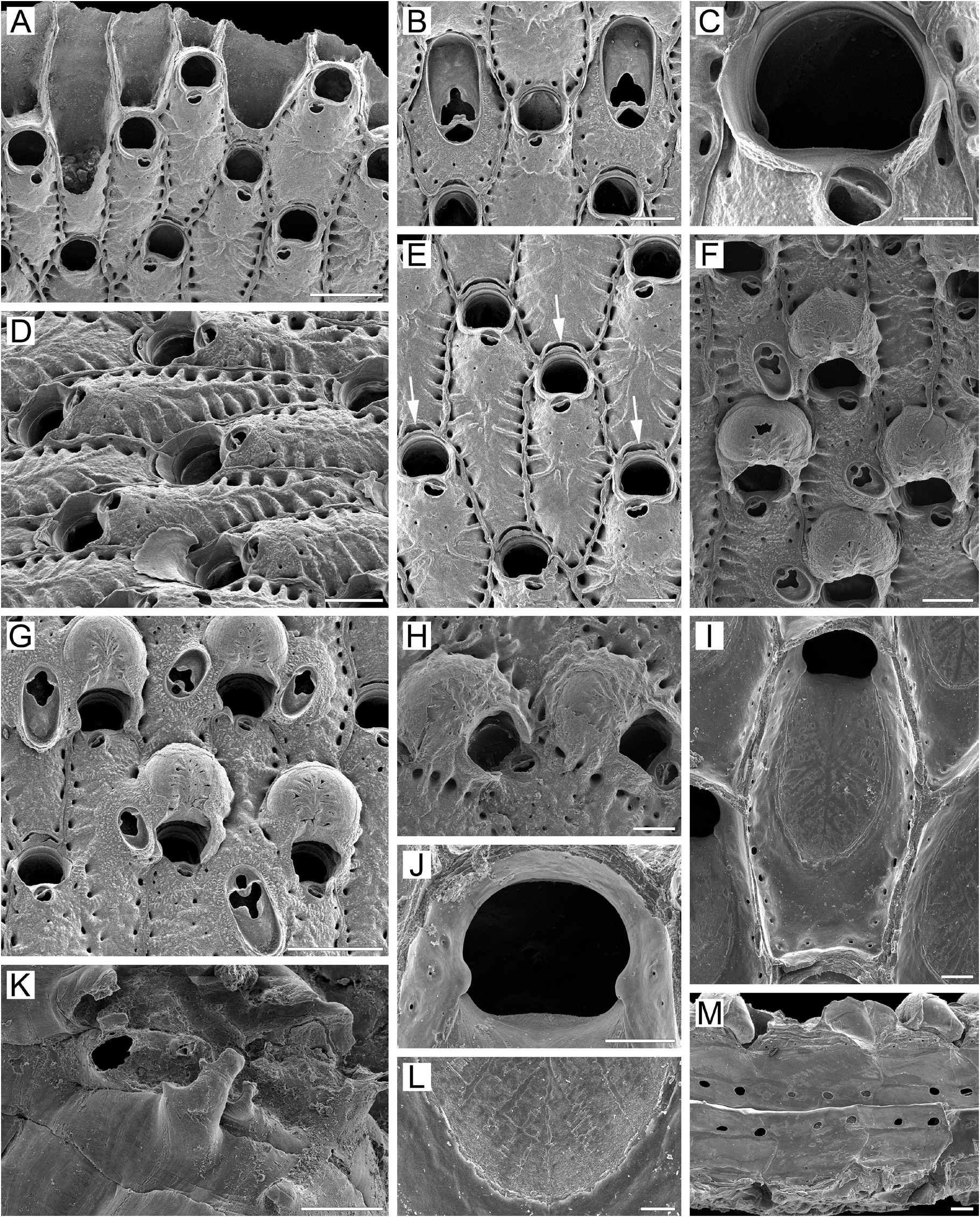
( Figs 8 View FIGURE 8 , 30E View FIGURE 30 )
Porella pacifica O’Donoghue, 1925, p. 20 , pl. 2, figs 7, 8.
Rhamphostomella View in CoL n. sp. 2: Hirose 2010, p. 92, pl. 156a–d.
Material examined. Neotype: ZIRAS 1 /50124, one colony fragment, KIENM Collection, RV Nazarovsk, Stn 238, 11 June 1988, Kronotsky Gulf , eastern Kamchatka Peninsula, Pacific Ocean , 54°53.0ʹ N, 162°15.0ʹ E, depth 122 m, rock dredge, collector A. V. Rzhavsky. GoogleMaps
NHMUK 2010.2 About NHMUK .9.4, one colony fragment , KIENM Collection, RV Nazarovsk, Stn 119, 24 May 1988, Avacha Gulf , eastern Kamchatka Peninsula, Pacific Ocean , 52°54.0ʹ N, 160°01.0ʹ E, depth 141 m, rock dredge, gravel, collector A GoogleMaps . V. Rzhavsky . NHMUK 2010.2 About NHMUK .9.7, one colony fragment , KIENM Collection, RV Nazarovsk, Stn 170, 9 May 1988, Avacha Gulf , eastern Kamchatka Peninsula, Pacific Ocean , 53°22.0ʹ N, 160°07.0ʹ E, depth 136 m, rock dredge, collector A GoogleMaps . V. Rzhavsky .
Additional material. 30 specimens. KIENM Collection (1988) Stn 114; IMB Collection (2011) Stns 3/1, 16/12, 18/14, 20/16, 24/19, 42/37, 43/38, 48/42, 60/50 (see Appendix 1 for details).
Measurements. ZIRAS 1/50124, Kronotsky Gulf, eastern Kamchatka, Pacific Ocean ( Figs 8A–M View FIGURE 8 , 30E View FIGURE 30 ). ZL, 0.82–1.57 (1.14 ± 0.19). ZW, 0.42–0.75 (0.59 ± 0.10). ZD, 0.52–0.83 ( n = 2). OrL, 0.17–0.22 (0.19 ± 0.01). OrW, 0.20–0.27 (0.24 ± 0.02). OeL, 0.32–0.43 (0.39 ± 0.03). OeW, 0.45–0.58 (0.51 ± 0.03). Av(s)L, 0.07–0.12 (0.10 ± 0.02). Av(ad)L, 0.20–0.90 (0.53 ± 0.18). Av(vic)L, 0.55–0.62 ( n = 2). P(m)N, 13–21 (17). P(oe)N, 9–15 (12).
Description. Colonies initially encrusting,multiserial, unilaminar ( Fig.8A View FIGURE 8 ),giving rise to extensive, meandering, erect bilamellar expansions attaining 41 × 38 mm in size. Dry colonies pink to light yellow. Bilaminate parts of colony up to 1.55 mm thick, adjoining layers not fully adherent, sometimes with narrow slit-like spaces in between ( Fig. 8M View FIGURE 8 ). Directions of growth of zooids in adjoining layers not wholly coinciding, with angles of up to 10° between main axes of opposing layers. Zooids large, elongate-hexagonal to oval, arranged in more or less regular straight, oblique rows, demarcated by fine undulating sutures between lateral and transverse walls. Boundaries between zooids clearly visible in young peripheral zooids ( Fig. 8A–G View FIGURE 8 ), gradually becoming indistinct, completely obliterated in oldest parts of colony ( Fig. 8H View FIGURE 8 ).
Frontal shield umbonuloid ( Fig. 8A, I View FIGURE 8 ), initially convex, smooth in young zooids, becoming flatter with granulated surface (fine to coarse) in older zooids. Small, round or elongate areolae along margins, separated by narrow, elongate interareolar ridges; in distal third to half of frontal shield, these ridges connecting with avicularian cystid visible in young parts of colony ( Fig. 8A, E View FIGURE 8 ); ridges sometimes fusing along zooidal midline in proximal part of frontal shield ( Fig. 8D View FIGURE 8 ). Thickening of frontal shield in older parts of colony resulting in smaller areolae as well as broadening of interareolar ridges, with ridges becoming less distinct and often disappearing. Umbonuloid component extensive, occupying about 70% of length of frontal shield (68% in one measured zooid), with fine parallel lineation and accretionary banding ( Fig. 8I, L View FIGURE 8 ). Ring scar discrete ( Fig. 8F View FIGURE 8 ), forming regular boundary between umbonuloid exterior wall and extra-umbonuloid interior wall microstructure.
Primary orifice ( Fig. 8C, J View FIGURE 8 ) broadly semicircular to bell-shaped; distal and lateral margins formed by upper terminal part of distal transverse wall bearing ill-defined rim ( Fig. 8A, C View FIGURE 8 ); rounded condyles laterally ( Fig. 8C, J View FIGURE 8 ). Distal margin of orifice round, proximal margin more or less straight or with very weak median convexity, proximolateral corners gently rounded. No oral spines.
Secondary orifice ( Fig. 8A–E View FIGURE 8 ) transversely elongate-semicircular to irregularly oval, cormidial, proximally and laterally restricted by frontal shield developing two low arch-like lobes that merge with distal part of avicularian cystid ( Fig. 8B, E View FIGURE 8 ). In ovicellate (and older) zooids, two taller peristomial lappets fusing with two corresponding lobes of the secondary calcification overgrowing ooecium ( Fig. 8F–H View FIGURE 8 ). Distolateral curvature of secondary orifice formed by vertical walls of distal and lateral zooids.
Suboral avicularium with cystid occupying one-quarter to about half of zooidal frontal shield ( Fig. 8C, E View FIGURE 8 ), convex in young zooids, less prominent in older zooids, with finely granulated surface and 2–8 minute communication pores on surface. Frontal surface of avicularium (rostral/postmandibular areas) situated on left or right slope of cystid, overlapping or out of zooidal midline, facing distolaterally. Rostrum broadly semicircular, blunt, directed proximomedially and obliquely frontally. Palatal foramen semicircular, conforming to shape of rostrum, opesia small, oval, bordered by extensive cryptocyst. Crossbar complete, with prominent ligula.
Adventitious avicularia varying greatly in size, located centrally to proximally on frontal shield ( Fig. 8F, G View FIGURE 8 ); cystid broad, inflated, oval to rounded, coarsely granulated. Avicularian frontal surface nearly parallel to frontal shield. Rostrum directed proximomedially to proximally, occasionally distally, spatulate or lingulate, blunt, palate of similar shape. Palatal foramen trifoliate, cryptocystal shelf extensive, opesia oval, bordered by well-developed cryptocyst. Crossbar complete, with conspicuous ligula.
Largest adventitious avicularia sometimes occupying significant area of frontal shield of adjacent zooids ( Fig. 8G View FIGURE 8 ); in ovicellate parts of colony, ooecia often flanked by two or three adventitious avicularia ( Fig. 8F, G View FIGURE 8 ).
Very large vicarious avicularia sometimes present near growing edge of colony ( Fig. 8B View FIGURE 8 ). Avicularian frontal surface facing frontally. Rostrum spatulate, slightly broader distally, directed distally; palate of similar shape. Palatal foramen Y-shaped with extensive cryptocystal shelf, opesia roundly triangular. Crossbar complete, with large ligula.
Ovicells hyperstomial, with ooecium gradually becoming covered by secondary calcification encroaching from frontal shields of daughter and neighbouring zooids. Although secondary calcification sometimes covers most of the ooecium, ovicells remain prominent ( Fig. 8F–H View FIGURE 8 ). Surface of secondary calcification finely granulated, with divergent sutures subdividing overgrowth originating from different zooids ( Fig. 8I, L View FIGURE 8 ). Ooecium formed by distal autozooid around shallow, arch-like concavity with communication pore at bottom, situated in proximalmost part of its frontal shield just immediate to distal margin of maternal primary orifice ( Fig. 8A, B, D, E View FIGURE 8 ). Ooecium with slightly concave proximal margin and smooth ectooecium bearing tiny slit-like pseudopores radially arranged in proximal half of ovicell roof ( Fig. 8F–H View FIGURE 8 ). Most pseudopores in old ooecia occluded by secondary calcification ( Fig. 8H View FIGURE 8 ). In ovicellate zooids, secondary calcification proceeding from frontal shields of neighbouring zooids forms bilaterally symmetrical peristome around zooidal orifice. Peristome consists of two lateral vertical lobes, connecting with proximal corners of ooecium ( Fig. 8F–H View FIGURE 8 ).
Zooids interconnected by 2–3 mural pore chambers in each distolateral wall ( Fig. 8M View FIGURE 8 ) and two multiporous septula in basal half of transverse wall, corresponding to two recesses with medial buttress ( Fig. 8A View FIGURE 8 ).
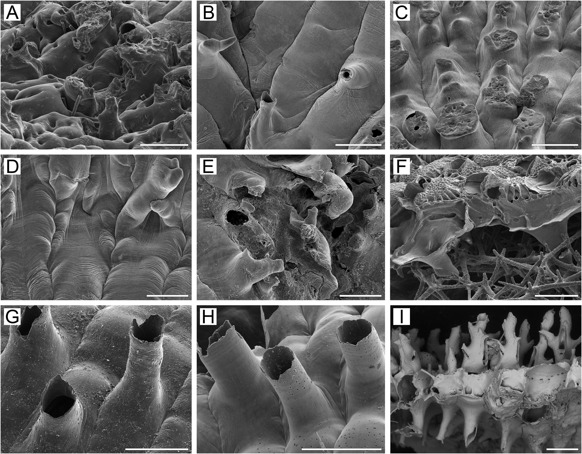
Basal surface of zooids ( Fig. 8K View FIGURE 8 ) fully calcified, with numerous irregular, sometimes bifurcate protuberances ( 0.12–0.39 mm in diameter) and transverse parallel folds on surface ( Fig. 30E View FIGURE 30 ). Boundaries between zooids recognizable basally by deep sinuous incisions. Basal areas of massive erect colonies may contain only heavily calcified kenozooids, very irregular in form and arrangement; these occasionally united into large clusters with indistinguishable boundaries between. Frontally budded zooids, with normal or reversed polarity, also frequently present in older parts of colony, including some with orifices sealed by closure plates.
Ancestrula and early astogeny not observed.
Remarks. O’Donoghue (1925) originally placed this species in Porella Gray, 1848 , but noted the unusual combination of several characters: the stout bilamellar colony-form, the presence of adventitious avicularia, and the atypical position of the suboral avicularium. Concerning the last, he mentioned, “The avicularium is always situated nearer one side, never exactly median, and its semicircular mandible is directed postero-medially at angle of about 45–60 degrees to the hinder end of the aperture”. Based on the generic diagnosis given by Hayward & Ryland (1979), Porella species lack adventitious and vicarious avicularia, and ooecia are imperforate or have a single central pseudopore. The combination of characters in R. pacifica that justify its inclusion in Rhamphostomella includes: 1) the oblique position of the suboral avicularium, situated asymmetrically nearer one side of the orifice, never exactly medially, with the rostrum at an angle of about 15–30° to the median line of the zooid; 2) ooecia with numerous slit-like pseudopores; and 3) the presence of adventitious and vicarious avicularia.
In some aspects, R. pacifica resembles R. commandorica n. sp.. However, it can be distinguished from the latter by the position of the primary orifice, and the form of the avicularian palatal foramen and overall colony morphology, as described above (see Remarks for R. commandorica n. sp.).
The characters of the two colonies from Sagami and Tokyo Bays, Honshu, Japan, described and illustrated by Hirose (2010) as Rhamphostomella n. sp. 2, fall within the range of morphological variation observed for R. pacifica .
Ecology. Rhamphostomella pacifica has been found at 97–490 m depth on mixed bottoms, including pebbles overlying silty sand. Substrata include pebbles and broken mollusc shells.
Distribution. In his original description, O’Donoghue (1925) stated: “These specimens are simply labelled Albatross. NW Pacific with no further data”. Hirose (2010) reported the species from Hatsushima Island, western Sagami Bay and also Yokohama, western shore of Tokyo Bay, Honshu, Japan, at 150 m depth. Our material came from 122–176 m in Kronotsky and Avacha gulfs, eastern Kamchatka Peninsula, and along the Pacific and Sea of Okhotsk sides of the middle to southern Kuril Islands between Simushir and Kunashir at 97– 490 m. Accordingly, R. pacifica is a Pacific Asian boreal, sublittoral to upper bathyal species, extending to the edge of the subtropics.
| RV |
Collection of Leptospira Strains |
| V |
Royal British Columbia Museum - Herbarium |
No known copyright restrictions apply. See Agosti, D., Egloff, W., 2009. Taxonomic information exchange and copyright: the Plazi approach. BMC Research Notes 2009, 2:53 for further explanation.
|
Kingdom |
|
|
Phylum |
|
|
Class |
|
|
Order |
|
|
SubOrder |
Flustrina |
|
SuperFamily |
Lepralielloidea |
|
Family |
|
|
Genus |
Rhamphostomella pacifica ( O’Donoghue, 1925 )
| Grischenko, Andrei V., Gordon, Dennis P., Taylor, Paul D., Kuklinski, Piotr, Denisenko, Nina V., Spencer-Jones, Mary E. & Ostrovsky, Andrew N. 2022 |
Rhamphostomella
| Hirose, M. 2010: 92 |
Porella pacifica O’Donoghue, 1925 , p. 20
| O'Donoghue, C. H. & O'Donoghue, E. 1925: 20 |