
Chelymorpha reimoseri Spaeth, 1928
|
publication ID |
https://doi.org/ 10.11646/zootaxa.3949.4.3 |
|
publication LSID |
lsid:zoobank.org:pub:C819893E-2416-4E3A-A659-37947A0AF6BF |
|
DOI |
https://doi.org/10.5281/zenodo.5686902 |
|
persistent identifier |
https://treatment.plazi.org/id/0389260C-FFC8-EB5D-F1FA-0637946FCBFD |
|
treatment provided by |
Plazi |
|
scientific name |
Chelymorpha reimoseri Spaeth, 1928 |
| status |
|
Chelymorpha reimoseri Spaeth, 1928 View in CoL
Egg ( Figs 1, 2 View FIGURES 1, 2 , 27, 28 View FIGURES 27 – 31 ). Measurements (n=6; [mm]). Length: 1.60, 1.40, 1.40, 1.50, 1.51, 1.50 width (respectively): 0.60, 0.50, 0.48, 0.50, 0.50, 0.51.
Chelymorpha reimoseri makes complex ootheca ( Figs. 1–2 View FIGURES 1, 2 ). The female deposits from 30 to 110 eggs in each ootheca. Preserved in alcohol ootheca is yellow and in its structure resembles honeycomb ( Figs 1, 2 View FIGURES 1, 2 ). Eggs are elongate-oval ( Fig. 27 View FIGURES 27 – 31 ), preserved in alcohol are uniformly yellowish-white. Surface of the chorion is smooth without punctation or any distinct sculpture ( Fig. 28 View FIGURES 27 – 31 ).
First instar larva ( Figs 8–17, 25 View FIGURES 25, 26 , 29–64 View FIGURES 27 – 31 View FIGURES 32 – 39 View FIGURES 40 – 45 View FIGURES 46 – 51 View FIGURES 52 – 58 View FIGURES 59 – 64 ). Measurements are presented in Table 1 View TABLE 1 . Length was measured without head, from anterior border of pronotum to the base of supra-anal processes; width of body was measured across metanotum without lateral scoli.
Body flattened dorso-ventrally, oval, moderately narrowed posteriorly, widest across meso- and metathorax ( Figs 8, 9 View FIGURE 9 , 32 View FIGURES 32 – 39 ). Body of larva preserved in alcohol yellowish-brown with light brown patches on pronotum, light brown lateral scoli, supra-anal processes, legs, spiracles and head.
Body with 14 pairs of lateral scoli and a single pair of supra-anal processes ( Figs 8, 32 View FIGURES 32 – 39 ). Meso- and metanotum on each antero-lateral side with distinct protuberance, each protuberance with two long club-like setae and one short apically pointed seta ( Fig. 9 View FIGURE 9 ). Protuberances are in place where in other cassidoid Cassidinae species occur scoli of 5th and 7th pair ( Świętojańska, 2009). All lateral scoli without lateral branches, but covered with few long club-like setae ( Figs 8, 9 View FIGURE 9 , 38 View FIGURES 32 – 39 ). Apex of scoli with one very long club-like seta ( Fig. 39 View FIGURES 32 – 39 ). Supra-anal processes very long, about two times longer than body length, without any lateral processes, basal 1/3 covered with asperites, apex without any sensilla, club-like in shape ( Figs 8, 32 View FIGURES 32 – 39 ).
Nine pairs of spiracles: one on thorax and 8 on abdomen. Spiracles distinctly elevated, their diameter almost of the same size, very slightly decreasing posterad ( Figs 8, 9 View FIGURE 9 , 35 View FIGURES 32 – 39 , 44 View FIGURES 40 – 45 ).
Dorsal and ventral surface of the body distinctly granulate ( Figs 8–10). Tergites and sternites with minute setae at anterior border and long club-like setae except for sternites of thorax and abdominal sternites I and II medially, which are covered with long pointed setae ( Figs 8–10, 40–51 View FIGURES 40 – 45 View FIGURES 46 – 51 ). Club-like setae of tergites gradually shortened posteriorly: the longest are setae of pronotum, the shortest of abdominal tergite VIII. Setae of head and legs in similar size to setae of thoracic tergites ( Figs 9 View FIGURE 9 , 11, 12 View FIGURES 11 – 17 , 25 View FIGURES 25, 26 ). The longest setae of body are on tergites of thorax, on abdominal sternites medially and at the top of lateral scoli ( Figs 9 View FIGURE 9 , 10 View FIGURE 10 , 38, 39 View FIGURES 32 – 39 , 40–43 View FIGURES 40 – 45 , 46 View FIGURES 46 – 51 –49).
Pronotum on each side with campaniform sensillum placed antero-medially and with 11 long club-like setae ( Figs 9 View FIGURE 9 , 40–42 View FIGURES 40 – 45 ). Anterior border of meso- and metanotum with a pair of minute setae placed medially and two minute setae on each side laterally. Meso- and metanotum also with four long club-like setae antero-medially ( Fig. 43 View FIGURES 40 – 45 ); 10 long club-like setae in row running along width of tergite posteriorly; one long club-like seta on each antero-lateral side. Anterior border of first abdominal tergite with a pair of minute setae placed medially and two minute setae on each side laterally ( Figs 9 View FIGURE 9 , 44, 45 View FIGURES 40 – 45 ). Abdominal tergites II–VIII with a pair of minute setae close to anterior border medially and single minute setae on each side laterally. One minute setae and one long club-like seta on each abdominal tergite laterally, close to spiracle. Moreover first abdominal tergite with row of 6 long clublike setae running along width anteriorly and two pairs of long club-like setae postero-medially; abdominal tergite II–VIII on each side with two pairs of long setae: first placed antero-laterally and the second postero-medially.
Anterior border of pro-, meso- and metasternum with four minute setae ( Fig. 10 View FIGURE 10 ). Two minute setae at base of each leg. Pro-, meso- and metasternum in the middle with two pairs of pointed setae ( Fig. 46 View FIGURES 46 – 51 ). Anterior margin of all abdominal sternites with a pair of minute setae. Abdominal sternites I–III with 6 long, pointed or club-like setae in the middle of segment, two club-like setae on each postero-lateral side and one short blunt seta on each anterolateral side ( Figs 10 View FIGURE 10 , 49, 51 View FIGURES 46 – 51 ). Abdominal sternites IV–VII with 4 club-like setae in the middle of segment, two clublike setae on each postero-lateral side and one short blunt seta on each antero-lateral side ( Figs 10 View FIGURE 10 , 50 View FIGURES 46 – 51 ). Abdominal sternite VIII with four club-like setae.
Head well sclerotized, hypognathous, retracted into pronotum ( Fig. 33 View FIGURES 32 – 39 ). Median suture complete, connected with fronto-clypeal suture. Clypeus distinct, wider than long, with a pair of setae and a pair of campaniform sensilla ( Figs 11, 12 View FIGURES 11 – 17 , 52, 53 View FIGURES 52 – 58 ).
Six stemmata on each side of head ( Fig. 34 View FIGURES 32 – 39 ).
Frontal surface of head with four small, vertical, pointed setae (V 1–4), and five frontal rows of club-like setae: row Fa with three setae, Fb with five (sometimes six) setae, Fc with three setae, Fd with single seta, Fe with two setae and one campaniform sensillum between setae Fc1 and Fe1 ( Fig. 11 View FIGURES 11 – 17 ). Temporal surface of head with three setae slightly shorter and pointed than setae of frons (T 1–3) and campaniform sensillum ( Fig. 12 View FIGURES 11 – 17 ).
Antennae two-segmented, set in membranous ring ( Figs 36, 37 View FIGURES 32 – 39 ). Both segments stout, more or less as long as wide. First antennomere with four campaniform sensilla, second with one small seta at base, and with a group of 6 peg-like sensilla at the apex: one prominent (sensory appendix) and five smaller.
Labrum free, wider than long, anterior margin emarginate ( Figs 13, 14 View FIGURES 11 – 17 , 59, 60, 62 View FIGURES 59 – 64 ). Anterior margin with 6 small setae placed medially and 6 setae on each side (visible from dorsal as well as ventral view) Dorsal side of labrum with four long setae and two pairs of campaniform sensilla medially and two setae at anterior margin. Mid part of ventral surface (epipharyngeal area) with a pair of small setae, 6 campaniform sensilla, and 16 small sensilla in two groups (each with 8 sensilla). Central and lateral parts of ventral side of labrum armed with numerous small spines.
Mandibles heavily sclerotized, palmate, with four distinct, triangular, apical teeth in one row, two middle teeth longer than reminder, pointed, dorsal tooth stout, ventral the smallest. Dorsal side of mandibles at base with two setae and two campaniform sensilla ( Figs 15, 16 View FIGURES 11 – 17 , 62 View FIGURES 59 – 64 ).
Maxillae and labium connate ( Figs 17 View FIGURES 11 – 17 , 59–64 View FIGURES 59 – 64 ). Each stipes (st) with three long pointed setae. Mala (mal) and lacinia (lac) not distinctly bordered from palpifer (pp). Palpifer with two setae and two campaniform sensilla placed ventrally. Lacinia in form of protuberance covered with spines placed on palpifer dorsally ( Figs 63, 64 View FIGURES 59 – 64 ). Mala bears ( Fig. 17 View FIGURES 11 – 17 ): 7 long pointed setae, one blunt seta, one very short blunt seta (or peg like sensillum?) and two campaniform sensilla. Maxillary palp two-segmented ( Figs 17 View FIGURES 11 – 17 , 63 View FIGURES 59 – 64 ): first segment (mpI) with two setae and one campaniform sensillum, second segment (mpII) with a group of sensilla at apex, and with campaniform sensillum and one seta below the apex. Labial palp (lp) one-segmented with a group of sensilla at the apex and one campaniform sensillum below the apex ( Figs 17 View FIGURES 11 – 17 , 61 View FIGURES 59 – 64 ). Hypopharynx (hyp) covered with numerous spines, and with four campaniform sensilla at base. Prementum (pre) with two long and two short setae, and four campaniform sensilla. Postmentum (post) with 6 setae ( Fig. 17 View FIGURES 11 – 17 ).
Legs elongated, consist of three segments: coxa, femur and tibiotarsus ( Figs 25 View FIGURES 25, 26 , 29–31 View FIGURES 27 – 31 ). Internal side of coxa with setae arranged in three groups: first group with two short pointed setae (placed at border between coxa and body); second with two short pointed setae and one long apically blunt seta; third with three short pointed setae and one long club-like seta. Externally coxa with one long club-like seta and three short pointed setae. Femur with 11 long setae (one of them very long, two times longer than remainder setae of femur, is placed ventrally) and one short seta placed dorsally close to the base. Basally on internal side of femur a group of five campaniform sensilla and one short pointed seta; at base ventrally one campaniform sensillum; basally on external side two campaniform sensilla. Tibiotarsus apically with heavily sclerotized, short and curved, single and simple claw armed basally with a pointed seta. Claw and pointed seta surrounded by a complex of 6 blunt and club-like setae ( Figs 29–31 View FIGURES 27 – 31 ). One short pointed seta and two campaniform sensilla above claw, two long club-like setae placed dorsally on tibiotarsus, and one pointed or club-like seta ventrally.
Fifth instar larva (Figs 3–5, 18–24, 26, 65–107). Measurements are presented in Table 1 View TABLE 1 . Length was measured without head, from anterior border of pronotum to the base of supra-anal processes; width of body were measured across metanotum without lateral scoli.
Body flattened dorso-ventrally, oval, widest across metathorax, slightly narrowed posteriorly (Figs 3–5). Body of larva preserved in alcohol light brown with brown spiracles and top of head. Coxae, femora and tibiotarsi brown dorsally, light brown ventrally. Meso-, metasternum and abdominal sternites I–III with small brown patch placed medially.
Body with 14 pairs of short lateral scoli and a pair of short supra-anal processes (Figs 3–5). Lateral scoli covered with short lateral branches each apically armed with blunt seta ( Fig. 89 View FIGURES 89 – 94 ). Apex of scoli armed with one long pointed seta ( Fig. 90 View FIGURES 89 – 94 ). Supra-anal processes short bent dorsally (Figs 4, 75, 76).
Setae of head and legs more or less of the same length, long, club-like or blunt, each placed on small tubercle ( Figs 18, 19 View FIGURES 18 – 24 , 26 View FIGURES 25, 26 , 77–80 View FIGURES 77 – 82 , 95 View FIGURES 95 – 101 View FIGURES 102 – 107 –102). Setae of tergites short, club-like each placed on prominent tubercle ( Figs 65–74 View FIGURES 65 – 70 View FIGURES 71 – 76 ). Long pointed setae on sternites of thorax and abdominal sternites I–III medially ( Figs 77 View FIGURES 77 – 82 , 83, 84 View FIGURES 83 – 88 ). Club-like setae each on prominent tubercles on abdominal sternites IV–VIII ( Figs 85–88 View FIGURES 83 – 88 ).
Pronotum with numerous club-like setae ( Figs 65, 66, 68 View FIGURES 65 – 70 ). The most numerous setae are around and in the middle of tergite, along body axis. Meso-, metanotum and each abdominal tergite with two irregular rows of clublike setae connected together on sides ( Figs 67, 69, 70 View FIGURES 65 – 70 ). Numerous club-like setae occur on lateral side of meso- and metanotum and on lateral side of abdominal tergites around each spiracle. Each abdominal tergite with two irregular rows of club-like setae connected together on sides ( Figs 71–74 View FIGURES 71 – 76 ).
Pro-, meso-, and metasternum with numerous pointed setae distributed in groups: two numerous groups at anterior border medially, two less numerous posteriorly and one numerous at base of each leg. Setae in groups at base of legs as numerous as setae in the groups at anterior border of sternites. Each abdominal sternite with numerous setae distributed regularly on entire surface ( Figs 84–86 View FIGURES 83 – 88 ). Number of setae on abdominal sternites decreases posteriorly. The longest and the most numerous are pointed setae placed in the middle of sternites I–III ( Figs 83, 84 View FIGURES 83 – 88 ).
Nine pairs of spiracles: one pair on thorax and 8 on abdomen ( Figs 91–94 View FIGURES 89 – 94 ). Spiracle of segment VIII very small. Inner wall of spiracles covered with fine asperites, entrance is closed by long filamentous structures ( Figs 91–94 View FIGURES 89 – 94 ). Edge of each spiracle opening partially with finger-like structures distributed regularly one by another ( Figs 92, 93 View FIGURES 89 – 94 ).
Head well sclerotized, hypognathous, retracted into pronotum. Median suture complete, connected with fronto-clypeal suture. Clypeus distinct, wider than long, with a pair of setae and a pair of campaniform sensilla ( Figs 18, 19 View FIGURES 18 – 24 , 95–102 View FIGURES 95 – 101 View FIGURES 102 – 107 ).
Six stemmata on each side of head ( Figs 96, 98 View FIGURES 95 – 101 ).
Head with numerous club-like setae regularly distributed on frons ( Figs 18 View FIGURES 18 – 24 , 95, 98–102 View FIGURES 95 – 101 View FIGURES 102 – 107 ), each side of vertex with four minute setae ( Fig. 97 View FIGURES 95 – 101 ), temporal side with four club-like setae ( Fig. 19 View FIGURES 18 – 24 ).
Antennae two- segmented, set in membraneous ring. First segment transverse, wider than second, with four campaniform sensilla. Second segment stout slightly longer than wide, with one small seta below the apex and with a group of 6 peg-like sensilla at the apex: one prominent—sensory appendix, and five smaller.
Labrum free, wider than long, anterior margin distinctly emarginate ( Figs 20, 21 View FIGURES 18 – 24 , 102, 103 View FIGURES 102 – 107 ). Anterior margin with 8 short setae placed medially in emargination (not visible from dorsal view) and five setae on each side (visible from dorsal as well as ventral view). Dorsal side of labrum with four setae and two pairs of campaniform sensilla medially and two setae at anterior margin. Mid part of ventral surface (epipharyngeal area) on each side with small seta, three campaniform sensilla, and two groups of four small sensilla. Central and lateral parts of ventral side of labrum armed with numerous small spines.
Mandibles heavily sclerotized, palmate, with four distinct, triangular, apical teeth in one row ( Figs 22, 23 View FIGURES 18 – 24 ). Dorsal side of mandibles at base with two setae and two campaniform sensilla.
Maxillae and labium connate ( Figs 24 View FIGURES 18 – 24 , 102 View FIGURES 102 – 107 ). Each stipes (st) with three long pointed setae. Mala (mal) and lacinia (lac) not distinctly bordered from palpifer (pp). Palpifer with two setae and two campaniform sensilla placed ventrally. Lacinia in form of protuberance covered with spines placed on palpifer dorsally ( Figs 105, 106 View FIGURES 102 – 107 ). Mala bear ( Fig. 24 View FIGURES 18 – 24 ): 7 long pointed setae, one blunt seta, one very short blunt seta (or peg like sensillum?) and two campaniform sensilla. Maxillary palp two-segmented ( Figs 24 View FIGURES 18 – 24 , 106, 107 View FIGURES 102 – 107 ): first segment (mpI) with two setae and one campaniform sensillum, second segment (mpII) with a group of sensilla at apex, and with campaniform sensillum and one seta below the apex. Labial palp (lp) one-segmented with a group of sensilla at the apex and one campaniform sensillum below the apex ( Figs 24 View FIGURES 18 – 24 , 104 View FIGURES 102 – 107 ). Hypopharynx (hyp) covered with numerous spines, and with four campaniform sensilla at base. Prementum (pre) with two long and two short setae, and four campaniform sensilla. Postmentum (post) with 6 setae ( Fig. 24 View FIGURES 18 – 24 ).
Legs three segmented, consist of: coxa, femur and tibiotarsus ( Figs 26 View FIGURES 25, 26 , 77–82 View FIGURES 77 – 82 ). All segments covered with numerous, mostly club-like setae placed on distinct tubercle. Femur additionally with one very long pointed seta on ventral side. Setae of femur, distributed regularly. Basally on internal side of femur a group of five campaniform sensilla and one short seta, one campaniform sensillum in a distance. Basally on external side of femur, two campaniform sensilla. Tibiotarsus apically with heavily sclerotized, curved, single and simple claw with one pointed seta at base. Above claw two campaniform sensilla. Claw and pointed seta surrounded by numerous blunt and club-like setae ( Figs 79–82 View FIGURES 77 – 82 ). Setae of ventral side of tibiotarsus and at base of claw in opposite to setae placed dorsally are not placed on tubercle.
Pupa ( Figs 6, 7). Measurements (n=6; [mm]). Length of body: 10.00, 8.80, 10.00, 10.20, 10.50, 8.00; width across second abdominal segment, without lateral scoli (respectively): 6.40, 6.10, 6.40, 6.30, 6.20, 5.00. Length of pronotum: 3.00, 2.80, 2.90, 2.90, 3.20, 2,70; width of pronotum (respectively): 6.40, 5.90, 6.40, 6.20, 6.50, 5.00. Body short-oval, yellowish-brown with brown spiracles ( Figs 6, 7).
Pro-, meso-and metanotum without lateral scoli. Anterior margin of pronotum on each side with two short processes. Abdominal segments I–V with distinct lateral scoli which are gradually very slightly shortened towards posterior part of body. All scoli covered with numerous short setae each placed on tubercle. Abdominal segments VI–VIII with short processes on each side. Processes of segment VIII the shortest. Abdominal segments IX with pair of processes directed posteriorly.
Tergites covered with short setae, each placed on tiny tubercle.
Abdominal segments I–V with a pair of spiracles; diameter of spiracles of each pair decreases posteriorly. Larval skins of last instar are retained by the pupa.
Biological remarks. Chelymorpha reimoseri is distributed in Argentina (Catamarca, Cordova, Entre Rios) and Brazil (Sao Paulo), its host plant is Ipomoea carnea fistulosa (Vasconcellos-Neto, 1988; Vasconcellos-Neto & Jolivet, 1988, Borowiec & Świętojańska 2014). In Brazil, the adults emerge from the soil, after a long diapause, which starts in the late autumn (May) and ends with the spring beginning (September). In the first month after diapause, the adults start to feed on I. carnea fistulosa leaves, with no mating behavior. Eggs are laid on both sides of leaves, rather on the abaxial surface. The larvae exhibit cycloalexy behavior, as mentioned by Vasconcellos- Neto, (1988) and Vasconcellos-Neto & Jolivet (1988). The diapause-emerged adults die when the eggs begin to hatch. This cycle persists from September to May. The fifth instar larva disperses to pupate on the trunk base of the host plant or on neighboring plants. The eggs remain on the leaf for about 8 days until hatching. The first instar lasts about 2,25±0,05 days, second instar 4,0 6 ± 0,10, third instar 2,53 ± 0,0 0 8, fourth instar 2,41 ± 0,0 6, fifth instar 5,78 ± 0,16, and pupae 5,26 ± 0,0 8 days. The best to collect are the larvae from the first hatch, since the parasitoids and pathogens did not appear yet.


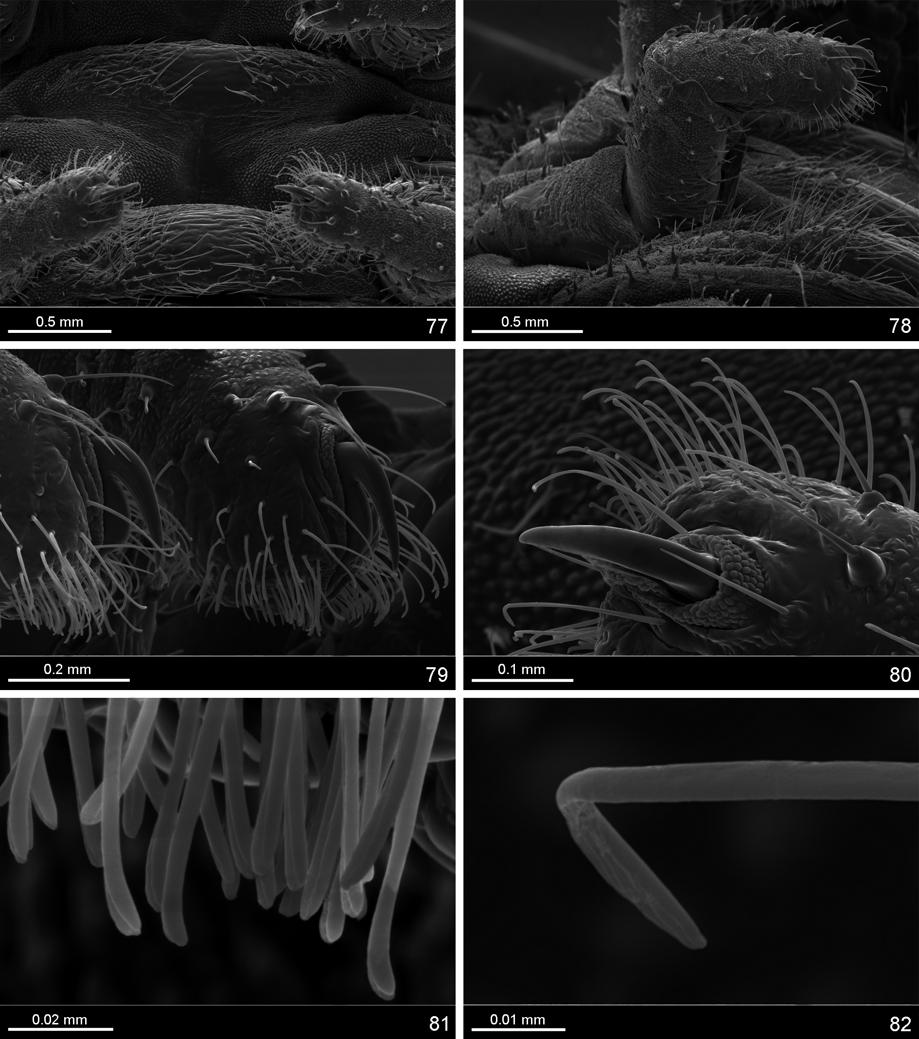
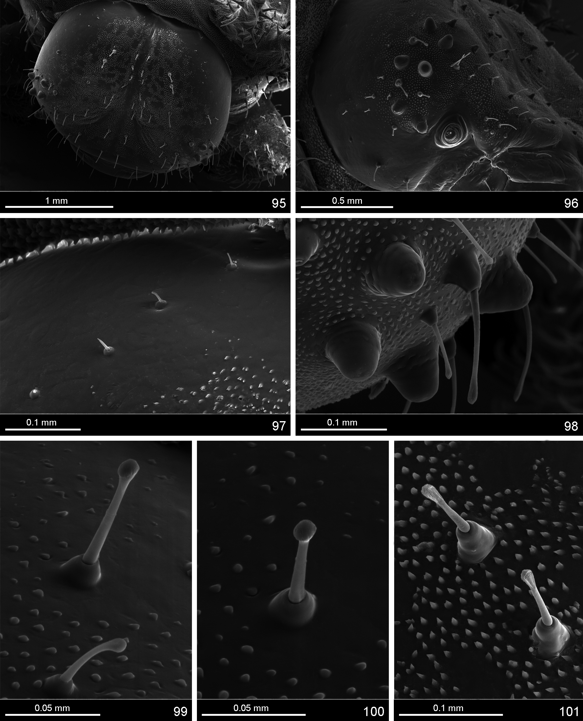

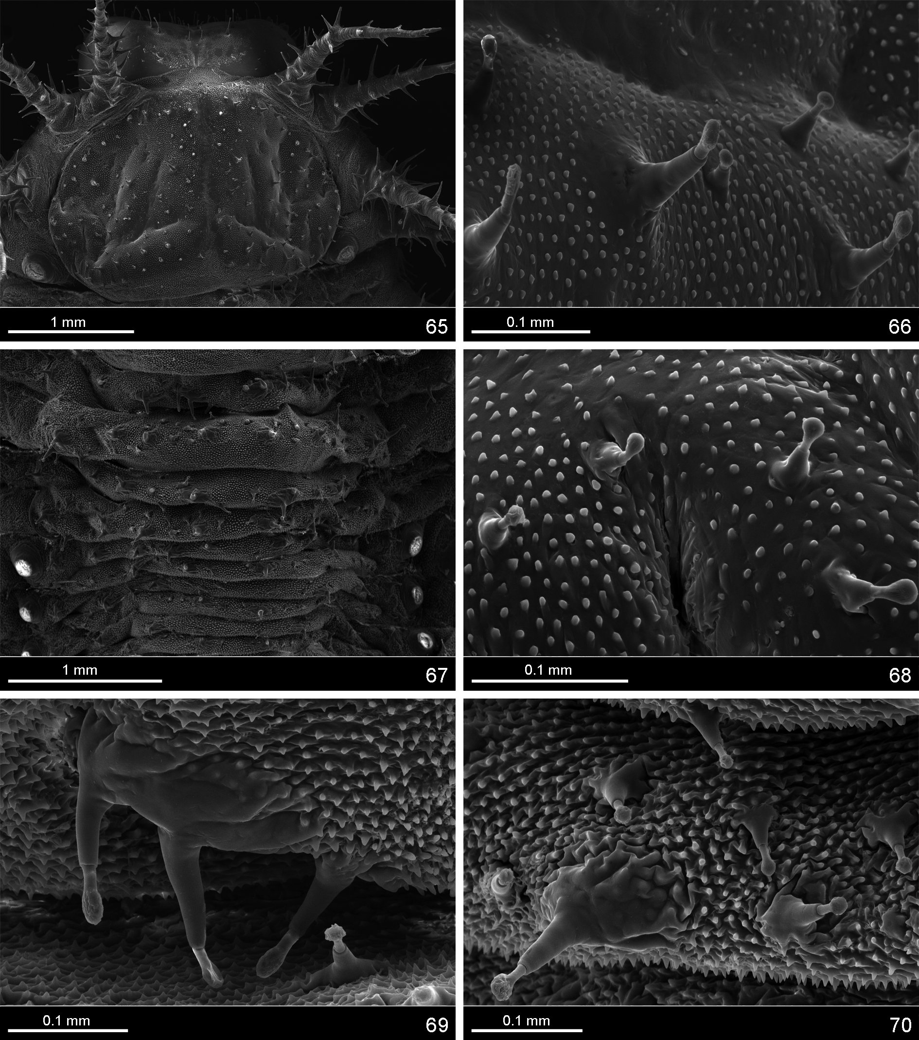

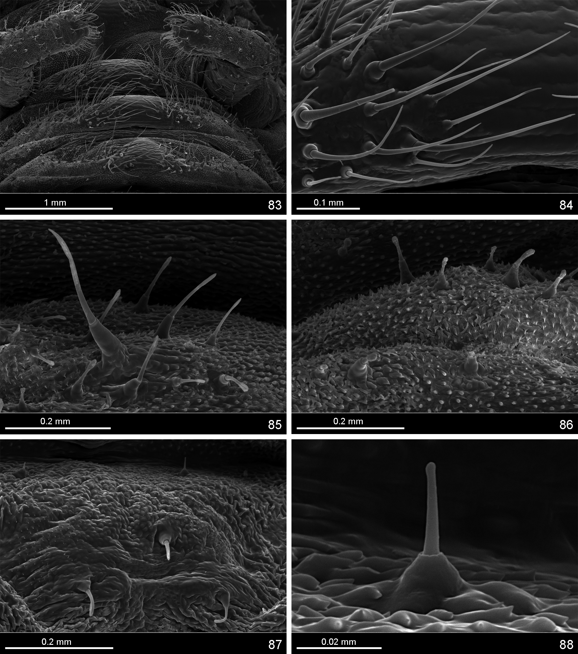
TABLE 1. Measurements of examined immature instars of Chelymorpha reimoseri.
| length of body | width of body across length of supra-anal width of head | |
|---|---|---|
| [mm] | metanotum [mm] processes [mm] [mm] | |
| first instar | ||
| 1. | 0.62 | 0.52 2.62 0.50 |
| 2. | 0.67 | 0.52 2.50 0.45 |
| 3. | 0.70 | 0.50 2.60 0.45 |
| 4. | 0.70 | 0.52 2.50 0.45 |
| 5. | 0.72 | 0.52 2.50 0.45 |
| 6. | 0.75 | 0.50 2.60 0.45 |
| 7. | 0.75 | 0.52 2.25 0.50 |
| 8. | 1.25 | 0.67 2.73 0.47 |
| 9. | 1.25 | 0.62 3.10 0.47 |
| 10. | 1.43 | 0.96 3.00 0.50 |
| 11. | 1.50 | 0.72 2.28 0.50 |
| 12. | 2.25 | 0.95 2.75 0.50 |
| fifth instar | ||
| 1. | 6.00 | 3.80 3.00 1.90 |
| 2. | 7.00 | 4.10 2.90 1.90 |
| 3. | 7.70 | 3.50 3.00 1.80 |
| 4. | 7.80 | 3.90 3.00 1.90 |
| 5. | 7.80 | 3.50 2.90 1.80 |
| 6. | 8.00 | 4.20 3.00 2.10 |
No known copyright restrictions apply. See Agosti, D., Egloff, W., 2009. Taxonomic information exchange and copyright: the Plazi approach. BMC Research Notes 2009, 2:53 for further explanation.