
Pseudocephennium maximum Jałoszyński
|
publication ID |
https://doi.org/ 10.11646/zootaxa.4088.4.7 |
|
publication LSID |
lsid:zoobank.org:pub:59D52D3C-3FD7-42A0-BDE9-8FC58DA38787 |
|
DOI |
https://doi.org/10.5281/zenodo.6068502 |
|
persistent identifier |
https://treatment.plazi.org/id/03898790-666B-FFA8-AFF0-FBCABA41F95F |
|
treatment provided by |
Plazi |
|
scientific name |
Pseudocephennium maximum Jałoszyński |
| status |
|
Pseudocephennium maximum Jałoszyński View in CoL
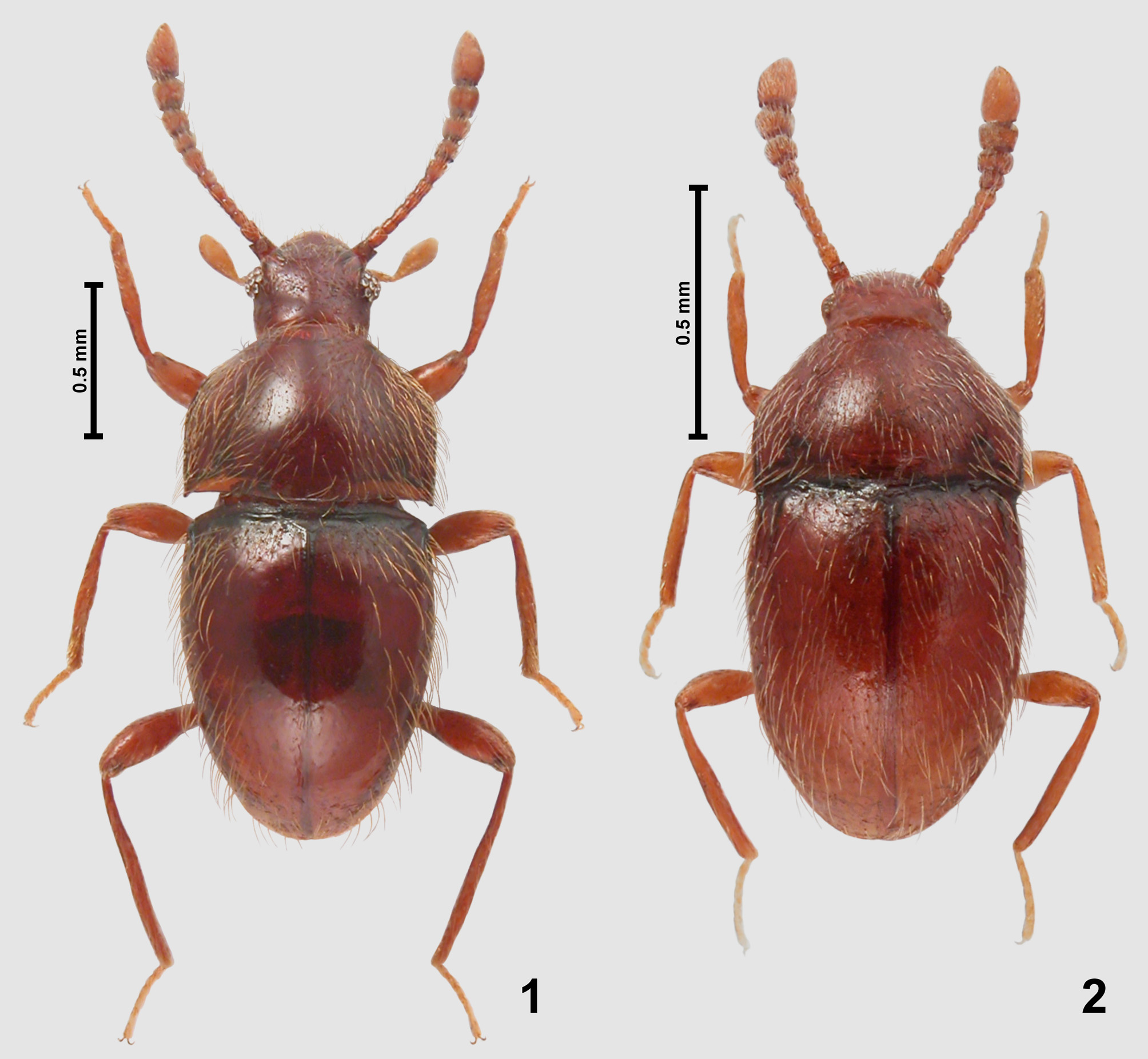
( Figs 1 View FIGURES 1 – 2 , 3–6, 15–18, 22)
Pseudocephennium maximum Jałoszyński, 2012: 467 , figs 10E–G, 11J–K.
Material studied: VENEZUELA (Aragua State): 6 ♂♂, 2 ♀♀, Rancho Grande (i.e., Henri Pittier National Park), near Maracay, presumably spring 1982 (see Materials and methods), leg. H. Franz (NHMW, cPJ).
Remarks. This species has been known so far from a single holotype male collected in the same area (Jałoszyński 2012). The aedeagi of newly collected specimens show some degree of variability, restricted to apical structures. Closer examination revealed that the aedeagus of the holotype (Figs 5–6) was fully erected, with the median projection (mep) rotated dorsally until it touched the dorsal wall of median lobe, lateral apical projections (lap) slightly spread apart, and membranous projections (mmp) extended laterally and inflated. In repose (Figs 3– 4), the median projection (mep) is pointing dorsodistally, the apical portions of lateral projections (lap) overlap at middle, and the membranous projections (mmp) are parallel to the long axis of the aedeagus and touching mesally, forming loosely folded membranous sacks. In the erected condition the aedeagus looks so different that initially newly found specimens with the endophallus in repose were taken for a new species. However, during preparation it was found that the median projection was movable and could rotate dorsally, and analysis of all apical structures revealed that they were movable but in all studied specimens their shape was the same. The only exception are the membranous projections, which are irregular in shape and can be inflated to various extent; in the holotype (Figs 5–6) one projection is inflated to a greater extent than the other.
No known copyright restrictions apply. See Agosti, D., Egloff, W., 2009. Taxonomic information exchange and copyright: the Plazi approach. BMC Research Notes 2009, 2:53 for further explanation.