
Vultocinus anfractus, Ng, Peter K. L. & Manuel-Santos, Marivene R., 2007
|
publication ID |
https://doi.org/ 10.5281/zenodo.178296 |
|
DOI |
https://doi.org/10.5281/zenodo.6250890 |
|
persistent identifier |
https://treatment.plazi.org/id/03898795-FFCD-FFA9-EEA2-FCC69ABDFD2E |
|
treatment provided by |
Plazi |
|
scientific name |
Vultocinus anfractus |
| status |
sp. nov. |
Vultocinus anfractus View in CoL , new species
Figs. 1–5 View FIGURE 1 View FIGURE 2 View FIGURE 3 View FIGURE 4 View FIGURE 5 , 8 View FIGURE 8 A, 9A, 10A, 11A, 12A, 13A
Material examined. Holotype: male (cl 19.5 mm, cw 20.3 mm) ( NMCR 19114); station M27, Santiago, Maribojoc Bay, Panglao, Bohol, Philippines, 9°0.043'N 123°0.051'E, coll. 23 Jun 2004. Paratypes: 1 male (cl 19.9 mm, cw 22.0 mm) ( ZRC), station P1, 90– 200m, Maribojoc Bay, Panglao, Bohol, Philippines, 9°36.1'N 123°45.0'E, coll. fishermen with tangle nets, 27 Jun 2004; 1 male (cl 27.3 mm, cw 25.3 mm) ( ZRC, ex NMCR 19115), Maribojoc Bay, Panglao, Bohol, Philippines, in tangle nets, ca. 200m, coll. J. Arbasto, Oct 2004 – Feb 2005; 1 male (cl 27.4 mm, cw 29.0 mm) ( MNHN), Maribojoc Bay, Panglao, Bohol, Visayas, Philippines, in tangle nets, 80–140m, coll. J. Arbasto, between 2004 and 2005. Non-type: male (cl 15.6 mm, cw 16.8 mm) ( ZRC), station DB16, sandy slope with coral patches, Tutuba off Santo, Vanuatu, in tangle nets, ₁₅․₃₆ ° S 167.16°E, 32̄ 40 mm depth, coll. J. Arbasto, 14 Sep 2006.
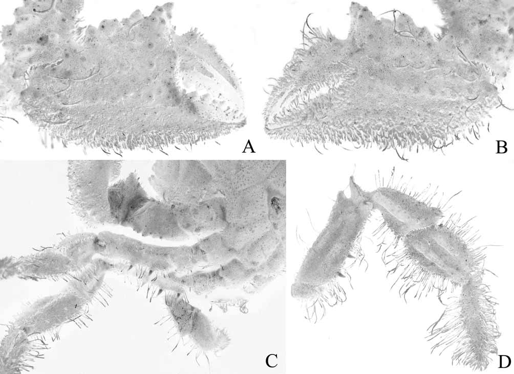
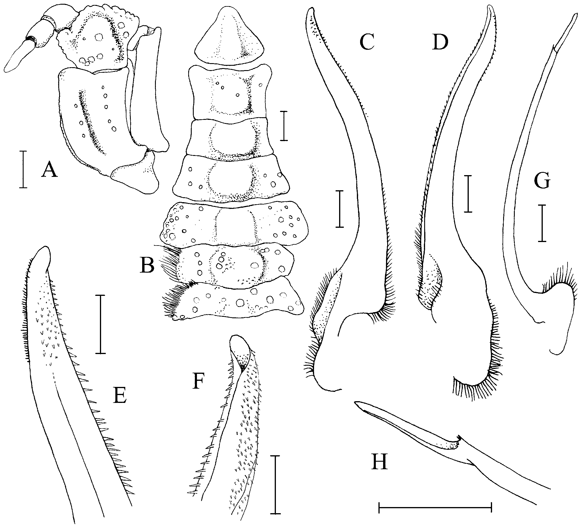
Description of male holotype. Carapace subquadrate, squarish to slightly broader than long; dorsal surface relatively flattened with low convex surfaces; regions well defined, separated by deep, prominent grooves, anterior part more convex than posterior part, marked by prominent areolae, covered with numerous, densely packed very short, stiff, black setae, velvet-like, partially obscuring surface but not completely obscuring margins; 1F divided into 2 cone-shaped lobes; 2F complete, surface uneven; 1M complete; 2M with median depression; 3M not continuous; 4M laterally long, distinct; 1P with small areolae; 2P long, narrow; 2L triangular in shape; 3L elongated, triangular in shape; 6L clearly demarcated, with tubercles of varying sizes; 3R with small areolae ( Figs. 1 View FIGURE 1 , 2 View FIGURE 2 , 3 View FIGURE 3 A, D). Frontal region almost one-third width of carapace, with a distinct notch formed by 2 conical extensions ( Figs. 2 View FIGURE 2 , 3 View FIGURE 3 A, D). Orbits rounded, eyes fitting tightly into cavity; margins areolate with small flat granules on rim, separated into 3 parts by deep sulci ( Fig. 3 View FIGURE 3 A, D); supra-orbital border tuberculated, each tubercle with smaller granules on surface, with prominent inner orbital tooth and lower external orbital tooth, with 2 smaller teeth between which are separated by a deep narrow fissure, one tooth separated from inner orbital tooth by a cleft, the other separated from outer orbital tooth by a deep narrow fissure ( Fig. 3 View FIGURE 3 A, D); infra-orbital border more or less elevated, with 3 deep notches ( Fig. 3 View FIGURE 3 A, D). Anterior dorsal and distal parts of eyestalks fringed with long thick and thin setae. Antennal basal segment almost reaching small ventral prolongation of orbital hiatus; flagellum slender, long, about twice length of diameter of orbit ( Fig. 3 View FIGURE 3 D). Antennules folding obliquely ( Fig. 3 View FIGURE 3 D). Suborbital and pterygostomial surfaces tomentose but smooth underneath; subhepatic region tomentose with small scattered granules. Anterolateral margin with 4 teeth (excluding external orbital tooth), first tooth smallest, areolate, with 3 small flat granules on rim; second tooth bigger, areolate, ringed with small flat granules; third tooth longest, more conical, with 1 flat granule, directed anteriorly; fourth tooth low, rounded, ringed by small flat granules ( Figs. 1 View FIGURE 1 , 2 View FIGURE 2 , 3 View FIGURE 3 A). Posterolateral margin almost straight, gently converging towards posterior carapace margin, lined with rather 5 large rounded granules that form 3 granulated crests ( Figs. 1 View FIGURE 1 , 2 View FIGURE 2 , 3 View FIGURE 3 A). Posterior carapace margin gently convex, median part slightly concave ( Figs. 3 View FIGURE 3 A, 11A).
Third maxilliped almost completely covered by very short tomentum; merus quadrate, antero-external angle expanded, 3 flat granules lining mid-anteroexternal angle towards upper margin of merus; merus medially concave with a deep sulcus near lateral portion, a shallower sulcus near inner margin; ischium subquadrangular, with a deep median sulcus; exognath straight, inner subdistal angle with prominent triangular tooth, with distinct flagellum that extends beyond width of merus ( Figs. 3 View FIGURE 3 B, C, 5A, 8A); ischium and basis separated by prominent suture ( Fig. 5 View FIGURE 5 A). Epistome appears sunken; posterior margin divided into 4 lobes, median lobes rectangular, separated by deep narrow fissure, lateral lobes separated from median lobes by uneven cleft ( Fig. 3 View FIGURE 3 B, D). Endostome with long, prominent ridge running from distal edge of median lobe of posterior margin of epistome to near mandibles, interrupted subdistally, just as epistomal lobe ends.
Both chelipeds large, subequal, laterally flattened, almost entirely covered with dense heavy tomentum ( Figs. 2 View FIGURE 2 , 3 View FIGURE 3 D, 4A, B). Merus of cheliped with 2 large granules on proximal portion, 2 spinules on distal part, forming 2 granulated crests; carpus with 2 large tubercles, several nodules, granules on outer dorsal surface. Ischio-merus fused but suture still discernible. Chela somewhat flattened laterally; outer surface covered with numerous granules and tubercles of various sizes, with one median longitudinal ridge on outer surface evident, inner subventral surface with a curved row of sharp, orange-tipped tubercles; fingers shorter than palm; propodal finger with 3 nodules on inner margin, immovable finger with lower margin covered with dense thick tomentum, elevated, forming stiff fringe; dactylus with 2 deep sulci filled with short thick tomentum; dark brown pigmentation present only on distal part of fingers and cutting edges ( Fig. 4 View FIGURE 4 A, B).
Ambulatory legs relatively long, relatively stout, thickly fringed by dense, stiff setae; P3 longest, P5 shortest, distinctly upturned ( Figs. 1 View FIGURE 1 , 2 View FIGURE 2 ). Merus of P2–P4 with 2 longitudinal ridges (anterior one larger), distinctly meeting near distal segment on outer surface, margins fringed by dense stiff thick setae, margins somewhat carinate, dorso-subdistal area with deep cleft that forms prominent subdistal tooth with rounded tip and larger sharp sublamellate distal tooth which joins distinctly carinate proximal margin, area appears cup-like; merus of P5 with 1 longitudinal bony ridge, cleft, 2 teeth, cup-like part on dorso-subdistal area less prominent ( Figs. 2 View FIGURE 2 , 3 View FIGURE 3 A, 4C, D); ventral surface of merus of P2–P4 with 2 or 3 long perpendicular spines on posterior margin, scattered granules on anterior margin; ventral surface of merus of P5 without spines, only granules on posterior margin, scattered much smaller granules on anterior margin ( Figs. 2 View FIGURE 2 , 3 View FIGURE 3 A, 4C, D). Carpus with prominent median ridge lined with several flat granules ( Figs. 2 View FIGURE 2 , 3 View FIGURE 3 A, 4C, D). Propodus of P2–P5 with 2 prominent median granular ridges ( Figs. 2 View FIGURE 2 , 4 View FIGURE 4 D). Dactylus long, as long as merus, covered by dense thick setae which hides tip, distal part not pigmented ( Figs. 2 View FIGURE 2 , 4 View FIGURE 4 C, D).
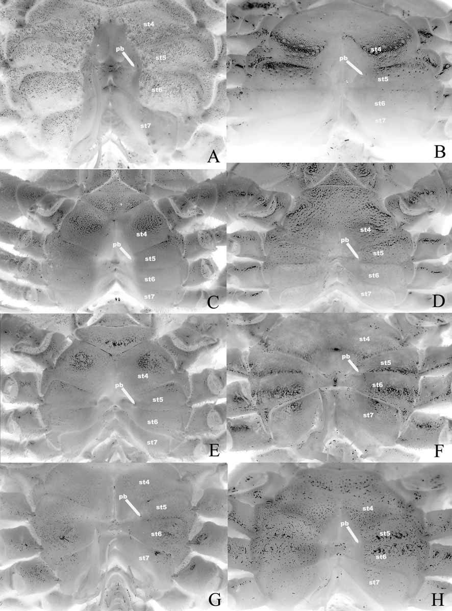
Thoracic sternum relatively narrow transversely, entire surface covered with very short tomentum, small granules ( Figs. 3 View FIGURE 3 B, C, 9A, 10A, 12A). Sternite 3 with shallow but distinct median longitudinal groove; sternite 4 as long as sternites 5 and 6 combined; sternite 7 very narrow longitudinally, posterior part tapering abruptly ( Fig. 10 View FIGURE 10 A), with posterior part of episternite 7 prolonged posteriorly to form spur which just touches coxa of P5 and covers penis ( Fig. 13 View FIGURE 13 A, B); sternite 8 not exposed when abdomen closed ( Fig. 10 View FIGURE 10 A). Longitudinal line running through median part of sternites 7 and 8, those on sternites 4 and 6 incomplete, that of sternite 5 undiscernible ( Fig. 12 View FIGURE 12 A). Suture 1/2 absent; suture 2/3 complete; suture 3/4 distinct with median part relatively shallower; sutures 4/5 and 5/6 medially interrupted ( Fig. 12 View FIGURE 12 A); suture 6/7 appears complete but median portion very shallow ( Fig. 12 View FIGURE 12 A); suture 7/8 distinctly complete with median part deep ( Fig. 12 View FIGURE 12 A). Episternites 4–7 not prominently expanded laterally ( Figs. 9 View FIGURE 9 A, 10A, 12A). Sterno-abdominal cavity relatively narrow transversely, almost reaches anterior edge of sternite 4 ( Figs. 9 View FIGURE 9 A, 12A). Press button of abdominal locking mechanism distinct, on posterior outer edge of sternite 5 of sterno-abdominal cavity ( Fig. 12 View FIGURE 12 A). Penis soft, effectively coxal, exits at base of coxa of P5 but covered by posterior prolongation of episternite 7 ( Figs. 13 View FIGURE 13 A, B).
Abdomen relatively narrow transversely, outer surfaces mostly covered with small low granules, very short, dense tomentum ( Figs. 5 View FIGURE 5 B, 9A, 11A). Telson triangular with gently concave lateral margins, rounded apex ( Figs. 5 View FIGURE 5 B, 9A); somite 1 longitudinally and transversely broad, trapezoidal, exposed, not covered by posterior carapace margin ( Fig. 11 View FIGURE 11 A), somites 2–6 relatively narrow transversely, somite 3 broadest transversely; somite 2 with convex lateral margins, subrectangular in shape; somite 3 with gently convex lateral margins, trapezoidal; somites 4 larger than somite 5, both trapezoidal with gently concave lateral margins; somite 6 as long as broad, lateral margins concave ( Figs. 9 View FIGURE 9 A, 10A); sutures between all somites and telson distinct, but somites 3 and 4 immovable, somites 4 and 5 slightly mobile ( Figs. 5 View FIGURE 5 B, 9A, 11A).
Penis effectively coxosternal although it exits clearly from coxa, enters directly into sterno-abdominal cavity, not protected by sternal plates except for posterior part of sternite 7 which forms a spur-like structure that covers it ( Figs. 13 View FIGURE 13 A, B). G1 relatively long, slender, with the rounded tip reaching suture of sternite 4, distal surfaces lined with numerous small spinules, tip open ( Figs. 5 View FIGURE 5 C–F). G2 as long as G1, very slender, long, with tip reaching beyond mid-portion of suture of sternite 5, distal segment ca. 0.2 times length of subdistal segment, base of distal segment forming cup-like structure lined with several spinules ( Figs. 5 View FIGURE 5 G, H). Females not known.
Colour. The carapace and pereiopods are white without any distinct pigmentation, but this is barely discernible as the dark brown or orangish-brown tomentum and setae covers most of the animal ( Fig. 1 View FIGURE 1 ). The mandibles are a dark brown, like dark-coloured chitin, and this colour stays even after many months and many changes of alcohol ( Fig. 8 View FIGURE 8 A). The posterior margin of the epistome is lined with small orange and white granules when fresh. Some of the the larger granules on the carapace and pereiopods (especially those on the chelipeds) are orange.
Etymology. The name is derived from the Latin anfractus for having many turns, alluding to the complex sculpture on the carapace.
Remarks. The unusual carapace and pereiopods of V. anfractus are unique in the Goneplacoidea as presently understood. The complex labyrinth-like setal pattern is reminiscent of pilumnids like Pilumnus labyrinthicus Miers, 1884 , or pseudoziids like Planopilumnus spongiosus ( Balss, 1933) although they are clearly unrelated (see discussion above). Certainly none possess such a complex setal pattern and the ambulatory legs are not armed with spines. The dense carapace and pereiopod setation of V. anfractus superficially resembles those of the mathildellid, Intesius pilosus Guinot & Richer de Forges, 1981, but in the latter species, the setae are more scattered and the carapace lacks the dense setae and distinct setal arrangement of V. anfractus (see also Crosnier & Ng 2004).
The holotype male of V. anfractus was collected from the rocky shoreline outside a Nipa mangrove near a river. It was crawling along on the shore in the afternoon when it was caught by the second author. We believe it had been actually caught by fishermen with tangle nets in the waters offshore as they had been observed cleaning their nets there earlier. In addition, there is a small piece of net stuck at the base of the chela of the specimen. It was almost certainly thrown onto the shore just before it was collected. The larger paratype male was collected from about 200m depth hiding inside a piece of sunken wood about 0.7 m in length and 0.3 m in diameter which was stuck in a tangle net (J. Arbasto, pers. comm.). The colour of the body and legs (dark brown) matches the wood substrate where the animal was found and it uses the spiniform ventral margins of the ambulatory meri to help anchor itself to the wood (J. Arbasto, pers. comm.). Freshly caught specimens were very dirty, being covered with detritus and mud, clearly a consequence of its setose body and pereiopods. This probably aids in camouflaging them against the wood as well.
The single male specimen of Vultocinus anfractus collected from Vanuatu was from a tangle net set at about ₃₂ ɭΟ ɬ0 m depth. The specimen was obtained alive but we have no indication if it had been living on wood. This specimen differs from the type series of the species in having an orangish-brown body (versus dark brown), somewhat rounder carapace, and proportionately slightly longer ambulatory legs ( Figs. 1 View FIGURE 1 C, 2C). It agrees with the type material, in almost all other aspects, and we prefer to regard it as conspecific with the Vultocinus anfractus material from the Philippines for the moment. Although mature, it is the smallest specimen of Vultocinus available, and the differences observed may well be due to size and habitat. A larger series of specimens from Vanuatu or other areas will be needed to ascertain this. The specimen of V. anfractus was observed alive for several hours before being preserved. It moved slowly and was not aggressive when handled, but that may have been due to it being tangled in the nets for many hours. When placed against a hard substrate, the ambulatory legs invariably clamp backwards, confirming what we believe the spines on the meri are used for.
It is unfortunate that all the specimens collected were males, so key female characters could not be determined.


No known copyright restrictions apply. See Agosti, D., Egloff, W., 2009. Taxonomic information exchange and copyright: the Plazi approach. BMC Research Notes 2009, 2:53 for further explanation.