
Gebiacantha fortispinata, Komai, 2020
|
publication ID |
https://doi.org/ 10.11646/zootaxa.4881.2.5 |
|
publication LSID |
lsid:zoobank.org:pub:7E570EDA-1A01-45AB-8ED2-79F8B8A6B9FE |
|
DOI |
https://doi.org/10.5281/zenodo.4328019 |
|
persistent identifier |
https://treatment.plazi.org/id/005FFE26-CBEC-4A8C-A4CB-584B39D3480E |
|
taxon LSID |
lsid:zoobank.org:act:005FFE26-CBEC-4A8C-A4CB-584B39D3480E |
|
treatment provided by |
Plazi |
|
scientific name |
Gebiacantha fortispinata |
| status |
sp. nov. |
Gebiacantha fortispinata View in CoL n. sp.
[New Japanese name: Toge-toge-anajyako]
( Figs 6–9 View FIGURE 6 View FIGURE 7 View FIGURE 8 View FIGURE 9 )
Material examined. Holotype: CBM-ZC 16176, female (cl 7.4 mm), Apo-gama, Manza, Onna Village , Okinawa Island , Ryukyu Islands , 30 m deep, 10 January 2010, yabby pump, coll. Naoki Shirakawa.
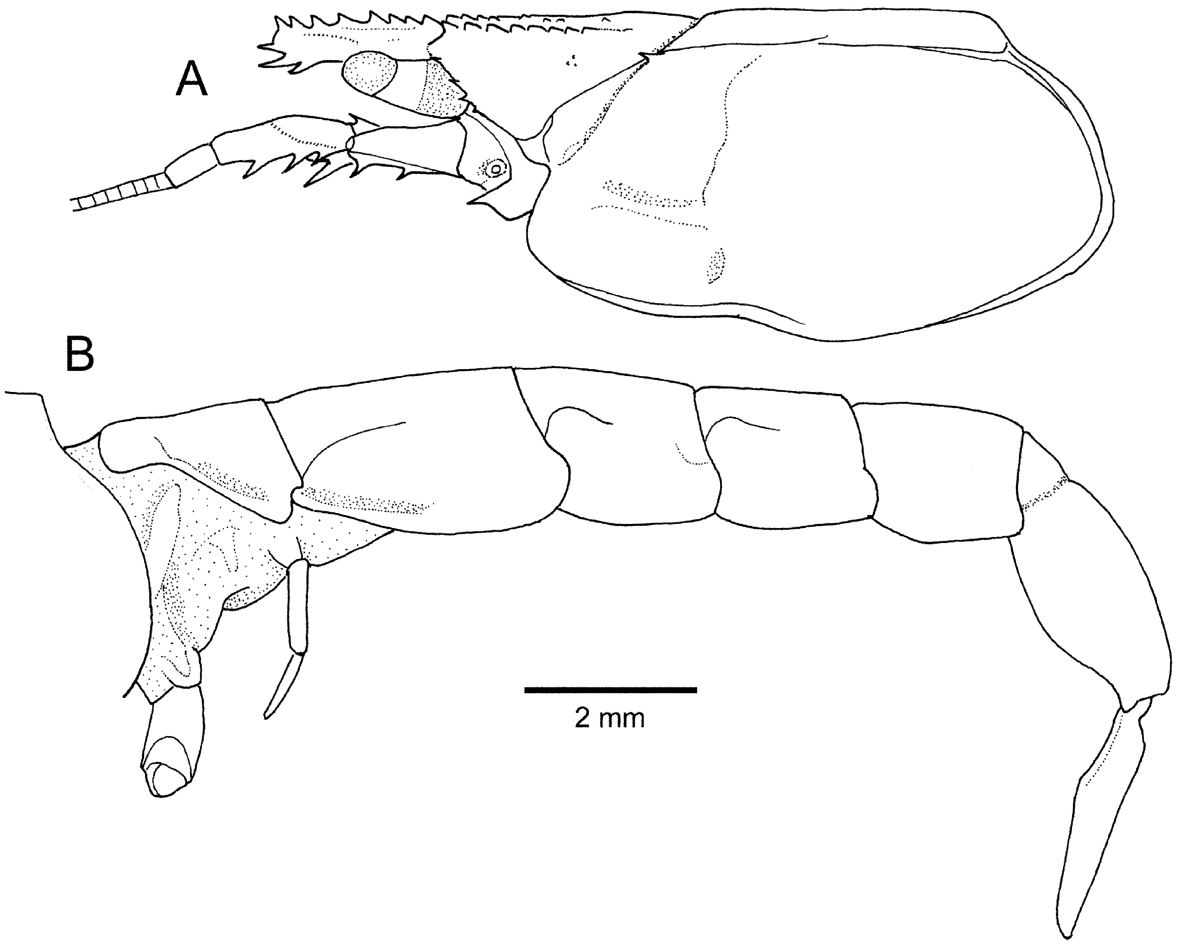
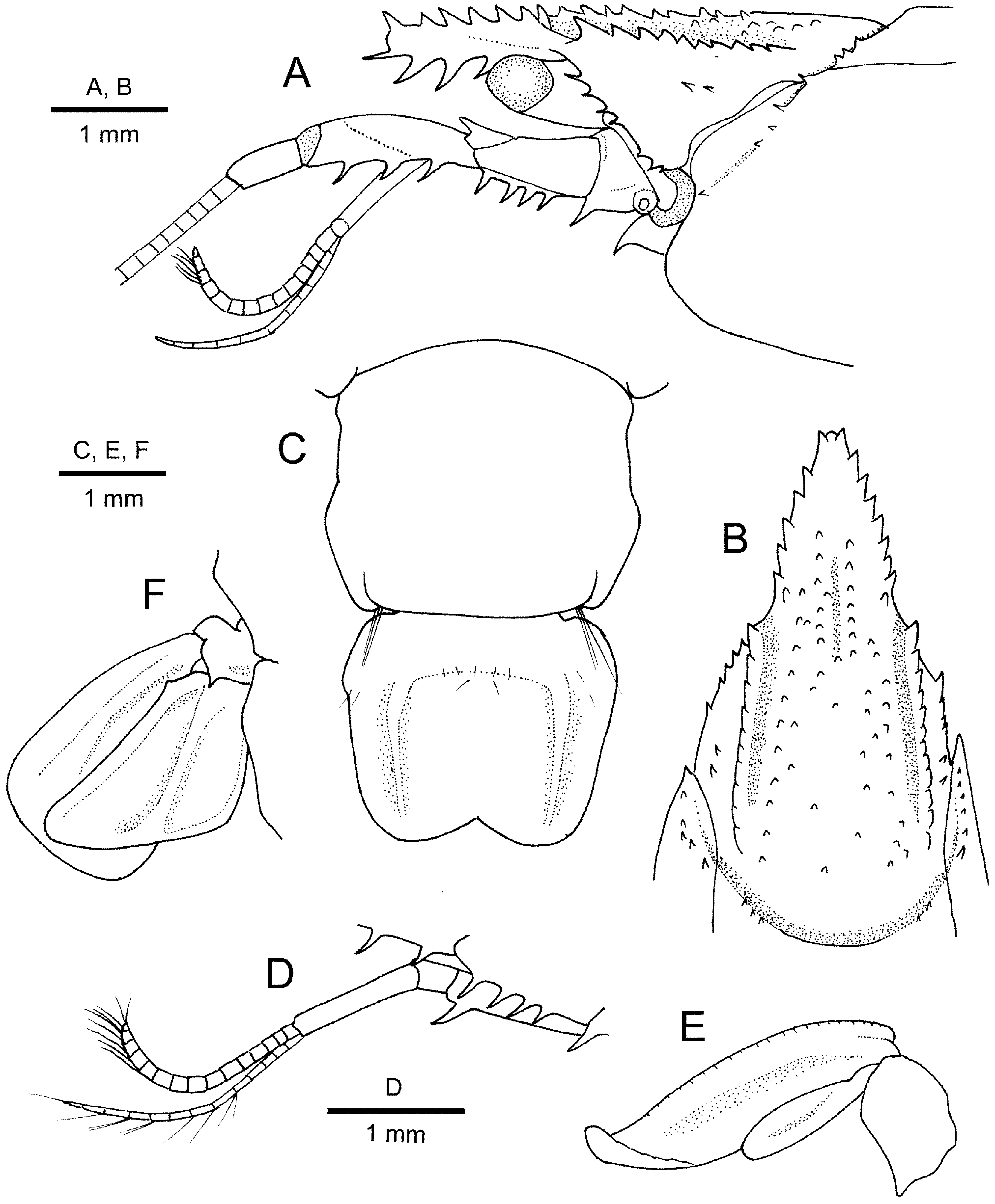
Description. Rostrum ( Figs 6A View FIGURE 6 , 7A, B View FIGURE 7 ) long, reaching beyond midlength of article 4 of antennal peduncle, subtriangular in dorsal view, 1.3 times as long as basal width, directed forward in lateral view, somewhat flattened dorsoventrally; dorsolateral margins with 7 (left) and 8 (right) narrowly spaced, relatively large spiniform tubercles, each tubercle directed anterodorsally, anteriormost pair terminal in position; dorsal surface setose, with shallow median sulcus proximally; ventral surface nearly straight, armed with 4 strong, moderately spaced spines in distal half, distalmost spine most slender, directed anteriorly; rostral apex roundly truncate in lateral view, overreached by dorsal terminal spines and distalmost ventral spine. Carapace ( Fig. 6A View FIGURE 6 ) somewhat compressed laterally; anterior part ( Fig. 7A, B View FIGURE 7 ) with lateral gastric ridges slightly diverging posteriorly, each terminating anteriorly in small spine and bearing row of 11 spiniform tubercles decreasing in size toward posterior; gastric region flanked by shallow longitudinal grooves running along gastric lateral ridges and having 2 irregular, longitudinal rows of small spiniform tubercles, extending anteriorly to rostral base on either side of midline; median part almost flat, unarmed; anterolateral margin with 7 spinules, including postocular spine; postorbital region with 2 spinules; pterygostomial margin rounded, unarmed; linea thalassinica extending from anterolateral notch to posterodorsal end of carapace, interrupted at midway between cervical groove and carapace posterior margin; posterior part with shoulder along cervical groove bearing row of spinules extending to anterolateral notch; branchiostegite poorly calcified.
Pleon ( Fig. 6B View FIGURE 6 ) fairly flattened dorsoventrally. Pleuron 1 narrow, ventrally rounded, demarcated from tergum by distinct longitudinal groove; pleuron 2 also narrow, anterior part demarcated from tergum by distinct longitudinal groove reaching to midlength of somite, ventral margin slightly upturned. Pleomeres 3–5 each with short curved groove on anterior part; pleural margins with row of seta directed laterally, each posterolateral margin rounded. Pleomere 6 ( Fig.7C View FIGURE 7 ) longest, 1.1 times as long as greatest width at 0.6 length of somite; lateral margins slightly sinuous, with low convexity at 0.6 length; posterior margin nearly straight, with small notch on each lateral part. Telson ( Fig. 1C View FIGURE 1 ) subrectangular, 1.2 times as long as greatest width at anterior 0.2; dorsal surface with clearly delimited longitudinal ridges laterally, proximal part slightly elevated, though not forming distinct transverse carina; lateral margins unarmed; posterior margin bilobed with deep median notch, fringed with fine long setae.
Ocular peduncle ( Fig. 7A View FIGURE 7 ) moderately stout, subcylindrical, unarmed, not reaching midlength of rostrum, slightly widened basally; cornea terminal, pigmented, corneal width subequal to diameter of eyestalk.
Antennular peduncle ( Fig. 7A, D View FIGURE 7 ) not reaching distal margin of antennal peduncle article 4. Article 1 subequal in length to distal 2 articles combined, armed with prominent ventrodistal spine directed anteriorly; statocyst lobe slightly inflated. Article 2 shortest, cup-like. Article 3 slightly widened distally, unarmed. Upper flagellum shorter than peduncle consisting of about 16 articles; lower flagellum much slenderer than upper flagellum, consisting of 12 articles.
Antennal peduncle ( Fig. 7A View FIGURE 7 ) article 1 with conspicuous, ventrally directed spine on ventrodistal margin. Articles 2 and 3 fused, unarmed on dorsolateral margin proximally; ventral margin armed with 4 evenly spaced spines. Article 4 with 3 subequal spines on ventral margin.Article 5 less than half-length of article 4, unarmed. Scaphocerite clearly demarcated basally, with 1 small spine dorsodistally. Flagellum longer than carapace, each article with thin setae of various length.
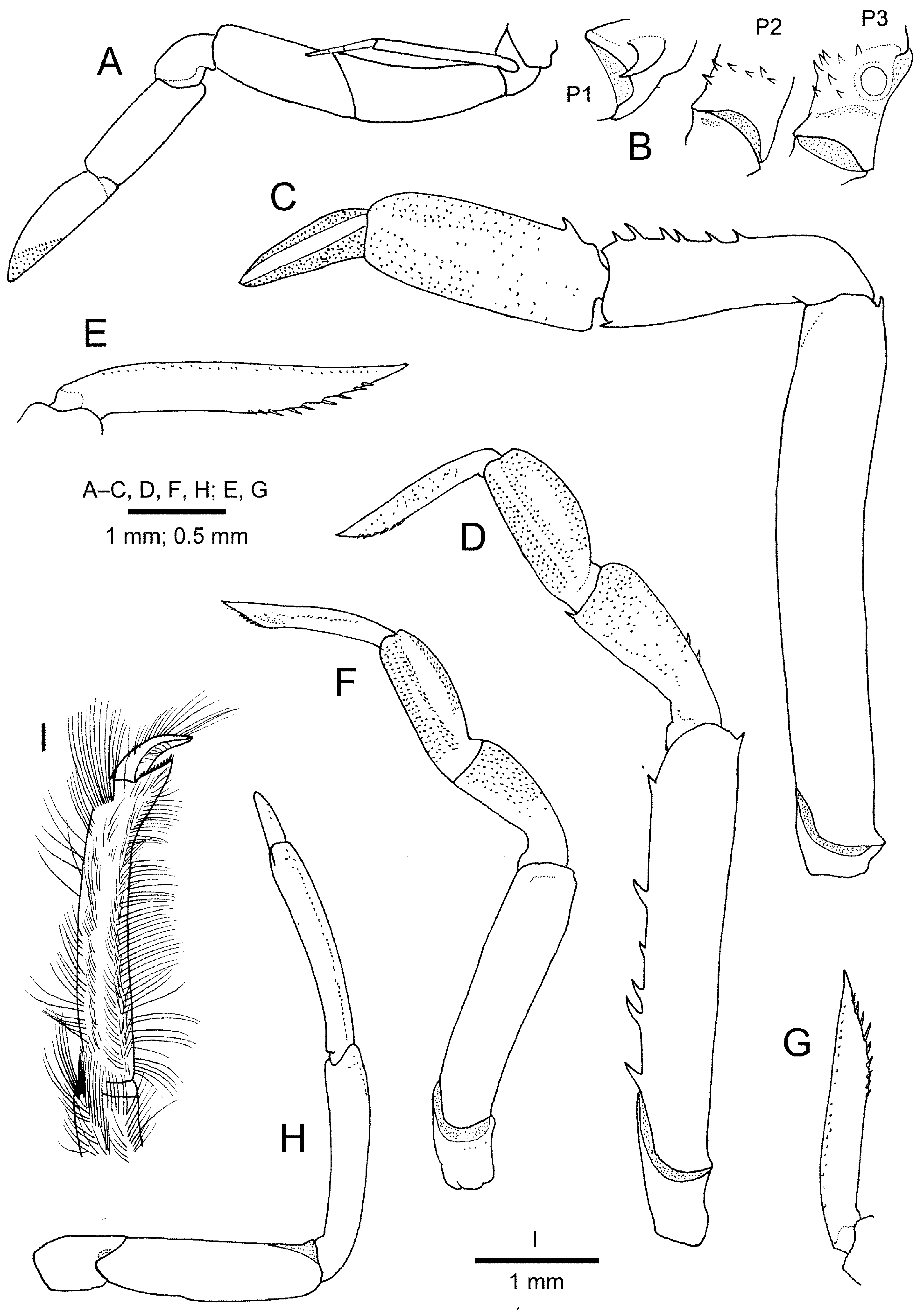
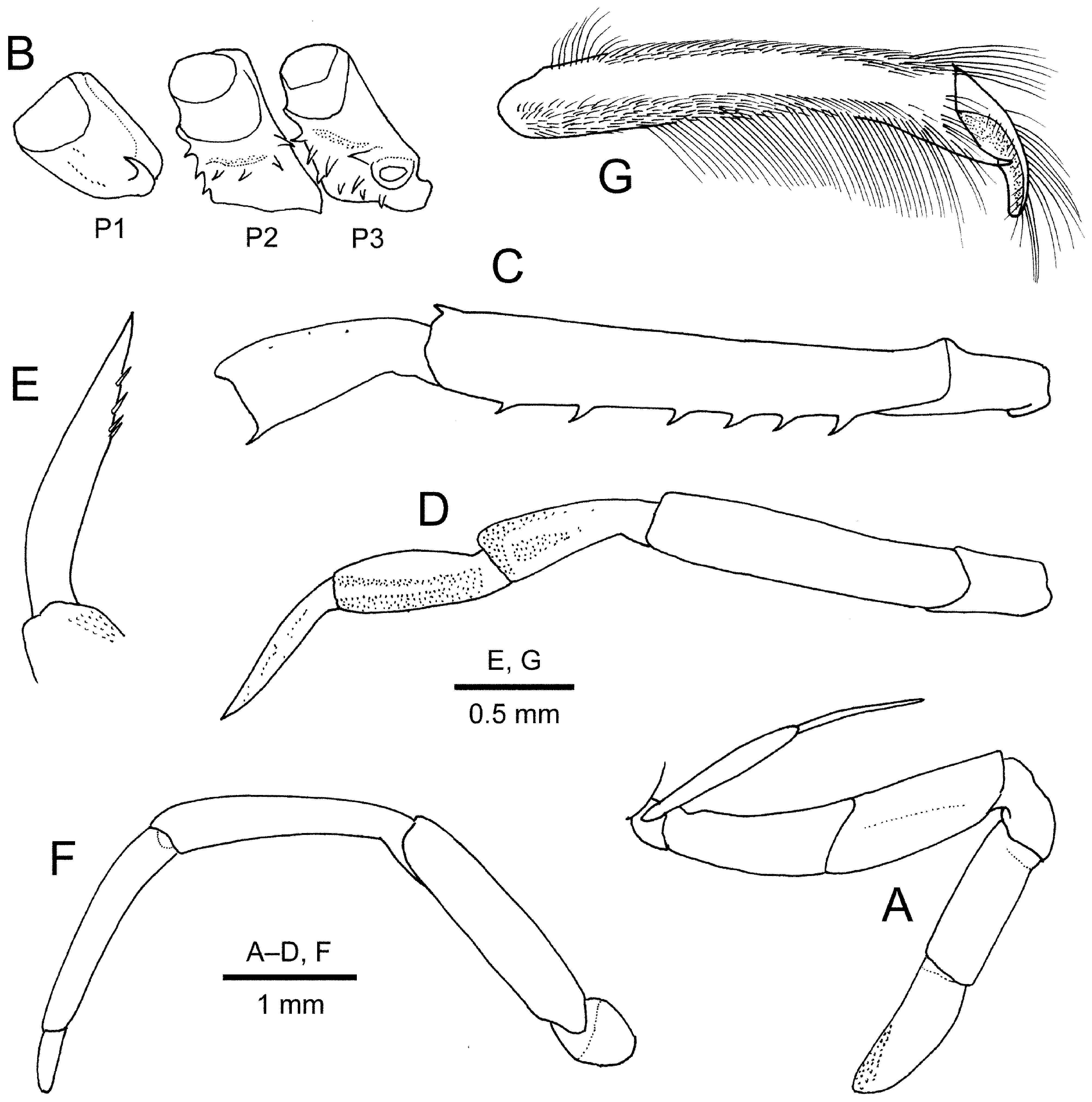
Mouthparts not dissected, though external observation made. Mandible without prominent tooth on occlusal margin of incisor process. Maxillule and maxilla without distinctive features. Maxilliped 1 without epipod. Maxilliped 2 without distinctive features. Maxilliped 3 ( Fig. 8A View FIGURE 8 ) with endopod moderately stout, extending as far as distal end of antennal peduncle; ischium to dactylus with thick long setae on lower (flexor) margins; ischium with 2 spinules on mesial face proximally; merus to propodus unarmed; dactylus nearly straight, subequal in length to propodus, terminating in blunt tip; exopod slender, not reaching dorsodistal margin of ischium, flagellum slender, simple; epipod small, leaf-like.
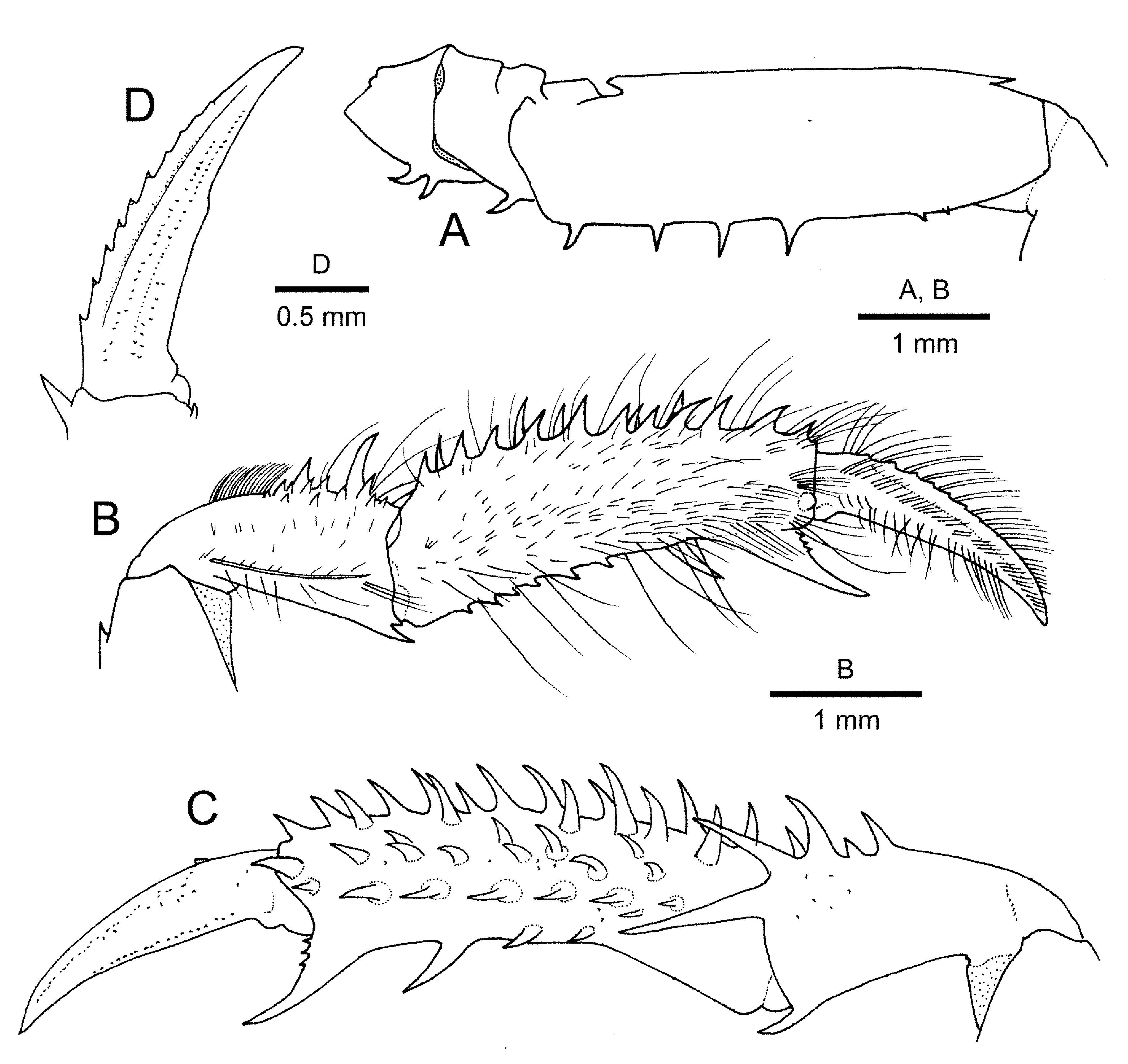
Pereopod 1 ( Fig. 9 View FIGURE 9 A–D) relatively slender, subchelate; setation of merus to carpus typical of upogebiids. Coxa ( Fig. 8B View FIGURE 8 ) with 1 conspicuous procurved spine on ventral surface. Ischium with 2 small hook-like spines on lower margin. Merus with row of 6 evenly spaced, erect spines in proximal 0.6 of lower margin and 2 additional spinules subdistally; upper margin nearly straight, armed with 1 small subdistal spine. Carpus cup-shaped, widened distally, slightly more than half length of palm; upper surface with spines of various size, arranged in 3 irregular rows (distomesial spine strongest); upper lateral surface with shallow longitudinal sulcus, lower lateral margin sharply carinate; mesial face with few small spines, distomesial margin with 2 prominent, elongate spines (lower spine extending proximal 0.25 of palm); lower margin with small subterminal spine. Carpus-propodus articulation with clockwise torsion of about 20° from perpendicular in right side. Palm gently arcuate, about 2.9 times as long as maximal height at proximal end; upper margin with row of 12 small to strong, slender spines and sparse moderately long setae over entire length (distalmost spine terminal); dactylar condyle on lateral face little developed; mesial face with about 30 small to moderately strong spines arranged in 4 irregular longitudinal rows (uppermost row adjacent to palm upper margin); lower margin gently concave, with 1 prominent spine proximal to base of fixed finger and row of 6 small spines in proximal half; setose ridge extending from lower proximal angle to about 0.3 length of palm; fixed finger less than half-length of dactylus, gently curved, occlusal margin with 3 small, acute teeth proximally. Dactylus 0.6 length of palm, slightly curved, terminating in minute corneous claw; upper (extensor) margin bearing about 10 minute denticles becoming obsolete distally and row of numerous setae increasing in length proximally; lateral face with longitudinal carina adjacent to upper margin and 2 rows of setae along midline; mesial face with double row of setae superior to midline; occlusal margin carinate in proximal 0.6, rounded in distal 0.4.
Pereopod 2 ( Fig. 8B View FIGURE 8 ) coxa bearing 5 spinules on ventromesial face; otherwise missing on both sides.
Pereopod 3 (only left preserved) ( Fig. 8C View FIGURE 8 ) with coxa having scattered spinules on mesial face, including 1 conspicuous spine anterolateral to gonopore ( Fig. 8B View FIGURE 8 ). Ischium unarmed. Merus with 1 small upper distal spine and row of 6 spines, widely spaced, on lower margin. Carpus widened distally; upper margin unarmed; lower margin with 1 tiny distal spine. Propodus and dactylus missing.
Pereopod 4 ( Fig. 8D View FIGURE 8 ) with coxa bearing 1 small spine on ventrodistal margin. Ischium to carpus unarmed; carpus cup-shaped. Propodus subequal in length to carpus, with gently arcuate upper margin; lower margin with row of minute denticles in proximal half. Dactylus ( Fig. 8E View FIGURE 8 ) subequal in length to propodus, faintly recurved distally, terminating in acute tip; lower (flexor) margin slightly sinuous, with row of 4 minute spiniform setae in distal 0.3.
Pereopod 5 ( Fig. 8F, G View FIGURE 8 ) subchelate, articles unarmed; no trace of gonopore on coxa. Carpus subequal in length to merus and propodus. Propodus slightly arcuate, with short fixed finger reaching beyond midlength of dactylus. Dactylus curved, terminating in blunt tip.
Arthrobranchs of type C (cf. Ngoc-Ho 1981), composed of deeply divided, narrow lamellae on either side of rachis.
Pleopods 1 ( Fig. 6B View FIGURE 6 ) of 2 articles. Pleopods 2–5 similar; endopods small and slender, about half-length of respective exopod; exopods relatively slender (cf. Fig. 7E View FIGURE 7 ).
Uropod ( Fig. 7F View FIGURE 7 ) with both endopod and exopod reaching posterior margin of telson. Protopod with small spine on posterior margin. Endopod subtrapezoidal with rounded distomesial angle and somewhat produced, rounded distolateral angle, 1.3 times as long as wide; inner margin nearly straight, outer margin slightly concave, with small knob-like tubercle proximally; dorsal surface with median carina, outer margin also bluntly carinate. Exopod widened distally, subtriangular, 1.4 times as long as wide, slightly exceeding beyond endopod, distal margin roundly truncate; upper surface with 2 longitudinal carina.
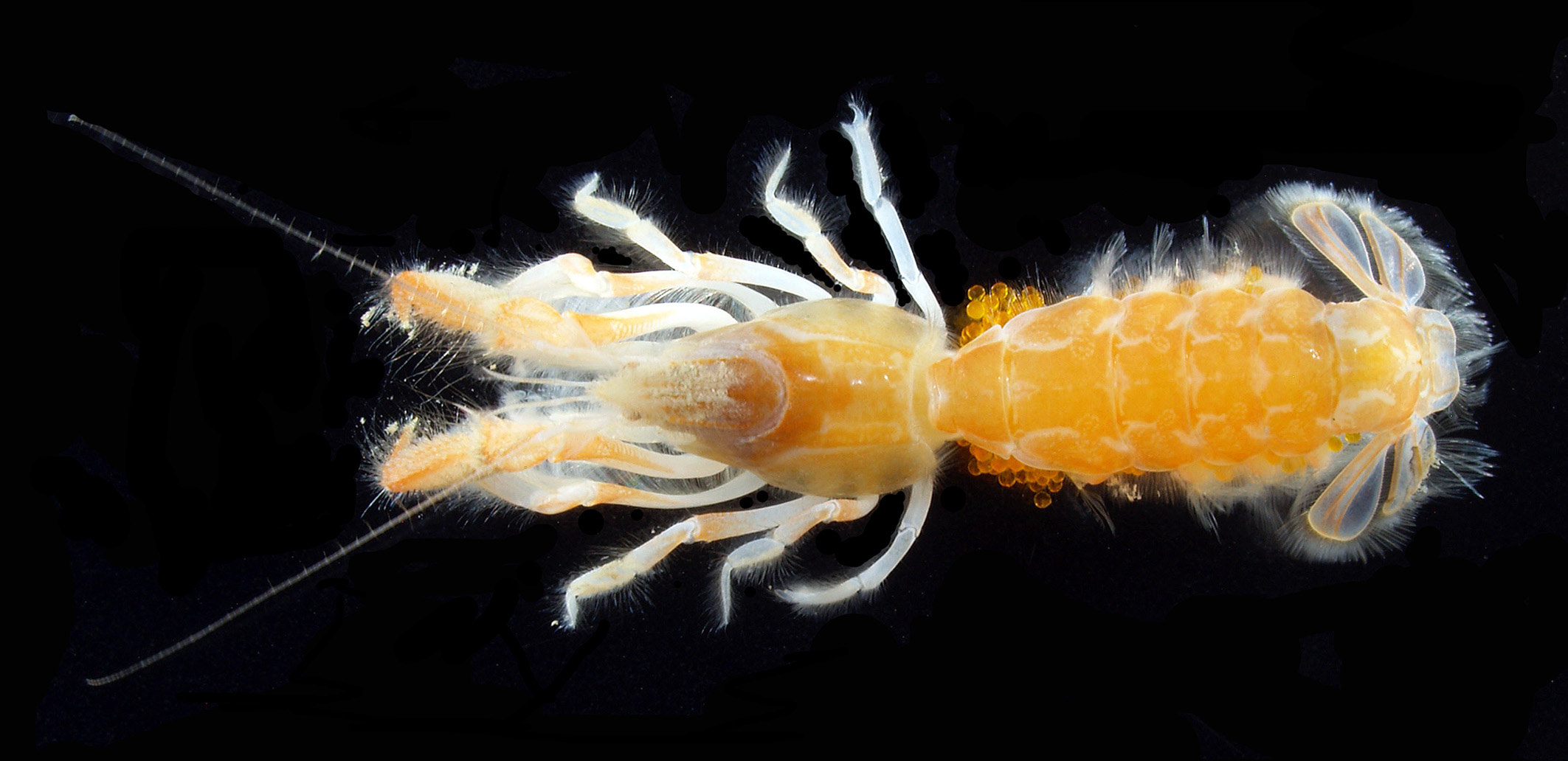
Colouration in life. Not known.
Remarks. The holotype of the new species is parasitized by an unidentified bopyrid, inhabiting the right branchial chamber.
As mentioned above, Gebiacantha fortispinata n. sp. is assigned to the species group characterised by a distally widened, subtriangular uropodal exopod (with a subtruncate distal margin). The new species appears closest to G. multispinosa in having a few spinules on the postocular region of the carapace, six or more spinules on the anterolateral margin of the carapace, many spinules on the shoulder along the cervical groove, 11 or more spines on the upper margin of the pereopod 1 propodus, more than 20 spines, arranged in four longitudinal rows, on the mesial face of the pereopod 1 palm and the presence of denticles on the occlusal margin of the pereopod 1 fixed finger (cf. Ngoc-Ho 1994a). Nevertheless, comparison with the original description of G. multispinosa reveals the following differences warranting the recognition of the new taxon: (1) four rostral ventral spines are rather widely spaced in the distal 0.7 length of the rostrum in G. fortispinata n. sp. ( Fig. 7A View FIGURE 7 ), whereas two or three ventral spines are clustered in the distal 0.3 in G. multispinosa (cf. Ngoc-Ho 1994a: fig. 2B); (2) the telson is relatively narrower with a more deeply notched posterior margin in G. fortispinata n. sp. than in G. multispinosa (1.2 times as wide as long versus 1.5 times: cf. Fig. 7C View FIGURE 7 and Ngoc-Ho 1994a: fig. 2G); (3) the cornea of the eye is relatively larger in G. fortispinata n. sp. than in G. multispinosa (cf. Fig. 7A View FIGURE 7 and Ngoc-Ho 1994: fig. 2B); (4) the pereopod 1 propodus bears six small spines on the proximal half of the lower margin in G. fortispinata n. sp. ( Fig. 9B View FIGURE 9 ), whereas there are about 10 spinules in the proximal two-thirds in G. multispinosa (cf. Ngoc-Ho 1994a: fig. 2C); (5) the pereopod 1 dactylus bears 10 more widely spaced small denticles on the upper margin in G. fortispinata n. sp. ( Fig. 9D View FIGURE 9 ) (versus about 15 denticles more narrowly spaced in G. multispinosa ; Ngoc-Ho 1994a: fig. 2C); (6) the pereopod 3 merus is armed with a small dorsodistal spine in G. fortispinata n. sp. ( Fig. 9A View FIGURE 9 ), while such a spine is absent in G. multispinosa (cf. Ngoc-Ho 1994a: fig. 2F); (8) the posterolateral angle of the uropodal endopod is rounded in G. fortispinata n. sp. ( Fig. 7F View FIGURE 7 ), rather than subacutely pointed in G. multispinosa (cf. Ngoc-Ho 1994a: fig. 2G).
Gebiacantha acanthochela is also similar to the new species in having relatively numerous spines on the upper margin and mesial face of the pereopod 1 propodus. Gebiacantha fortispinata n. sp. is easily distinguished from G. acanthochela by the following particulars: (1) dorsolateral tubercles on the rostrum are more acuminate in G. fortispinata n. sp. than in G. acanthochela (cf. Fig. 7A View FIGURE 7 and Fig. 3A View FIGURE 3 ); (2) four rostral ventral spines are relatively stronger, of which the posteriormost spine is located proximal to the midlength of the rostrum in G. fortispinata n. sp. ( Fig. 7A View FIGURE 7 ) (versus two or three ventral spines are clustered in the subterminal portion of the rostrum in G. acanthochela ; Fig. 3A View FIGURE 3 ); (3) the carapace postocular region is armed with two spinules in G. fortispinata n. sp. ( Fig. 7A View FIGURE 7 ), rather than unarmed or at most with a few minute granules in G. acanthochela ( Fig. 3A View FIGURE 3 ); (4) the shoulder bordering the cervical groove is armed with eight spinules in G. fortispinata n. sp. ( Fig. 7A View FIGURE 7 ), whereas with only one small spine at the crossing point with the linea thalassinica in G. acanthochela ( Fig. 3A View FIGURE 3 ); (5) the median notch of the telson posterior margin is deeper in G. fortispinata n. sp. than in G. acanthochela ( Fig. 7C View FIGURE 7 versus Fig. 3D View FIGURE 3 ); (6) the antennal peduncle article 1 is armed with one prominent spine on the ventrodistal margin in G. fortispinata n. sp. ( Fig. 7 View FIGURE 7 AO, while unarmed in G. acanthochela ( Fig. 3A View FIGURE 3 ); (7) the occlusal margin of the pereopod 1 fixed finger bears three small acute denticles proximally in G. fortitpinata n. sp. ( Fig. 9C View FIGURE 9 ), while unarmed in G. acanthochela ( Fig. 5C View FIGURE 5 ); (8) the pereopod 1 dactylus bears 10 spiniform denticles on the upper (extensor) margin in G. fortispinata n. sp. ( Fig. 9D View FIGURE 9 ), whereas having only four or five blunt denticles in G. acanthochela ( Fig. 5D View FIGURE 5 ).
Dworschak (2019) reported G. multispinosa from the Philippines with notes on variation in the number and shape of the rostral ventral spines, the number of the anterolateral and cervical spinules on the carapace and the armature of the article 5 of the antennal peduncle. Specimens illustrated by Dworschak (2019) do not agree with the original description and illustration by Ngoc-Ho (1994a) in the rounded posterolateral angle of the uropodal exopod. The above comparison between the new species and G. multispinosa relies only upon the original description of the latter species, considering the possibility that the Philippine specimens might be specifically distinct from G. multispinosa .
This study increases representatives of Gebiacantha known from Japanese waters to three. Gebiacantha sagamiensis is referred to the species group characterised by the oval-shaped uropodal exopod, and is notable in the genus for the absence of rostral ventral spines ( Komai 2017).
Etymology. From the combination of the Latin, fortis (= strong) and spinatus (spined), in reference to the relatively strong armature on the carapace and cheliped in the new species, used as an adjective.

No known copyright restrictions apply. See Agosti, D., Egloff, W., 2009. Taxonomic information exchange and copyright: the Plazi approach. BMC Research Notes 2009, 2:53 for further explanation.