
Fernandea Melichar, 1912
|
publication ID |
https://doi.org/10.11646/zootaxa.4139.1.6 |
|
publication LSID |
lsid:zoobank.org:pub:7174F684-FE9B-48B6-A446-838EC48A717F |
|
DOI |
https://doi.org/10.5281/zenodo.6088088 |
|
persistent identifier |
https://treatment.plazi.org/id/03899314-2438-7126-5BA5-FD56EBA9DAAF |
|
treatment provided by |
Plazi |
|
scientific name |
Fernandea Melichar, 1912 |
| status |
|
Genus Fernandea Melichar, 1912 View in CoL
Fernandea Melichar, 1912: 53 View in CoL ; Schmidt 1915: 349; Metcalf 1946: 48.
Type species. Fernandea conradti Melichar, 1912 ; by original designation.
Diagnosis. Fernandea can be distinguished by the following combination of characters: body ovoid, dorsally convex; head without distinct cephalic process; vertex narrow, with basal width much narrower than transverse diameter of eyes, lateral carinae strongly ridged and foliaceous, and median carina absent; frons with intermediate carinae gradually divergent ventrad and approaching beneath middle of eyes; frontoclypeal suture nearly straight; mesonotum with carinae very weak, lateral carinae nearly straight and abruptly incurved apically; forewings short, only slightly extending beyond abdomen apex, convex, coriaceous but translucent, veins with short setae, venation strongly reticulate with dense dendroid secondary veinlets among longitudinal veins on the whole surface, pterostigmal area absent; hindwings with anal area reduced, without secondary fold; legs elongate; fore coxae and femora greatly flattened, dilated, and foliaceous laterally, hind tibiae with seven apical spines; gonostyles with upper process dorsoventrally compressed; and male and female segment X with apical dorsal margin deeply excavated, so anal style not reaching beyond apical ventral margin of segment X.
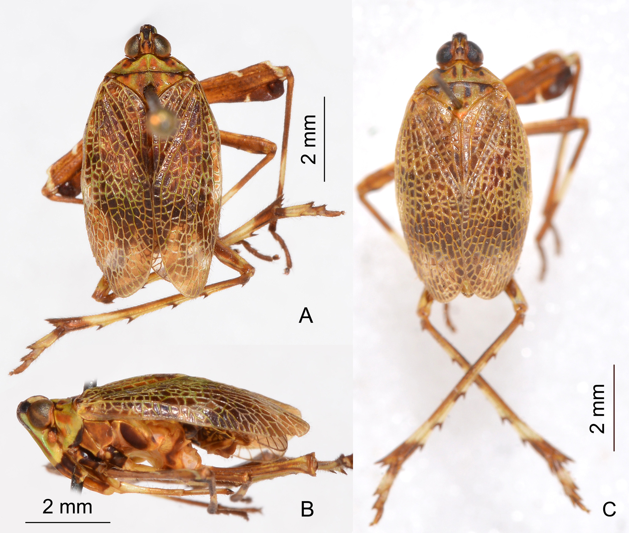
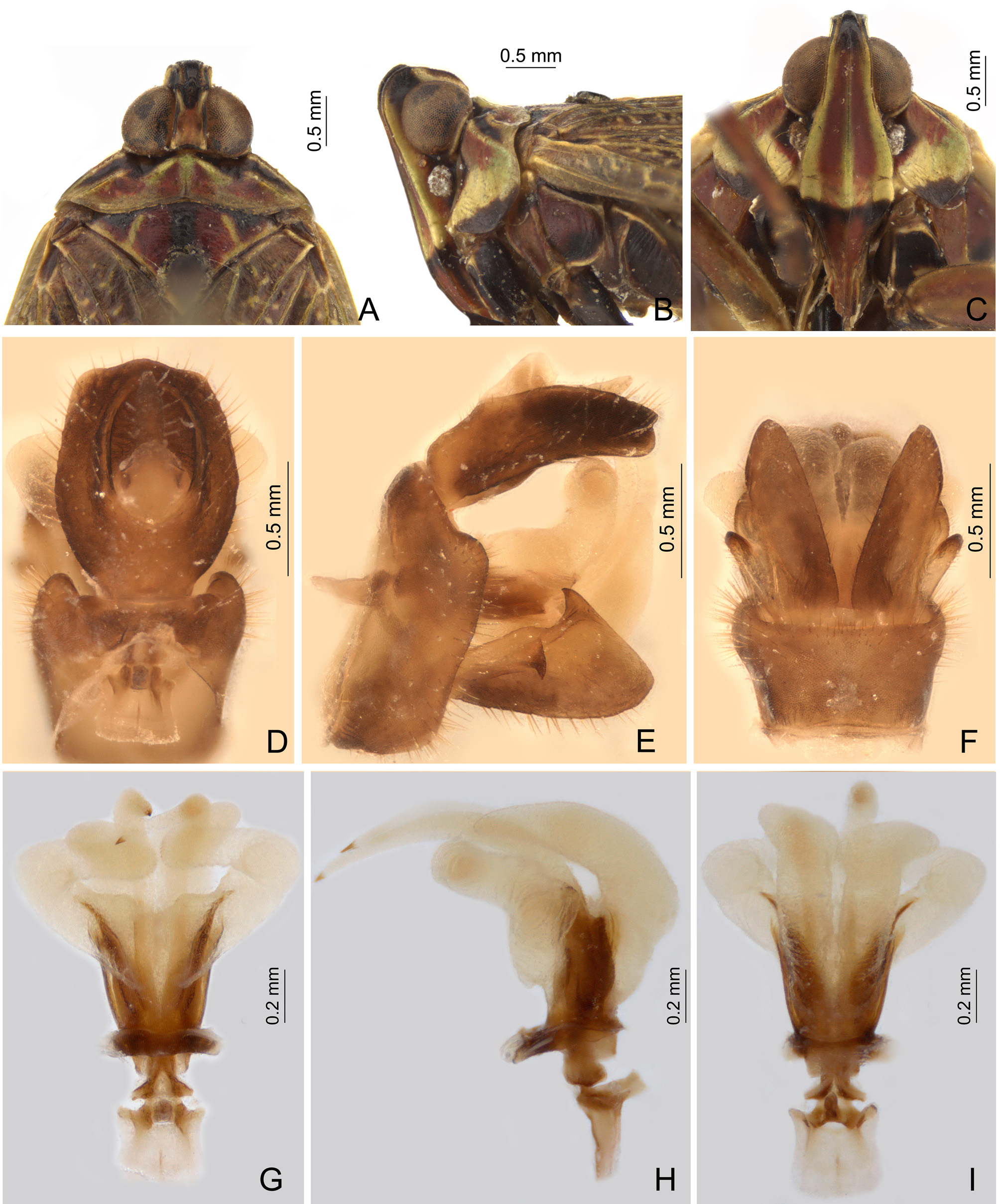
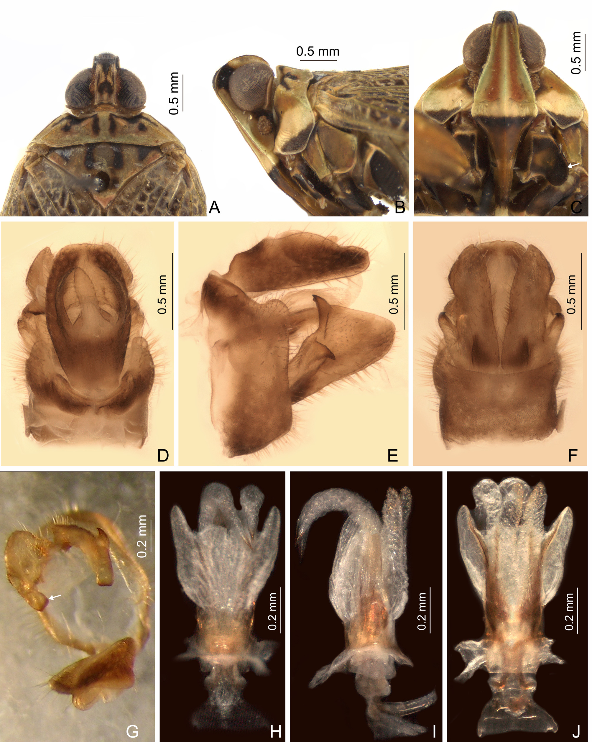
Description. Body ovoid, dorsally convex ( Fig. 1 View FIGURE 1 ). Head short, not produced in a distinct cephalic process. Vertex ( Figs 3 View FIGURE 3 A, 5A) with basal width much narrower than transverse diameter of eyes; lateral carinae strongly ridged, foliaceous, and parallel between eyes, jointed with lateral carinae of frons in perfect arc in profile ( Figs 3 View FIGURE 3 B, 5B); anterior margin angularly convex but more or less grooved centrally, just beyond anterior margin of eyes ( Figs 3 View FIGURE 3 A, 5A); posterior margin ridged and broadly concave; disc of vertex swollen at half length, medially furrowed, and median carina absent. Frons ( Figs 3 View FIGURE 3 C, 5C) slightly inflated and bulbous at apex, distinctly beyond apex of vertex; frons narrowest at apex, broadest at frontoclypeal suture; intermediate carinae not sharp, gradually divergent ventrad and approaching beneath middle of eyes; median carina distinctly ridged and complete. Frontoclypeal suture nearly straight. Postclypeus and anteclypeus convex medially, with distinct median carina. Rostrum long, extending nearly to middle of hind femora. Eyes very large and semiglobose, postocular swelling developed transversely. Antennae with scape very small; pedicel large and subglobose, with more than 50 distinct sensory plaque organs distributed over entire surface; flagellum long, setuliform.
Pronotum ( Figs 3 View FIGURE 3 A, 5A) relatively large and broad, anterior central margin angularly convex, lateral marginal areas straight and sloping with two longitudinal carinae on each side between eyes and tegulae, lower lateral carinae convex and visible in dorsal view ( Figs 3 View FIGURE 3 A, 5A); posterior margin nearly straight; disc flat, median carina weakly ridged, with a lateral pit on each side. Mesonotum ( Figs 3 View FIGURE 3 A, 5A) broad, short and flat, carinae very weak, lateral carinae nearly straight and abruptly incurved apically.
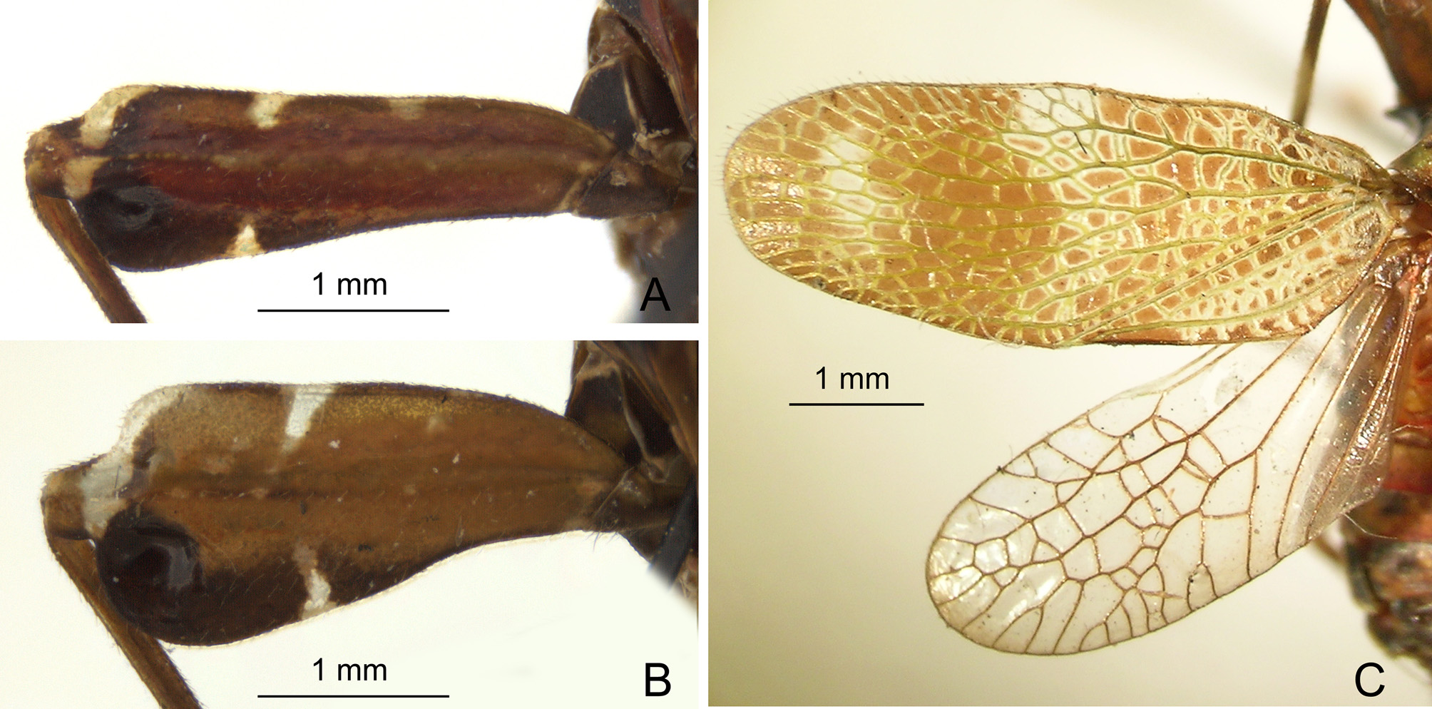
Forewings ( Fig. 2 View FIGURE 2 C) “brachypterous” in size, which corresponds to “koeliopterous” by Metcalf (1950) or “submacropterous” by Emeljanov (e.g. 2011). They are relatively short, slightly extending beyond abdomen apex, convex, coriaceous but translucent; veins with short setae. Venation patters variable in left/right wing; ScP+R, MP, and CuA bifurcated near basal 1/3, and branching successively accessory veins for several times; MP distinctly bifurcated posterior to CuA; venation strongly reticulate, dense dendroid secondary veinlets among longitudinal veins on the whole wings, including clavus; number of apical cells between R and CuA more than 25; pterostigmal area absent. Hindwings ( Fig. 2 View FIGURE 2 C) well-developed, but anal area reduced, without secondary fold; venation pattern also variable in left/right wing; numerous veinlets among longitudinal veins.
Legs elongate; fore coxae ( Fig. 5 View FIGURE 5 C) short, lateral carina strongly flattened and foliaceous; fore femora ( Fig. 2 View FIGURE 2 A, B) elongate, greatly flattened, dilated, and foliaceous laterally, subapical part with a large depression on the inner side ventrally, bulging dorsally; middle femora elongate, slightly flattened and dilated; hind tibiae with 5–6 lateral spines and seven apical spines; hind tarsomeres I with ten and tarsomeres II with eight apical spines, respectively; apical spines of tarsomeres with long setae instead of platellae.
Abdomen with pregenital segments short and broad, without distinct median and intermediate carinae dorsally. Tergites III–VI with a large depression near lateral margin on each side. Sternites III–VI tuberculate (trace of sensory pits), with 6–10 tubercles on each side.
Male terminalia. Pygofer with dorsal margin slightly excavated to accommodate segment X, dorso-lateral margins angularly produced posteriorly in dorsal view ( Figs 3 View FIGURE 3 D, 5D). Gonostyles ( Figs 3 View FIGURE 3 E, 5E) symmetrical, expanding towards apex, broadest apically, apex straight; upper process acute apically in profile ( Figs 3 View FIGURE 3 E, 5E), but actually compressed dorsoventrally in caudal view ( Fig. 5 View FIGURE 5 G). Aedeagus ( Figs 3 View FIGURE 3 G–I, 5H–J) with a pair of endosomal processes: membranous, distinctly inflated, apically acute, and directed dorsally ( Figs 3 View FIGURE 3 H, 5H); phallobase sclerotized and pigmented at base and laterally, with paired membranous and inflated lobes. Segment X ( Figs 3 View FIGURE 3 D, 5D) large and oval in dorsal view, apical dorsal margin deeply excavated to accommodate anal style; anal style large and elongate, but not extending beyond the apical ventral margin of segment X.
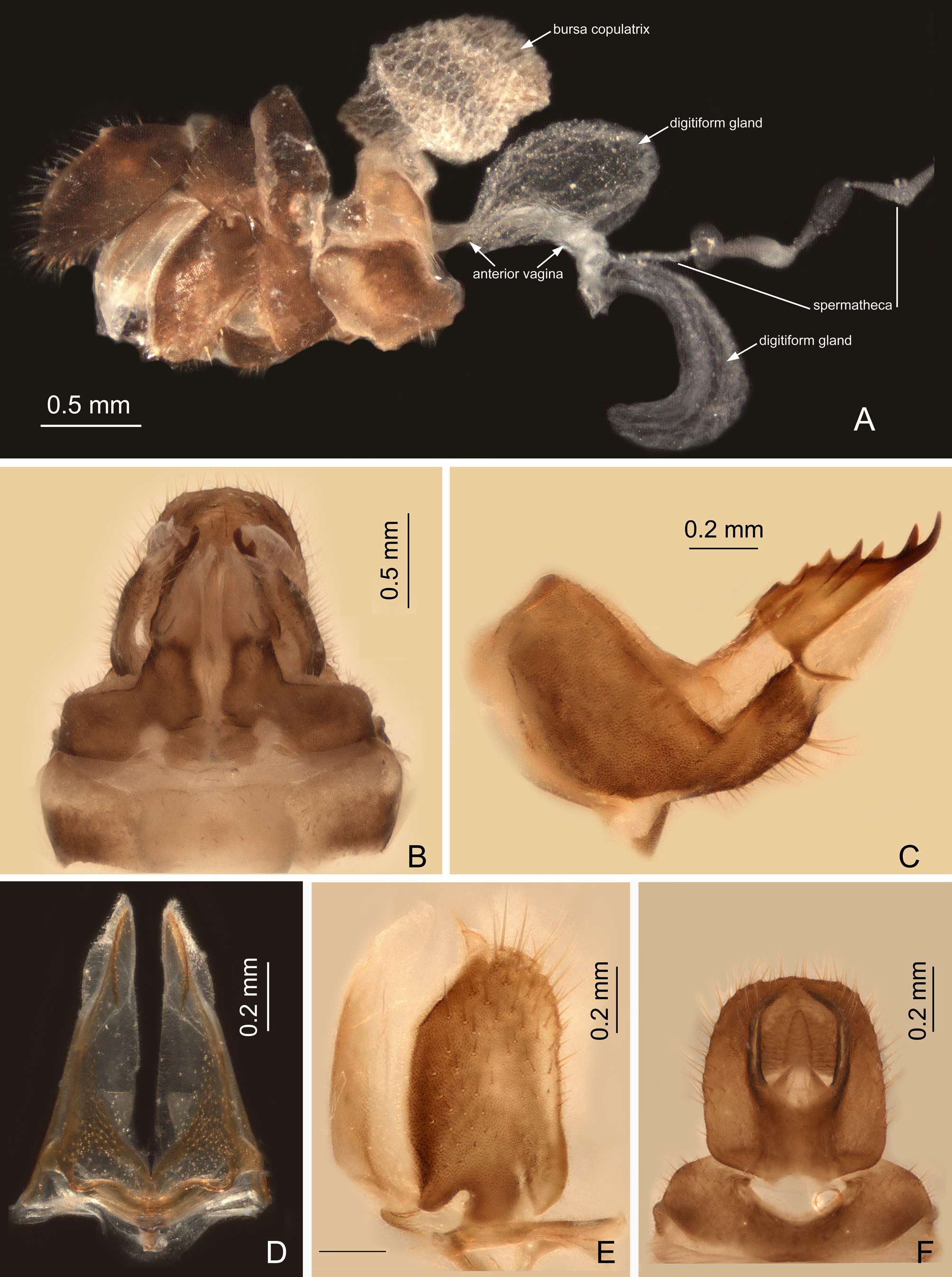
Female terminalia ( Fig. 4 View FIGURE 4 A, B). Gonocoxae VIII with two membranous and flattened endogonocoxal processes (Gxp) on endogonocoxal lobe: Gxp1 large and elongate, with a long sclerotized plate in it; Gxp2 smaller and shorter. Gonapophyses VIII (first valvulae) with anterior connective lamina large and sclerotized, with seven teeth of varying sizes and shapes in lateral view ( Fig. 4 View FIGURE 4 C). Gonapophyses IX (second valvulae) with posterior connective lamina triangular, symmetrical in ventral view ( Fig. 4 View FIGURE 4 D), fused with the intergonocoxal plate (iGxp) at base; iGxp extended cephalad into genital cavity, forming wall of gonospiculum. Gonoplacs (third valvulae) with two lobes homologous; lateral lobe large and moderately sclerotized, with long setae at apex; the posterior lobe membranous, containing long sclerotized plate ( Fig. 4 View FIGURE 4 E). Segment X large and broad in dorsal view ( Fig. 4 View FIGURE 4 F), apical dorsal margin deeply excavated to accommodate anal style; anal style large and elongate, but not extending beyond the apical ventral margin of segment X. Female ectodermal genital ducts ditrysian: oviporus opening exteriorly from posterior vagina and surrounded by gonapophyses VIII and IX, and copulaporus opening anterior to gonapophyses VIII between abdominal sternites VII and VIII. Bursa copulatrix ( Fig. 4 View FIGURE 4 A) superficially membranous, regularly gridded, without sclerotized ornamentations. A pair of large digitiform glands ( Fig. 4 View FIGURE 4 A) branched at anterior extremity of the anterior vagina on each side of the spermatheca. Spermatheca ( Fig. 4 View FIGURE 4 A) divided clearly into five parts: orificium receptaculi, ductus receptaculi, diverticulum ductus, pars intermedialis, and glandula apicalis.
Diversity and distribution. Fernandea comprises two species which are endemic to the Guineo-Congolian region of western Africa.
Remarks. Fernandea is similar to Macronaso Synave, 1960 , but can be distinguished from the latter by the head without distinct cephalic process, and the fore coxae and femora greatly flattened, dilated, and foliaceous laterally.
Fernandea View in CoL and Macronaso View in CoL are unique among African Orthopagini in sharing the following characters: body habitus ovoid, dorsally convex, forewings relatively short, only slightly extending beyond abdomen apex, convex and coriaceous, with dense dendroid secondary veins among longitudinal veins on the whole surface; hindwings with anal area reduced, without secondary fold. As modifications of the forewing size and venation are frequent among planthoppers and convergences have been documented in many unrelated groups ( Gnezdilov 2013; Bourgoin et al. 2015), the taxonomic status and a possible sister-group relationship of the two genera needs to be further confirmed by a phylogenetic analysis for the World Orthopagini .
No known copyright restrictions apply. See Agosti, D., Egloff, W., 2009. Taxonomic information exchange and copyright: the Plazi approach. BMC Research Notes 2009, 2:53 for further explanation.
|
Kingdom |
|
|
Phylum |
|
|
Class |
|
|
Order |
|
|
Family |
Fernandea Melichar, 1912
| Song, Zhi-Shun, Malenovský, Igor & Liang, Ai-Ping 2016 |
Fernandea
| Metcalf 1946: 48 |
| Schmidt 1915: 349 |
| Melichar 1912: 53 |