
Asbestopluma (Asbestopluma) maxisigma Ekins, Erpenbeck & Hooper, 2020
|
publication ID |
https://doi.org/ 10.11646/zootaxa.5293.3.2 |
|
publication LSID |
lsid:zoobank.org:pub:FE67E8C2-AFE5-491C-B673-2ECE82FA4D87 |
|
DOI |
https://doi.org/10.5281/zenodo.7961314 |
|
persistent identifier |
https://treatment.plazi.org/id/038A1C6B-FFA5-5253-8E87-FD6699208897 |
|
treatment provided by |
Plazi |
|
scientific name |
Asbestopluma (Asbestopluma) maxisigma Ekins, Erpenbeck & Hooper, 2020 |
| status |
|
Asbestopluma (Asbestopluma) maxisigma Ekins, Erpenbeck & Hooper, 2020 View in CoL View at ENA
Figures 8–9 View FIGURE 8 View FIGURE 9 , Table 4 View TABLE 4
Material Examined: Holotype of Asbestopluma (Asbestopluma) maxisigma: QM G 337488 off Jervis Bay, Station 56, New South Wales, Australia, -35.333003, -35.332000, 151.258000 – 151.214000, 2636- 2342 m, Beam Trawl, Coll. Merrick Ekins on RV Investigator, Cruise IN2017 _ V 03, Sample 56-236, 29/v/2017. QM G339376, Noddy Reef, Great Barrier Reef, Queensland, Australia, -13.51773023, 144.1015761, 815.495 m, Site: SO398, Sample: 130, ROV SuBastian, Coll. Martie McNeil on RV FALKOR, cruise FK200930, 15/X/2020.
Comparative Material: Asbestopluma(Abestopluma)biserialis ( Ridley&Dendy,1886) : NHMUK1887.5 View Materials .2.187, South Pacific -22.35, -150.283, 4361 m, Challenger St. 281, 6/X/1875 Lectotype ; NHMUK 1887.5 View Materials .2.190, South Pacific -39.2167, -118.81.67, 4115 m, Challenger St. 291, Paralectotype .
Distribution: This species is known from continental slope Queensland and New South Wales, Australia, at bathyal depth.
Description:
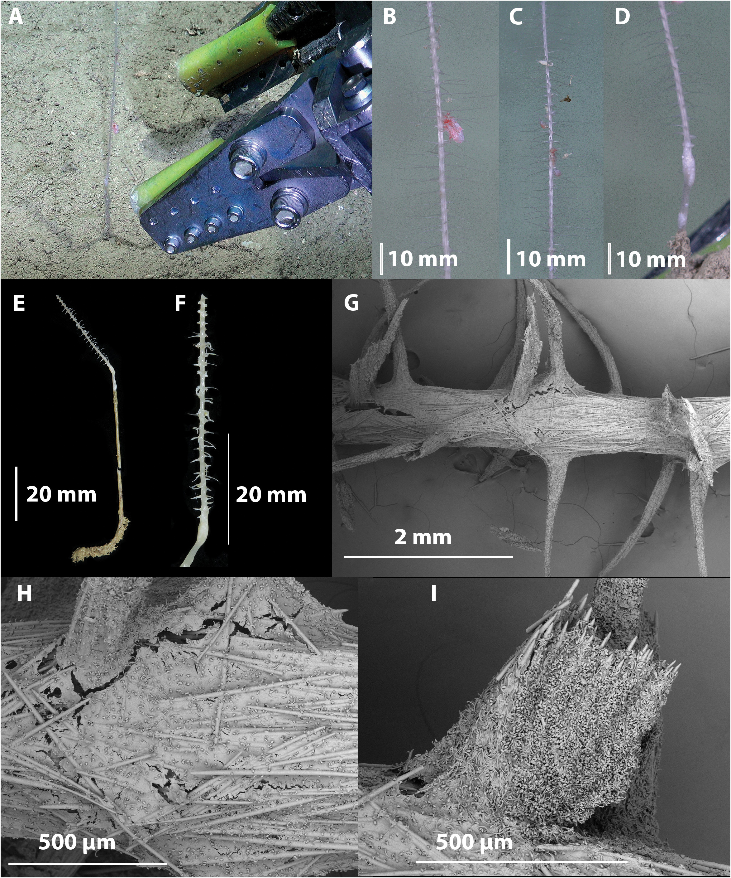
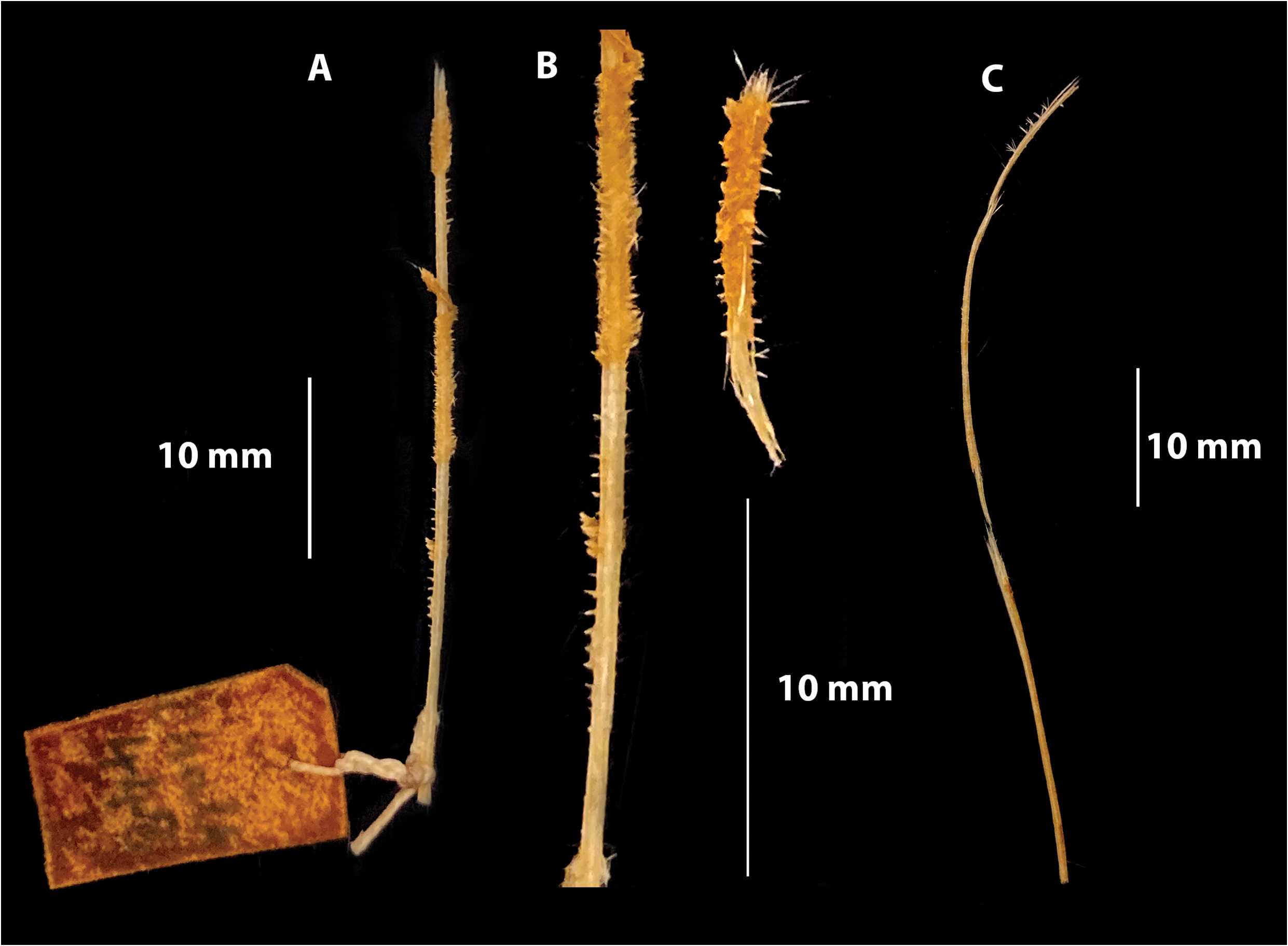
Growth form: The new specimen (QM G339376) consists of an erect columnar pedunculate sponge with pinnate filaments projecting at right angles to the stem ( Figs. 8 E, F View FIGURE 8 ). This specimen is 135 mm long, up to 2 mm wide. Ninety mm of this length comprises the below ground stem and basal root, whilst only 45 mm projects above the substrate, the top part of the specimen was lost during processing. The filaments of the preserved specimen are up to 7 mm in length, but possibly longer in vivo ( Figs. 8 A–D View FIGURE 8 ). They are 0.5 mm in width and project into four columns, with a right angle between them.
Colour: The above ground parts are white in situ and on the deck, whilst the below ground components are tan coloured.
Ectosomal skeleton: The ectosome of both the stem and the filaments consist of soft tissue encrusted with anisochelae and sigmas. The ectosome of the lower stem and roots is encrusted with the acanthostyles.
Endosomal skeleton: The axis of the stem and the filaments consists of tightly bound longitudinal tracts of mycalostyles. The mycalostyles are also arranged as buttresses providing support for the filaments that are also composed of the same styles and arise tangential to the stem, so that at their ends the mycalostyles converge onto the filament mycalostyles. In addition, there are supplementary smaller very fine and short filament columns composed of the subtylostyles projecting at right angles to the stem and similarly converging with buttressing mycalostyles. The subtylostyles are not present in the stem nor the roots, which is composed of the mycalostyles only.
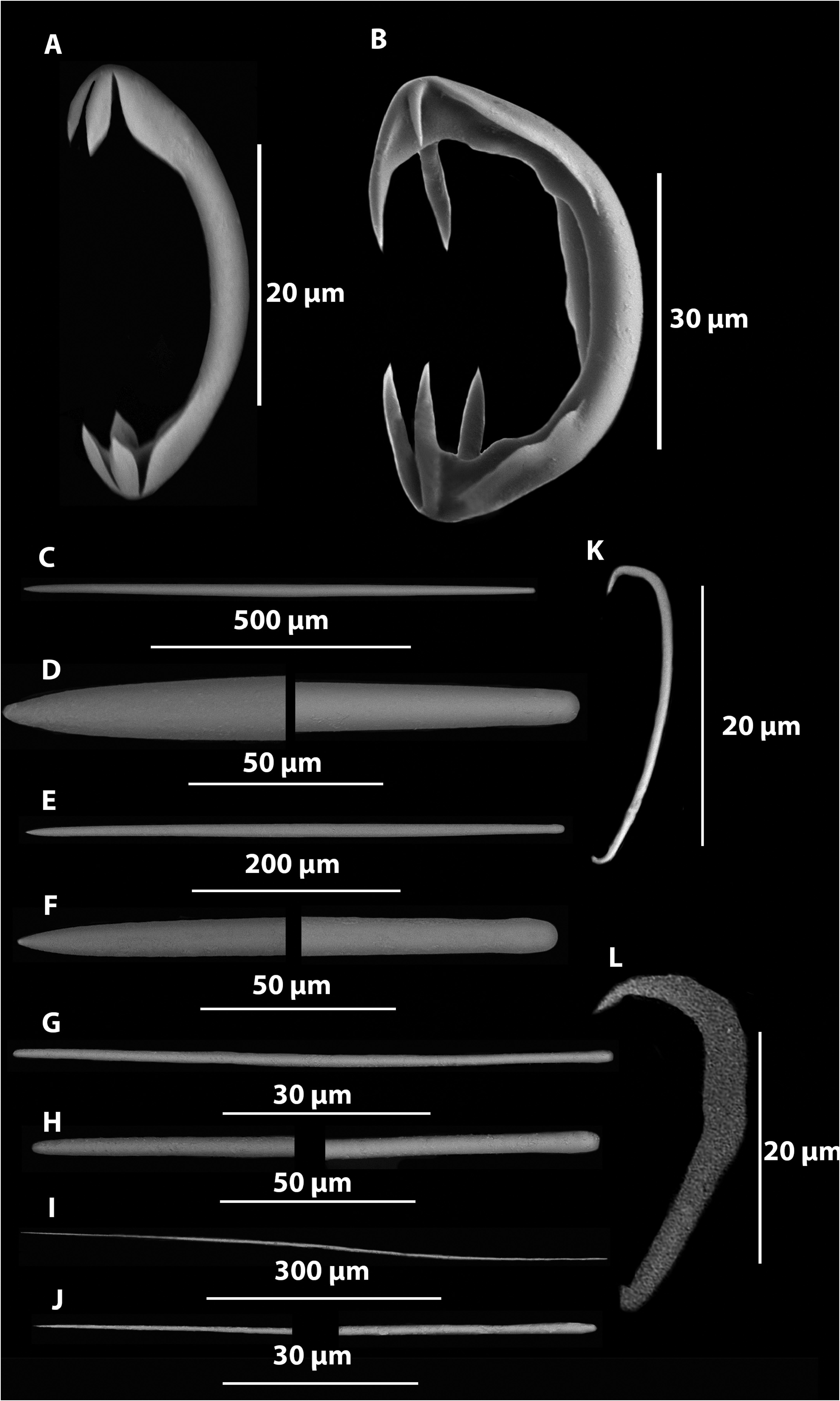
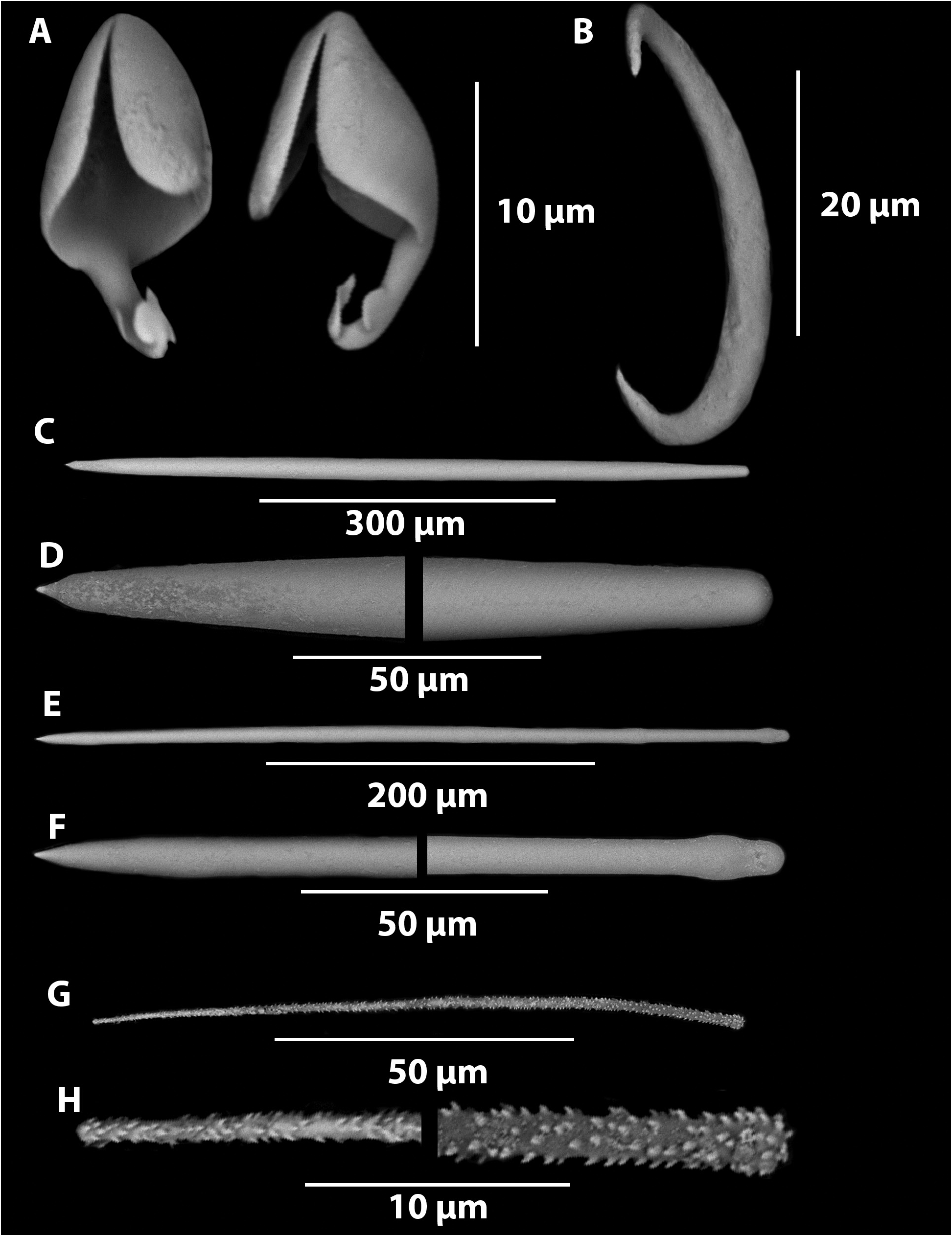
Megascleres: Styles of three types exist in different parts of the sponge. Large mycalostyles, thickest at the middle of the spicule and tapering at both ends (531–(730)–897 µm x 12.5–(20.3)–29.9 µm, n=59) occur in the filaments, the stem and the basal root like appendage ( Figs. 9 C, D View FIGURE 9 ). Smaller subtylostyles with slightly swollen bases and tapering to fine points (393–(509)–604 x 4.2–(9.8)–12.6 µm, n=22) occur in the filaments ( Figs. 9 E, F View FIGURE 9 ). Very thin long, often subtly bent fragile acanthostyles occur in the lower stem and root like appendage (66.8– (119.1)–246.0) x 0.8–(2.8)–6.6 µm, n=44) ( Figs. 9 G, H View FIGURE 9 ).
Microscleres: Palmate anisochelae ( Fig. 9 A View FIGURE 9 )., head with the lateral alae fully fused to the shaft and a large frontal alae significantly detached from lateral alae, foot with two fully fused nearly atrophied lateral alae and a single larger frontal ala with a tooth-like termination (Length 10.3–(12.8)–14.1 µm, large frontal alae width 3.2–(4.4)–6.5 µm, small lateral alae width 1.8–(2.2)–2.7 µm, n=24) Small sigmancistras ( Fig. 9 B View FIGURE 9 ), with an almost 90 o twist (28.2–(32.3)–36.0 µm, n=20) were found. The larger sigmas recorded in the holotype by Ekins et al. (2020), were not found in this specimen. It is possible these may have been contaminants from Chondrocladia (Chondrocladia) clavata Ridley & Dendy, 1886 , multiple samples that were collected from the same beam trawl station.
Remarks: As previously noted in Ekins et al. (2020a), As. (As.) maxisigma is most closely related to As. (As.) biserialis ( Ridley & Dendy, 1886) , known from the South Pacific ( Ridley & Dendy 1886), Kermadec Trench, ( Lévi 1964), Coral Sea off New Caledonia ( Lévi 1993), and the North Pacific, south of the Aleutian Islands ( Koltun 1970), from bathyal and abyssal depths (qv. Lopez et al. 2011; Ekins et al. 2020a). Both species have vaguely similar pinnate pedunculate morphologies but As. (As.) maxisigma has twice as many columns of filaments (i.e., four as opposed to two) and is round in cross-section rather than flattened ( Fig. 10 View FIGURE 10 ). This re-description of As. (As.) maxisigma with new material collected by ROV, clearly shows that only about one third of the sponge is above the ground i.e., the ‘snow’ layer ( Figs. 8A, E, F View FIGURE 8 ). The inclusion of the below-ground stem and the root like structures of QM G339376 from the Great Barrier Reef shows the presence of acanthostyles, which were not found on the holotype of As. (As.) maxisigma . Since this species is only known to live in soft sediment, it is likely the spined surface of the acanthostyles serve as anchors securing the sponge in the soft sediment. Another example of exaptation of spicules in carnivorous sponges. These acanthostyles are similar to those found in As. (As.) biserialis but were only reported in Lévi (1964) and Koltun (1970), not in the original description by Ridley & Dendy (1886) nor Lévi (1993). With the inclusion of new acanthostyles this species is even more closely aligned with As. (As.) biserialis ( Ridley & Dendy, 1886) . However, the species are kept separate as this distinction between the clearly biserial species As. (As.) biserialis , is also this distinction between it and another closely related species As. (As.) belgicae ( Topsent, 1901a) , which has 6–8 rows of filaments. Asbestopluma (As.) biserialis var. californiana de Laubenfels, 1935 is currently only distinguished by having slightly smaller chelae from a described single value of 6 µm, as opposed to the current species which begin at 10 µm. Re-examination of the type material of As. (As.) biserialis var. californiana , may reveal a greater size range of these microscleres.
In its gross morphology this species also resembles As. (As.) belgicae , (qv. Lopez et al. 2011, Hestetun et al. 2015; Goodwin et al. 2017). Although, As. (As.) maxisigma has acanthostyles, it has the following differences: fewer radial filaments, the absence of grooves, smaller mycalostyles, an absence of the strongyles. Asbestopluma (Asbestopluma) quadriserialis Tendal, 1973 , from the North Atlantic also has four rows of filaments, however, it can be clearly distinguished by the presence of two sizes of anisochelae. These closely related species are compared in Table 4 View TABLE 4 . Asbestopluma (As.) sarsensis Goodwin et al., 2017 is similar in spiculation to As. (As.) belgicae , and also differs from the present species for the same reasons given above, in addition to also having a very different growth form. Asbestopluma (Asbestopluma) obae Koltun, 1964 from Wilkes Land, Antarctica differs from the present species in lacking horizontal filaments.
As shown in Fig. 3 View FIGURE 3 of Ekins et al. (2020a), As. (As.) maxisigma does not have the same molecular sequence as any other Asbestopluma (Asbestopluma) spp. Unfortunately, none of the closely related species listed above have any molecular sequence data either. The only one of the closest morphologically is As. (As.) cf. belgicae , which has a different molecular profile.As new collections of species of this genus and their corresponding sequences become available, will result in better resolution within the genus.
| RV |
Collection of Leptospira Strains |
| V |
Royal British Columbia Museum - Herbarium |
| QM |
Queensland Museum |
No known copyright restrictions apply. See Agosti, D., Egloff, W., 2009. Taxonomic information exchange and copyright: the Plazi approach. BMC Research Notes 2009, 2:53 for further explanation.