
Polyandrocarpa arianae Monniot, F. 2016
|
publication ID |
https://doi.org/ 10.11646/zootaxa.4410.2.3 |
|
publication LSID |
lsid:zoobank.org:pub:7A46BD51-20A9-4FDA-81FB-D771BA9011FC |
|
DOI |
https://doi.org/10.5281/zenodo.6485088 |
|
persistent identifier |
https://treatment.plazi.org/id/038A4612-FF8D-FFC2-BEDE-FA4C50B4F9F3 |
|
treatment provided by |
Plazi |
|
scientific name |
Polyandrocarpa arianae Monniot, F. 2016 |
| status |
|
Polyandrocarpa arianae Monniot, F. 2016 View in CoL
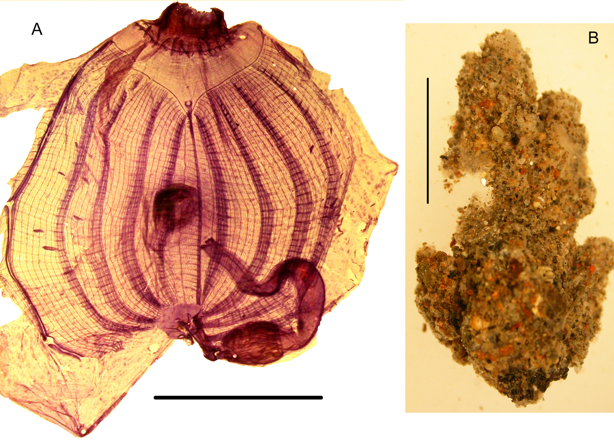
Fig. 6 View FIGURE 6
Station: AR 367 (MNHN S1 POL.A 58)
The zooids 1cm long are upright above a basal sheet and tightly linked together by their lateral side ( Fig. 6B View FIGURE 6 ). The tunic is thin transparent and entirely covered with sand and mud, without epibionts. The siphons are not protruding and have 4 lobes, the oral one apical and the atrial more dorsal. Each siphon has an internal velum spotted with crystalline inclusions. This structure is the same as in P. anguinea . The oral tentacles are in 2 orders of size. The prepharyngeal band in a single rod is distant from the tentacle ring and draws a wide V ( Fig. 6A View FIGURE 6 ). The prepharyngeal area has no papillae. The dorsal tubercle is button like and opens anteriorly in a C. The body wall is marbled ( Fig. 6A View FIGURE 6 ) and thin with a weak network of muscular fibres. There are no endocarps. The branchial tissue is thin ( Fig. 6A View FIGURE 6 ). The dorsal lamina is long and low with a smooth edge. There are 4 branchial folds on each side ( Fig.6A View FIGURE 6 ). The formula of one zooid on the right side is:
E-6 (10) 5 (14) 5 (14) 4 (12) 1-DL
As in the Mediterranean specimens there is always a single longitudinal vessel between the dorsal lamina and the first branchial fold of the right side ( Fig. 6A View FIGURE 6 ). This is the principal and constant difference with Polyandrocarpa anguinea which has several vessels in this place instead of one. The stigmata are crossed by parastigmatic vessels. The digestive tract occupies a small part of the left body side ( Fig. 6A View FIGURE 6 ). The stomach is olive shaped with longitudinal folds and without caecum; the intestine is isodiametric, the primary loop is widely open and the rectum is long. The anus has 8 to 12 lobes. The gut is loosely attached to the body wall by a few trabeculae. The gonads were not fully mature. In one specimen 9 polycarps are on the right side and 7 on the left not arranged in lines ( Fig. 6A View FIGURE 6 ). Each polycarp is elongated, the ovary on the internal side and several testis vesicles external in a double line. The polycaps hang between the branchial sac and the body wall attached by trabeculae.
All characters of the Martinique specimens are the same as those described from the Mediterranean Sea. The distribution of this species is presently limited to these two regions. A discussion about the different species of Polyandrocarpa was given in Monniot F. (2016a).
No known copyright restrictions apply. See Agosti, D., Egloff, W., 2009. Taxonomic information exchange and copyright: the Plazi approach. BMC Research Notes 2009, 2:53 for further explanation.