
Daphnia turbinata Sars, 1903
|
publication ID |
https://doi.org/ 10.11646/zootaxa.4658.2.6 |
|
publication LSID |
lsid:zoobank.org:pub:FDE56232-1363-45E1-BCED-AE6687A96BC9 |
|
persistent identifier |
https://treatment.plazi.org/id/038A879B-FFF6-630E-FF0F-5FA8FEA7FE3A |
|
treatment provided by |
Plazi |
|
scientific name |
Daphnia turbinata Sars, 1903 |
| status |
|
Daphnia turbinata Sars, 1903 View in CoL
( Figs 2 – 6 View FIGURE 2 View FIGURE 3 View FIGURE 4 View FIGURE 5 View FIGURE 6 )
Daphnia longispina var. turbinata Sars, 1903: 156 – 168 View in CoL , Pl. 4: Figs. 3, 3a – c View FIGURE 3 .
Daphnia turbinata Sars in Glagolev 1995: 58 View in CoL , Pl. 48: Figs 16 – 17; Ayushsuren et al. 2013: 125, Fig. 4 View FIGURE 4 .
not D. longispina turbinata Sars in Manujlova 1964 View in CoL : Fig. 36: fragment 21.
Type locality. “Lake Kurtu-Kol”, the basin of Teletskoye Lake , Altai Republic, Russia. It is difficult to find this water body in recent maps, this is a non-drainage lake in the basin of the Bashkaus River , affluent of the Chulymshan River inflowing Teletskoe Lake ( Golubtsov & Malkov 2007), most probably, this is a small lake locally named as “Kara-Kol” (N50.5619°, E87.8874°), from which the Kurtu-Kol River flows out GoogleMaps .
Type material. GOS F 9524 and GOS F 9524. These two slides with locality labelled as “ Altay ” represent, most probably, a type series, as Sars (1903) has the material from a single lake and never reported on any subsequent findings of D. turbinata from this region.
Material studied. Russia. Parthenogenetic females from un-named lake 2 near Kosh-Agach (N50.0105º, E88.61234º, 1745 m.a.s.l.), Altai Republic, coll. in 10.08.2015 by Y.R. Galimov, AAK M-3169 GoogleMaps . Parthenogenetic females from unnamed lake 1 near Kosh-Agach (N50.0175º, E88.60416º, 1754 m.a.s.l.), Altai Republic, coll. in 10.08.2015 by Y.R. Galimov,AAK M-3173 GoogleMaps . Parthenogenetic females from unnamed lake (N50.24367º, E89.46833º, 2322 m.a.s.l.), Tyva Republic, coll. in 11.08.2015 by Y.R. Galimov, AAK M-3170 GoogleMaps . Parthenogenetic females from unnamed pond 3, the Todzha Depression (N52.43º, E96.3º, 922 m. a.s.l.), Tyva Republic, coll. by E.I. Zuykova, EIZ, no number GoogleMaps . Mongolia. Parthenogenetic females from Urt Tsaramiin Nuur (Lake) (N47.68809º, E102.691º, 1352 m.a.s.l.) near Hatgal , Huvsgul Aimag, coll. in 02.08.2005 by Ch. Jersabek, AAK 2008-018 GoogleMaps . Males, ephippial & parthenogenetic females from Zhaakhan Nuur (Lake) (N48.334º, E96.0618º, 1984 m.a.s.l.), Zavkhan Aimag, coll. by Ch.Ayushsuren, AAK 2018-083 GoogleMaps . Parthenogenetic females from Khongor Nuur (Lake) (N48.50786º, E90.60475º, 2376 m.a.s.l.), Bayan-Ölgii Aimag, coll. in 24.07.2008 by D.P. Karabanov, AAK M-0774 GoogleMaps . Parthenogenetic females from Khargal Nuur (Lake) (N49.9375º, E102.7233º, 1066 m.a.s.l.), Bulgan-Orkhon Aimag, coll in 24.07.2009 by D.P. Karabanov, AAK M-1322 GoogleMaps . Parthenogenetic females from a pond (N51.08263º, E99.70533º, 1588 m.a.s.l.) SE Shar Nuur (lake) near Hanh, Huvsgul Aimag, coll. in 28.07.2005 by Ch. Jersabek, AAK 2008-013 GoogleMaps .
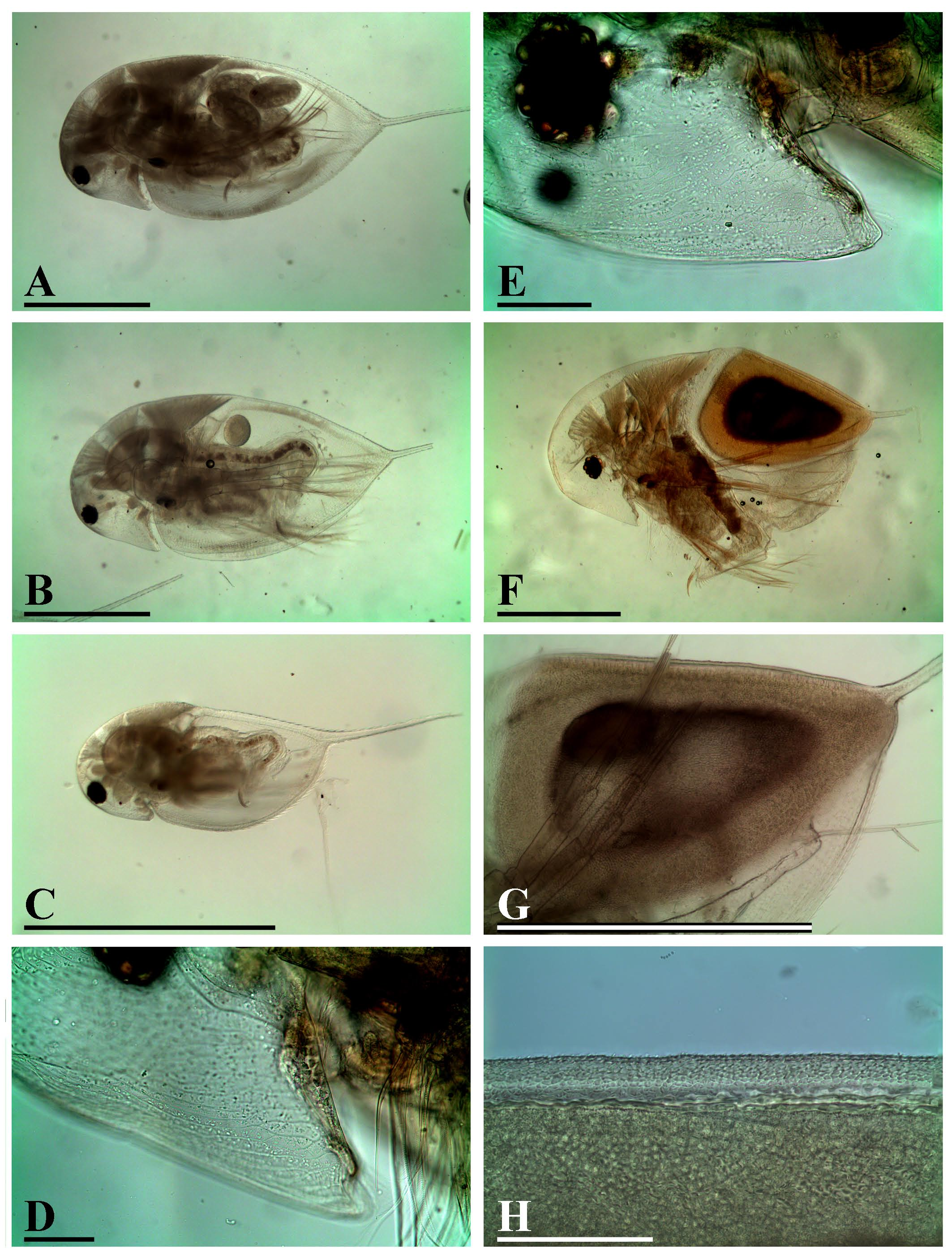
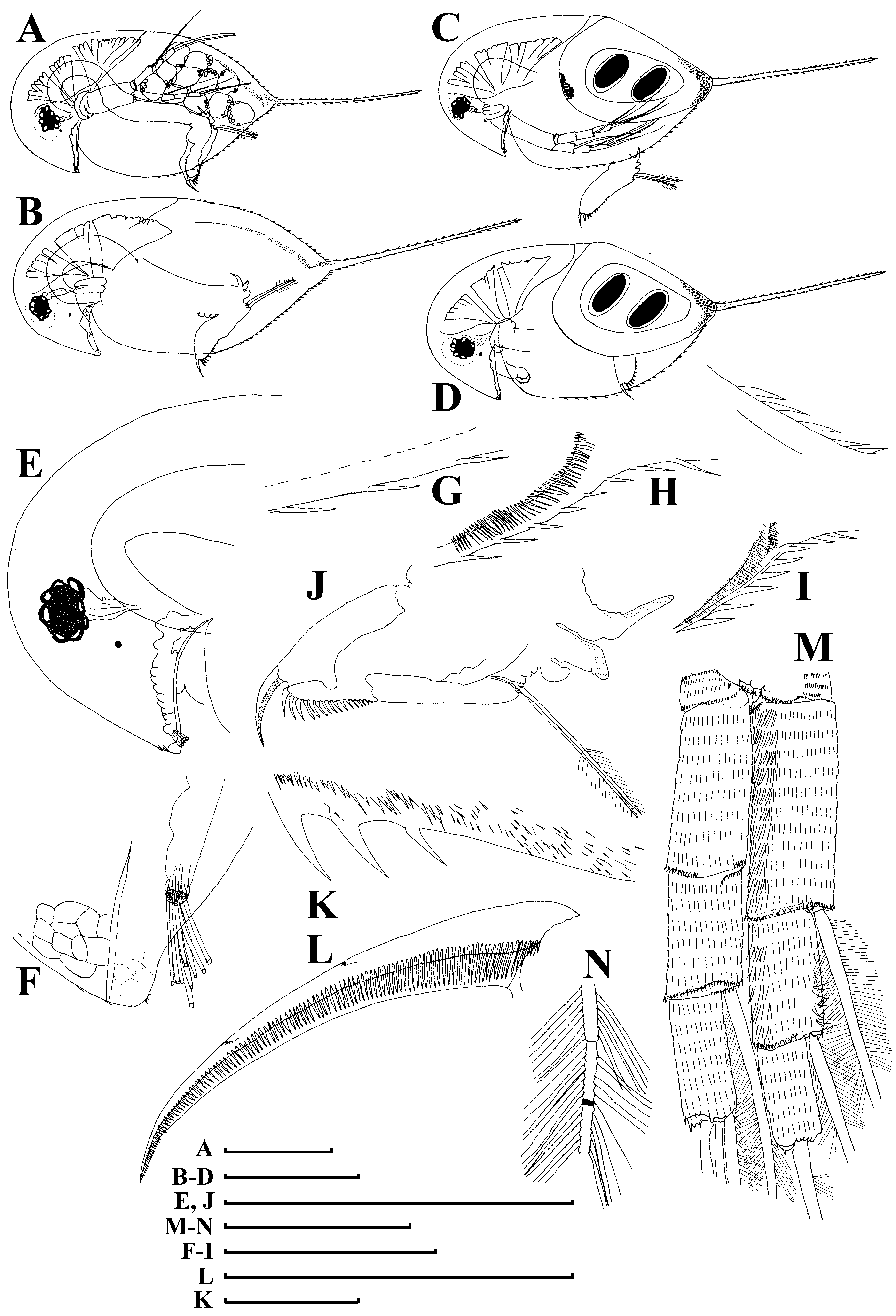
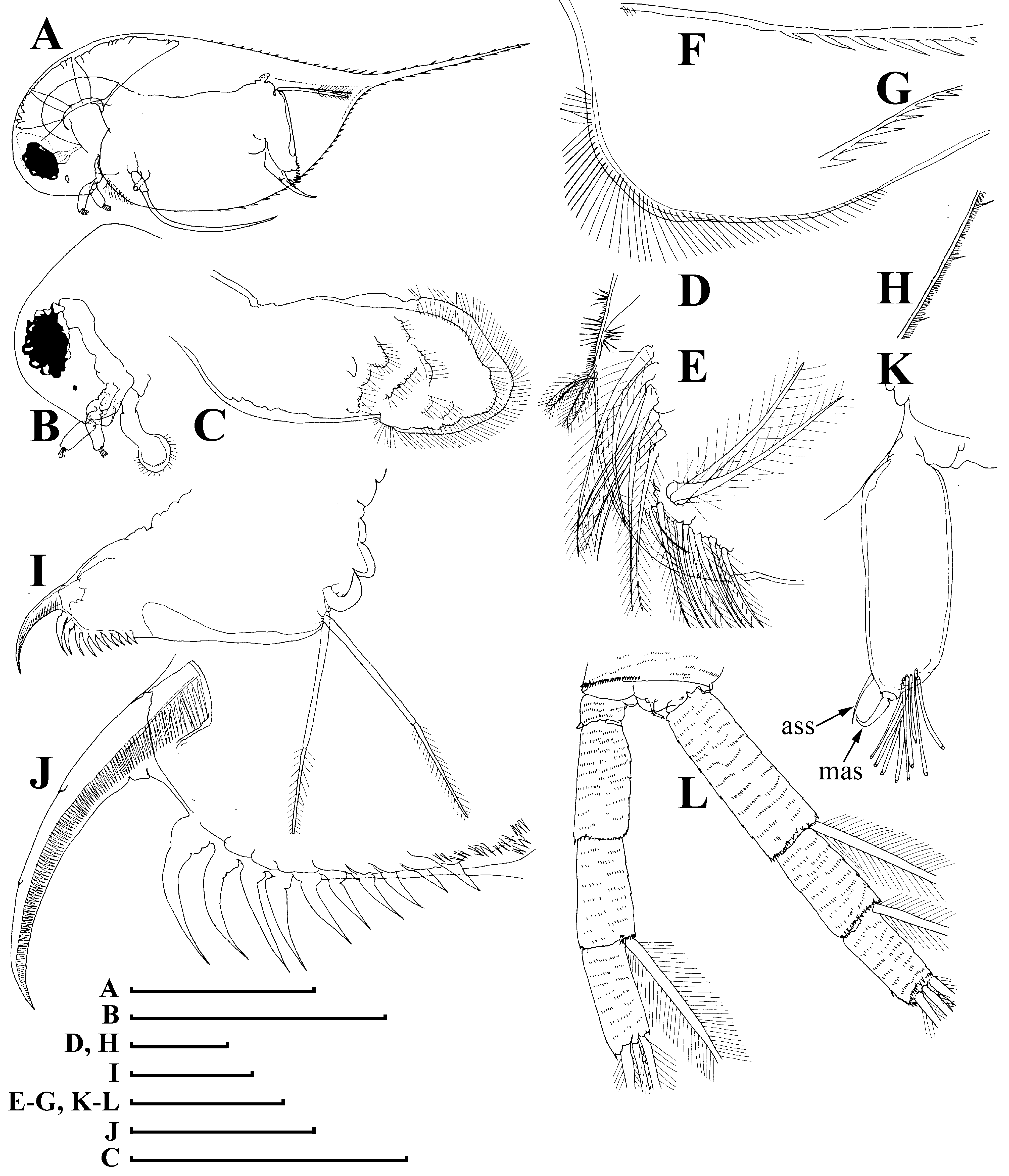
Redescription. Adult parthenogenetic female. Body pigmented in yellow-brown in different extension, and level of pigmentation varies even among specimens from same population ( Fig. 2A – B View FIGURE 2 ), in some samples all specimens were pigmented. Body subovoid in lateral view, maximum height of head approximately equal to maximum body height in middle of valves ( Fig. 3A – B View FIGURE 3 ). Dorsal margin of valves slightly and regularly convex, no depression between head and rest of body. Postero-dorsal body portion remarkably narrowing distally to postero-dorsal angle supplied with a very long caudal spine, ventral margin regularly convex. All surface of head and valves with a wellvisible, specially rough as compared to other longispina -like taxa, sculpture of anastomosing lines ( Fig. 2D – E View FIGURE 2 ).
Head very wide, with short rostrum, its tip not bent ( Fig. 3E, F View FIGURE 3 ); posterior margin of head short, with a very low prominence in middle ( Fig. 2E View FIGURE 2 ); ventral margin of head almost straight ( Fig. 2D View FIGURE 2 ) to regularly convex ( Fig. 2E View FIGURE 2 ) or even with a smooth angle in the antero-ventral surface ( Fig. 3B View FIGURE 3 ). Compound eye medium-sized, ocellus minute and located closer to eye than to base of antenna I. Labrum with a fleshy main body and a large, setulated distal labral plate.
Carapace subovoid, spinulae ( Fig. 3G View FIGURE 3 ) occupy the posterior half of dorsal margin and half to more than half of ventral margin. No setae at ventral margin, in posteriormost portion of ventral margin (on its inner face) a row of spinulae, but no setae ( Fig. 3 View FIGURE 3 H–I).
Abdomen relatively short, consisting of four segments. The first (basal most) abdominal segment with especially long (up to 2.5–2 times longer than postabdominal claw) process bent anteriorly; the second segment with a long process approximately as long as postabdominal claw; the third segment with a short process; the fourth segment without a projection; on all processes there are rows of minute setulae ( Fig. 3J View FIGURE 3 ).
Postabdomen elongated, tapering distally, with ventral margin slightly convex, lacking of setulae ( Fig. 3J View FIGURE 3 ). Preanal margin long (remarkably longer than anal plus postanal portions of postabdomen), almost straight, its region densely covered by minute setulae ( Fig. 3K View FIGURE 3 ). Pre-anal angle absent, postanal angle rounded. Numerous (up to 15) paired spines on postanal and anal portion, their size continuously increasing distally. Postabdominal seta longer than preanal margin. Postabdominal claw slightly and regularly bent, with a pointed tip ( Fig. 3L View FIGURE 3 ). On ventral (convex) side, two bunches of minute setulae; on dorsal (concave side), three successive pectens fluently turned each to other.
Antenna I body fully reduced ( Fig. 3F View FIGURE 3 ), nine aesthetascs (somewhat unequal in size) protruding immediately from head surface, their tips do not reach tip of rostrum, antennular sensory seta fine, arise immediately from head surface instead of mound of the antenna I.
Antenna II with coxal part possessing two short sensory setae of different length, its basal segment elongated, with a distal sensory seta on its posterior face and minute distal spine at its anterior face ( Fig. 3M View FIGURE 3 ). Antennal branches with series of minute setulae. Spines on apical segments rudimentary. Antennal formula: setae 0-0-1-3/1-1-3. Chitinous insertion within distal segment of each swimming seta relatively far from joint with proximal segment ( Fig. 3N View FIGURE 3 ).
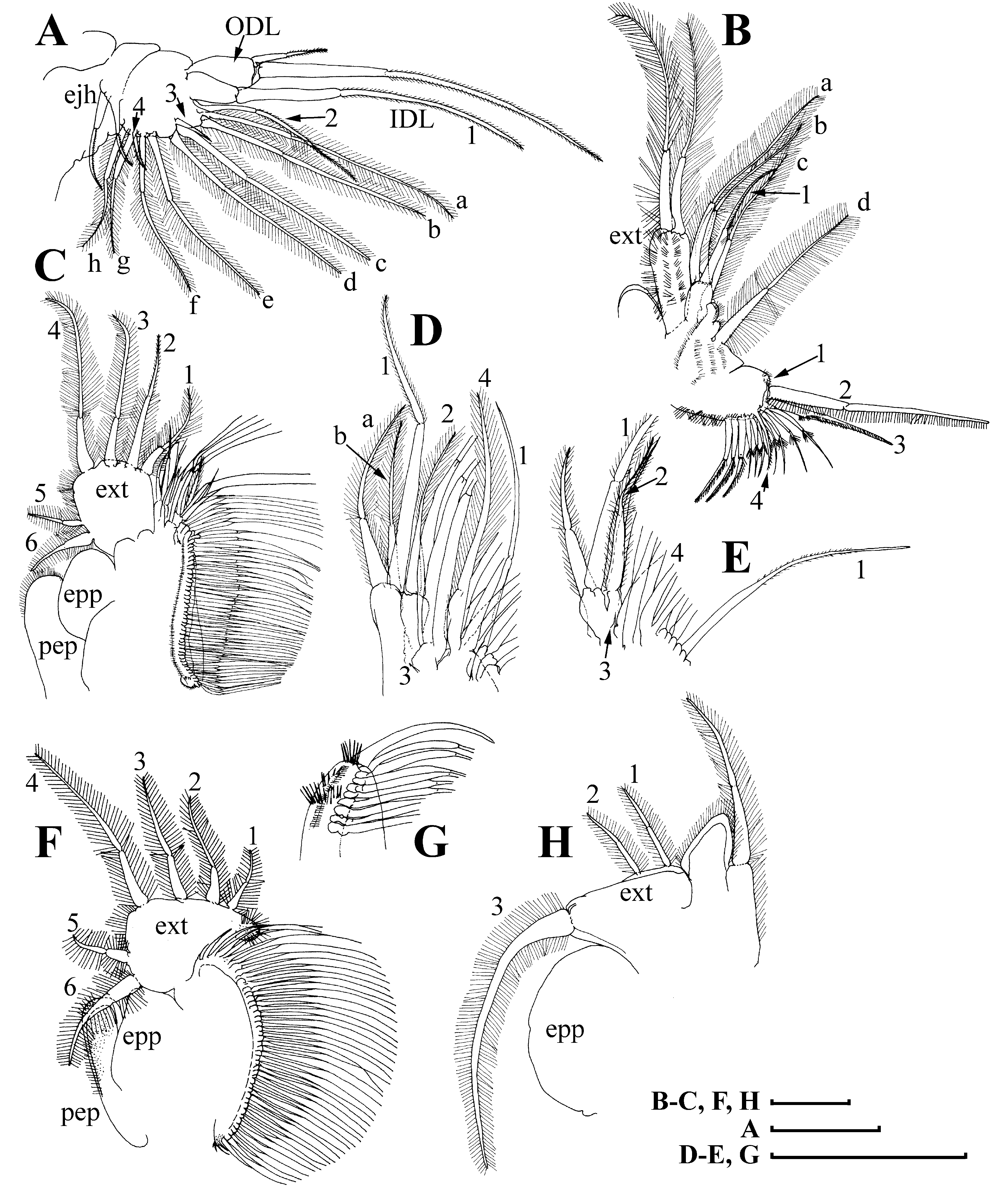
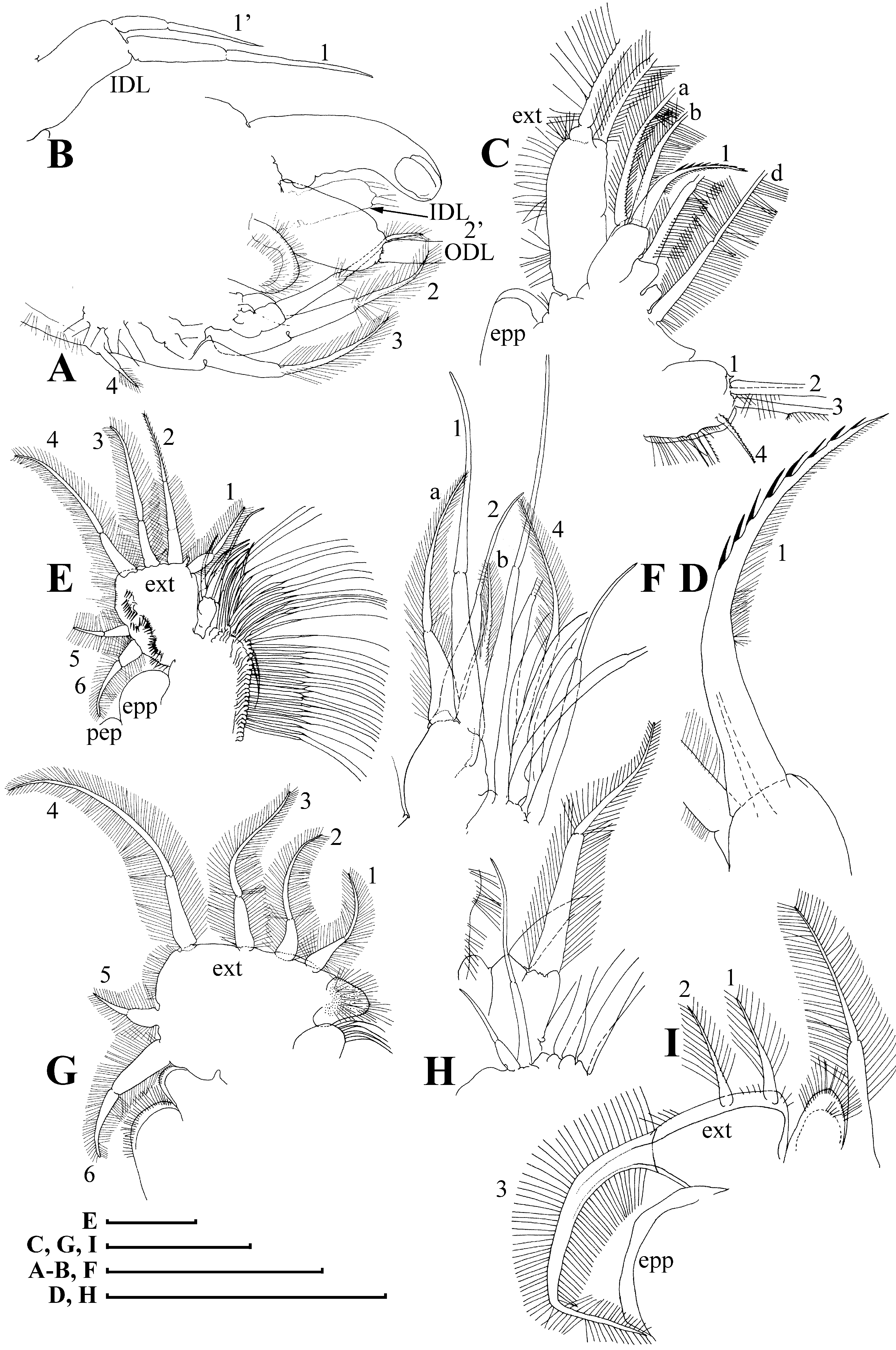
Limb I without accessory seta; outer distal lobe ( Fig. 4A View FIGURE 4 : ODL) with a long seta bilaterally armed distally with minute setulae, and a thin, short (length about 2–3 diameters of ODL) seta bilaterally setulated distally; inner distal lobe ( Fig. 4A View FIGURE 4 : IDL), or endite 4, with a single, long anterior seta ( Fig. 4A View FIGURE 4 : 1), bilaterally armed by minute setulae distally. Endite 3 with a relatively long anterior seta 2, armed with minute setulae, and two long posterior setae (ab). Endite 2 with a very short anterior seta 3, and two posterior setae (c–d). Endite 1 with a relatively small anterior seta 4 and four posterior setae (e–h). Two ejector hooks ( Fig. 4A View FIGURE 4 : ejh) of somewhat different length.
Limb II with distal portion as a large lobe (presumable exopodite, Fig. 4B View FIGURE 4 : ext) bearing a large, soft, distal seta, and a large, soft, lateral seta of similar length. Four endites bearing five setae; among them, a stiff, anterior seta ( Fig. 4B View FIGURE 4 : 1) about 2/3 length of each of two other setae on this endite, armed with fine setulae distally. Gnathobase with four anterior setae ( Fig. 4B View FIGURE 4 : 1–4) and about 10 posterior setae of gnathobasic ‘filter plate’ (their number is slightly varying among individuals).
Limb III with a setulated pre-epipodite ( Fig. 4C View FIGURE 4 : pep), subglobular epipodite ( Fig. 4C View FIGURE 4 : epp) and a flat exopodite ( Fig. 4C View FIGURE 4 : ext) bearing four distal ( Fig. 4C View FIGURE 4 : 1–4) and two lateral setae ( Fig. 4C View FIGURE 4 : 5–6). Inner-distal portion of limb with four endites: endite 4 with a specially long anterior seta ( Fig. 4 View FIGURE 4 C–D: 1) and a single posterior seta (a); endite 3 with a single anterior seta (2) and a single posterior seta (b); endite 2 with a small anterior seta (3) and two posterior setae; endite 1 with a large anterior seta (4) and four posterior setae. The rest of limb inner-distal portion as a singular large lobe, bearing numerous posterior soft setae, and a single, relatively long anterior seta ( Fig. 4 View FIGURE 4 D–E: 1), this limb part represents a modified gnathobase III.
Limb IV with a setulated pre-epipodite ( Fig. 4F View FIGURE 4 : pep), ovoid epipodite ( Fig. 4F View FIGURE 4 : epp) and wide, flat exopodite ( Fig. 4F View FIGURE 4 : ext), with protruding and setulated inner-distal angle, and bearing four distal ( Fig. 4F View FIGURE 4 : 1–4) and two lateral setae ( Fig. 4F View FIGURE 4 : 5–6). Inner-distal portion of this limb with completely fused endites, distally with two setae of unclear homology, the most part of limb inner margin is a gnathobase filter plate consisting of numerous posterior setae ( Fig. 4G View FIGURE 4 ).
Limb V with a globular epipodite ( Fig. 4H View FIGURE 4 : epp); triangular exopodite ( Fig. 4H View FIGURE 4 : ext) supplied with two small distal setae ( Fig. 4H View FIGURE 4 : 1–2) and a large lateral seta ( Fig. 4H View FIGURE 4 : 3). Inner limb portion as an ovoid flat lobe, with setulated inner margin and a single, large seta.
Juvenile female. Body more elongated, head relatively smaller, eye relatively large as compared to adult female ( Fig. 2C View FIGURE 2 ).
Ephippial female. Shape ( Fig. 2F – H View FIGURE 2 , 3C – D View FIGURE 3 ). In general body external shape as in parthenogenetic female, but dorsal carapace portion modified to ephippum pigmented in brown; egg locule region with especially dark pigmentation ( Fig. 2F – G View FIGURE 2 ); dorsal margin of carapace straight, dorsal wall of carapace additionally chitinised, forming a dorsal plate, covered by minute spinulae ( Fig. 2H View FIGURE 2 ). Ephippium with two resting eggs, axes of which sub-perpendicular to its dorsal margin; egg chambers separated from each other, most part covered with sculpturing of polygonal cells; postero-dorsal portion of valves with caudal spine incorporated into ephippium.
Adult male. Body low, elongated, narrowing posteriorly; dorsal margin of valves almost straight, not elevated above head; no depression between head and valves; postero-dorsal angle distinct, with a long caudal spine ( Fig. 5A View FIGURE 5 ).
Head. Posterior head margin straight, with a very low step-like prominence in proximal portion of margin ( Fig. 5B View FIGURE 5 ). Postero-ventral head angle as a rudimentary rostrum. Ventral margin of head almost straight, anterior-most extremity not occupied with optic vesicle, no supra-ocular depression posteriorly to it. Eye not specially large, anterior most head extremity not fully occupied with optic vesicle, ocellus very small. Labrum with a large distal labral plate ( Fig. 5C View FIGURE 5 ).
Valve with antero-ventral portion rounded; all ventral margin with numerous setae submarginally on inner face of valve ( Fig. 5A View FIGURE 5 , D–E). Posterior half of each dorsal and ventral margin with small marginal denticles ( Fig. 5 View FIGURE 5 F–G); in contrast to female, inner face of valve posterior margin with rare setae and small setulae between them ( Fig. 5H View FIGURE 5 ).
Abdomen with rudimentary mounds on first and second segment; low mound on third and fourth segments ( Fig. 5I View FIGURE 5 ).
Postabdomen shape and armature in general as in female, but preanal margin longer.About 10 paired teeth large (distalmost 2–3 times longer than claw diameter) strongly increasing in size distally ( Fig. 5J View FIGURE 5 ). Gonopore opens laterally, without a genital papilla. On outer surface of postabdominal claws, pectens consist of thin spinules only; two bunches of minute setulae on claw convex side.
Antenna I elongated, straight ( Fig. 5K View FIGURE 5 ); nine aesthetascs of different length located terminally, among them, largest aesthetasc as long as antenna I maximum diameter. Antennular sensory seta ( Fig. 5K View FIGURE 5 : ass) of moderate length (about 0.7 diameter of antenna I), located subterminally, projects beyond tip of antenna I body; male seta (flagellum) ( Fig. 5K View FIGURE 5 : mas) distally on antenna body on a low post-aesthetasc process, remarkably shorter than an aesthetasc, with curved tip.
Antenna II ( Fig. 5L View FIGURE 5 ) as in female.
Limb I ( Fig. 6 View FIGURE 6 A–B) with outer distal lobe large, cylindrical ( Fig. 6A View FIGURE 6 : ODL), bearing a small seta and a very large seta ( ODL is not illustrated here); inner distal lobe with a relatively long, bent copulatory hook, and two setae of different size ( Fig. 6B View FIGURE 6 : 1 and 1’); endite 3 with four setae (additional seta marked as 2’ in Fig. 6A View FIGURE 6 ), setae 2 and 2’ equally long, seta 3 remarkably larger than in female, seta 4 as in female.
Limb II ( Fig. 6 View FIGURE 6 C–D) with distalmost endite with a modified anterior seta 1, unilaterally armed by long, relatively thin setulae ( Fig. 6D View FIGURE 6 ); gnathobase seta 1 larger as compared to female, but also small.
Limb III ( Fig. 6 View FIGURE 6 E–F) in general as in female, but exopodite seta 2 longer as compared to female. Limb IV ( Fig. 6 View FIGURE 6 G–H) as in female. Limb V exopod with all setae armed by longer setulae, seta on inner face also supplied with longer setulae ( Fig. 6I View FIGURE 6 ).
Size. Parthenogenetic females 0.87–2.36 mm in Zhaakhan Nuur, 0.78–2.54 mm in Khargal Nuur (without caudal spine) (up to 2.4 mm without caudal spine according to Sars, 1903); adult males 1.78–1. 91 mm in Zhaakhan Nuur. Differential diagnosis. Sars (1903: 167) justifiably pointed that this taxon could be distinguished by “the unu- sually large and procumbent head and the form of the carapace, which rapidly tapers posteriorly, giving the whole animal, seen laterally, a somewhat turbinated form”. D. turbinata could be discriminated from any other longispina - like taxa due to its well-visible, rough reticulation.
But the strongest difference from other members of the D. longispina -group concerns the male morphology: the specially short and thick male seta (flagellum) in combination with a relatively large sensory seta of antenna I is a unique trait of D. turbinata among the longispina -like taxa.
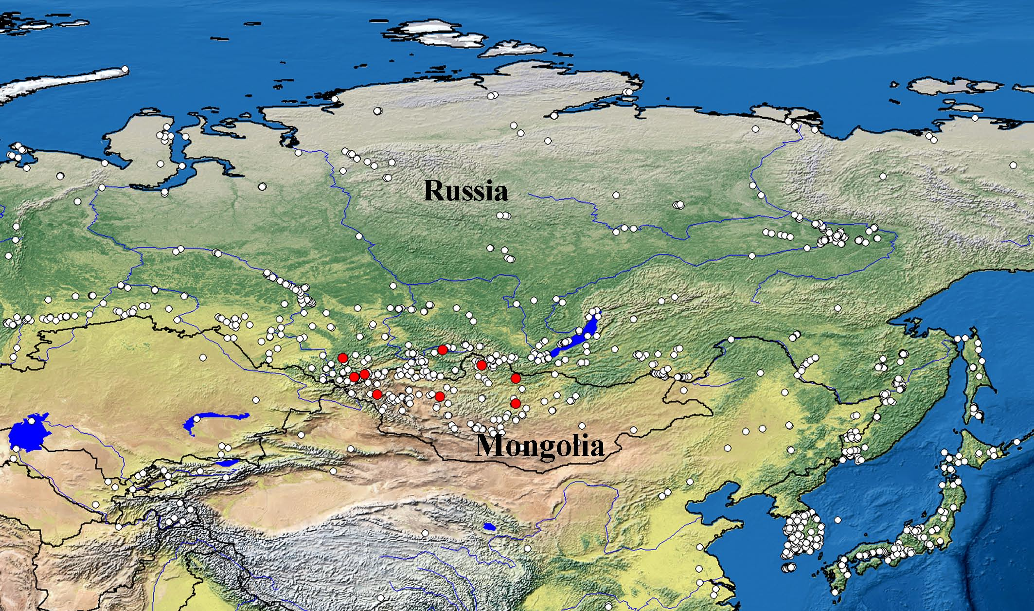
Distribution. D. turbinata has a relatively narrow distribution range as compared to other members of the D. longispina group. According to our samples, it is known from the Altai-Sayan mountain region (exactly, from Eastern Sayan, Altai, Mongolian Altai and Khangai mountains). There is a chance that it will be found in the mountains of some other countries of Middle Asia (first of all, Kazakhstan) and NW China. Records from Baikal region need to be checked, at least, we did not find D. turbinata in our samples from this region ( Fig. 1 View FIGURE 1 ). Unfortunately, previous literature sources are mainly useless for understanding of its distribution range, as the quality of identification is unknown. For example Manujlova (1964: Fig. 36, fragment 21) most probably dealt with other longispina -like taxon instead of D. turbinata . The latter apparently belongs to the mountain endemic faunistic complex according to Kotov (2016).
No known copyright restrictions apply. See Agosti, D., Egloff, W., 2009. Taxonomic information exchange and copyright: the Plazi approach. BMC Research Notes 2009, 2:53 for further explanation.
|
Kingdom |
|
|
Phylum |
|
|
Class |
|
|
Order |
|
|
Family |
|
|
Genus |
Daphnia turbinata Sars, 1903
| Zuykova, Elena I., Sheveleva, Natalia G. & Kotov, Alexey A. 2019 |
Daphnia turbinata Sars in Glagolev 1995: 58
| Ayushsuren, Ch. & Sheveleva, N. G. & Arov, I. V. 2013: 125 |
| Glagolev, S. M. 1995: 58 |