
Metarhabditis costai ( Martins, 1985 ) Sudhaus, 2011
|
publication ID |
https://doi.org/ 10.1080/00222933.2013.798702 |
|
DOI |
https://doi.org/10.5281/zenodo.5197897 |
|
persistent identifier |
https://treatment.plazi.org/id/038A87BE-FFE0-B85C-E512-787BEA60F9EE |
|
treatment provided by |
Felipe |
|
scientific name |
Metarhabditis costai ( Martins, 1985 ) Sudhaus, 2011 |
| status |
|
Metarhabditis costai ( Martins, 1985) Sudhaus, 2011
( Figures 1 View Figure 1 and 2 View Figure 2 )
Measurements
See Table 1.
Description
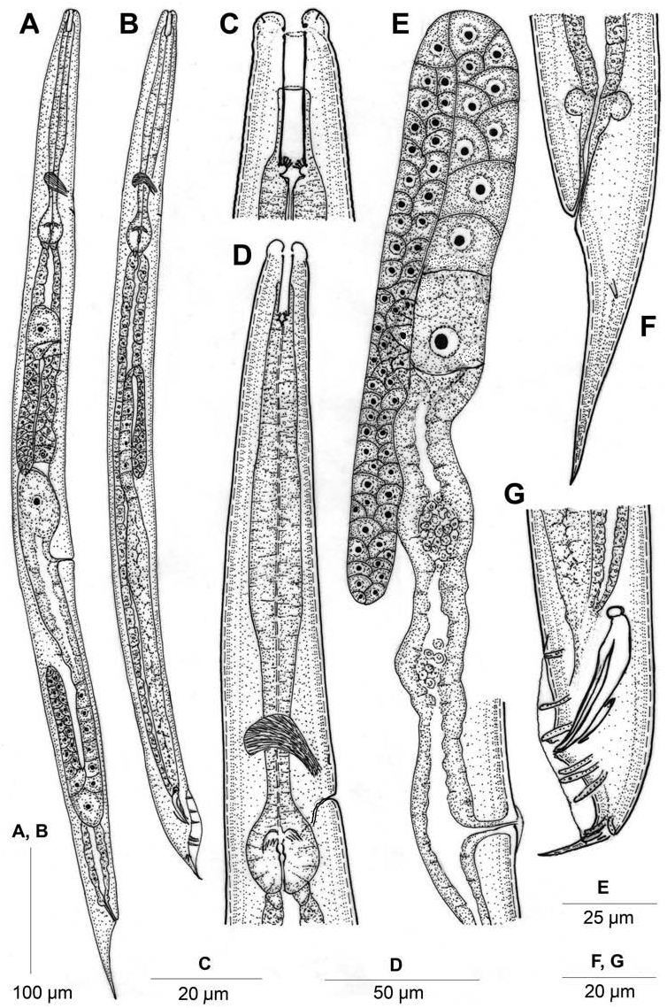
Female. Body straight or slightly arcuate, tapering at both extremities but more toward the posterior end. Cuticle with fine transverse striations. Longitudinal striations faint or inconspicuous. Lateral field with six longitudinal lines. Lip region slightly offset from adjoining body. Lips globular, usually expanded, grouped in doublets forming three sectors (one subdorsal and two subventral sectors) around a triradiate oral aperture. Amphids inconspicuous under light microscope. Stoma rhabditoid type, moderately cuticularized, 1.6–2.4 times the lip region diameter in length. Pharyngeal sleeve surrounding 55–58% of stoma. Cheilostom not cuticularized; gymnostom smaller than stegostom. Metastegostom isomorphic with each plate bearing four or five knobbed, setose denticles. Telostegostom isoglottoid. Pharynx differentiated into cylindrical, 105–135 µm long corpus; isthmus slightly narrower, 44–65 µm long and a round double-chambered, basal bulb of 25–29 × 20–25 µm dimension with a grinder. Isthmus slightly tapering posteriorly towards basal bulb. Nerve ring at 69–77% of pharyngeal length. Secretory–excretory pore slightly anterior to basal bulb, about 76–86% of pharyngeal length with anterior 6–10 µm of duct cuticularized and relatively straight; further posteriorly duct is convoluted towards the secretory cells. Hemizonid observed in most specimens at level of nerve ring. Pharyngeal corpus 1.3–1.6 times longer than postcorpus (isthmus and basal bulb together). Body at pharyngeal end 2.6–3.1 times the lip region diameter in length. Cardia conoid, 4–7 µm long. Intestinal cells with large nuclei, lining of intestinal lumen thick, refractive. Rectum length 1.1–1.5 times anal body diameter, with three rectal glands at its junction with intestine. Prerectal part not conspicuously differentiated. Reproductive system didelphic, amphidelphic. Ovaries dorsally reflexed; anterior ovary right and posterior on left side of intestine. Oviduct proximally connected to offset, ovoid spermatheca containing sperm. One to twelve intrauterine eggs, in different stages of embryonation, dimensions 55–60 × 26–34 µm. Vagina thick-walled, at right angle to longitudinal body axis, length about one-third of vulval body diameter. Vulva a wide transverse slit, covering ca. 80% of corresponding body diameter with epiptygma and distinct cuticular flap. Tail elongate conoid. Phasmids tubular, located about one anal body diameter posterior to anus.
Male. Similar to female in general morphology except in greater posterior curvature of body. Testis single, reflexed ventrally, located on left side of intestine. Vas deferens a broad tube without demarcation of seminal vesicle. Ejaculatory glands absent. Spicules robust, length ca.1.1–1.7 times anal body diameter, having round capitula, ventral triangular process and prominent ventral and dorsal arms. Gubernaculum broad, trough-shaped, 37–45% of spicule length. Tail spicate. Bursa fairly wide, anteriorly open, leptoderan type not enveloping small tail spike. Bursal cuticle usually transversely striated. Genital papillae eight pairs in 1 + 1/1/3 + 2 + P configuration. First (GP1) and second genital papillae (GP2) spaced, precloacal; GP3 adcloacal; out of six postcloacal pair – GP4 , GP5 and GP6 relatively close with GP5 dorsally oriented; GP7 , GP8 closely placed with former pointing dorsally. Phasmids very narrow tubes opening at terminal end of bursa. Copulatory muscles prominent in few specimens showing six pairs of thin bands.
Habitat and locality
Soil samples collected from a field with dumped farmyard manure at Sikandra Rao (Geographic coordinates 27 ◦ 41 l 17.04 ll N, 78 ◦ 23 l 10.83 ll E), Aligarh, India.
Voucher material
Seven females and six males on slide Metarhabditis costai NOSR /2–7 deposited in Nematode Collection, Department of Zoology , Aligarh Muslim University, Aligarh, India . One female and one male on slide Metarhabditis costai NOSR /1 deposited at the Laboratory of Nematology , Wageningen University and Research Centre ( WUR), 6700 ES Wageningen, the Netherlands .
Based on the morphological characteristics of the present population, a revised diagnosis of M. costai is given hereunder.
Emended diagnosis
Metarhabditis costai ( Martins, 1985) Sudhaus, 2011 is characterized by medium-sized oviviviparous females with six lateral lines; slightly offset lip region; globular lips in doublets forming three sectors; each metastegostomal plate bearing setose denticles; distinctly weak corpus followed by a tapering isthmus; one to twelve intrauterine eggs; females with conical tails and males with robust spicules having prominent capitula and prominent dorsal and ventral arms; trough-shaped gubernaculum; open leptoderan bursa with a very small uncovered tail spike and genital papillae in 1 + 1/1/3 + 2 + P configuration.
Remarks
The present population shows conformity to M. costai ( Martins, 1985) Sudhaus, 2011 in most morphological and morphometric characteristics except some minor differences, namely, slenderer individuals with relatively greater ‘a’ value in males (17.0–26.4 versus 14–17) as well as in females (18.2–22.7 versus 13.2–19.8). The present population also has relatively smaller (30–38 µm versus 33–50 µm) yet robust spicules and a smaller gubernaculum (12–16 µm versus 20–24 µm). Although the original population of M. costai was reported from the external auditory meatus of cattle suffering from otitis externa, the location of the present population does not rule out the connection to cattle as the sampling site had dumped farmyard manure.
The present population shows marked differences from the locally existing species M. freitasi ( Martins, 1985) Sudhaus, 2011 in having smaller males (608–1058 µm versus 1035–1405 µm) and females (791–978 µm versus 1253–1714 µm) with smaller ‘b’ values (3.6–5.5; 3.7–4.7 versus 6.7–9.1; 6.5–8.5) in both sexes; females with ovoid (versus elongate) eggs and with a smaller tail (60–93 versus 112–154 µm) ending in a conical (versus fine in M. freitasi apud Martins, 1985 ), attenuated hair-like terminus.
The present population shows marked differences from the locally existing species M. andrassyana in having relatively smaller ‘a’ (18.2–22.7 versus 20–26) and ‘ cl’ (2.8–5.1 versus 5–7) values; greater ‘c’ (9.3–13.6 versus 6.5–8.1) and ‘V’ (52.2–56.3 versus 48–52) values; lip region with expanded (versus not expanded) lips; large-sized eggs (55–60 × 26–34 µm versus 26–30 × 19–24 µm) and conoid tail (versus long attenuated in M. andrassyana apud Tahseen et al., 2004 ).
The present population also differs markedly from another species M. amsactae Ali et al., 2011 , reported from India, in having fewer lateral lines (6 versus 10–11); a longer pharyngeal collar (50–58% versus 30% of stoma length) and males with greater ‘c’ (20.7–33.2 versus 8.9–17.8); smaller ‘ cl’ (1.0–1.8 versus ∼ 2.5–3.5); a relatively wide and developed (versus narrow) bursa and a very small tail spike (versus large uncovered in M. amsactae apud Ali et al., 2011 ).

No known copyright restrictions apply. See Agosti, D., Egloff, W., 2009. Taxonomic information exchange and copyright: the Plazi approach. BMC Research Notes 2009, 2:53 for further explanation.
|
Kingdom |
|
|
Phylum |
|
|
Class |
|
|
Order |
|
|
Family |
|
|
Genus |